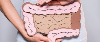
The large intestine is the final part of the digestive tract. In this section of the intestine, water is absorbed and feces are formed, which will then be excreted from the body. In addition, the large intestine is responsible for absorbing vitamins, amino acids, glucose, and electrolytes from food. This part of the digestive tract is susceptible to a variety of diseases - the development of tumors, inflammatory processes, impaired absorption of nutrients and peristalsis (motor and contractile activity of muscle tissue in the walls of the intestine, promoting the movement of its contents). Why are such pathologies dangerous? They directly affect the general condition of a person, and some of them (this is more true for oncology) are extremely dangerous to life and health if they are not identified in time and treatment is not started. One of the methods for diagnosing diseases of this part of the gastrointestinal tract is radiography of the large intestine.
How does the human large intestine work, why is it examined?
The main stages of digestion associated with the primary processing and digestion of incoming food occur in the stomach and small intestine. However, after the food bolus passes into the large intestine, the process of its processing does not end - it is the large intestine that is responsible for the further entry of remaining beneficial substances into the blood from processed food.
Content:
- How does the human large intestine work, why is it examined?
- What diseases can affect the large intestine?
- X-ray of the large intestine: irrigoscopy and irrigography
- Indications and contraindications for irrigoscopy
- How to properly prepare for a colon x-ray
- Irrigoscopy for adults and children: technique
- Possible risks and consequences of the procedure
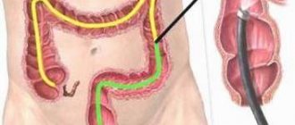
Anatomically, the department is represented by the cecum, colon, sigmoid and rectum. The colon, in turn, has three subsections - ascending, transverse and descending. The rectum has a wider part - the so-called ampulla of the rectum, and a tapering section located closer to the anus - the anus. Visually, the large intestine looks like a loop, the shape of which is similar to a square with an open contour. On average, its diameter reaches 6-6.5 centimeters, and its length is about two meters.
The large intestine contains a wide range of different beneficial bacteria. This specific microbiota contributes to the further processing of the food bolus and its transformation into feces. The beneficial nutrients that remained in the contents of the food bolus during its stay in the thick section are processed and converted by bacteria into vitamins, sugars and amino acids. They are then absorbed by the mucous membrane of the intestinal wall and enter the blood.
The remains of undigested food accumulate in the large intestine and leave the body during bowel movements.
The large intestine, as part of a well-functioning mechanism, performs its functions in conjunction with the rest of the digestive tract, so in the body of a healthy person, food passes a continuous path from the mouth to the rectum, through which unprocessed mass is excreted.
With pathological changes in the large intestine, the patient complains of characteristic symptoms, deterioration in health, weight loss, abdominal pain and other ailments. In such cases, it is necessary to prescribe diagnostic procedures, in particular, x-rays of the large intestine.
What should you expect during and after the study?
As the intestines fill with barium, the need to defecate arises. There is a feeling of pressure in the abdominal cavity or even slight cramps.
Most patients easily tolerate such discomfort. The tip of the tube, which remains in the rectum, helps the patient retain barium in the intestines. Any difficulties should be reported to the radiologist immediately.
During the examination, the doctor asks the patient to turn from side to side and change body position several times. Periodically, external pressure is applied to the patient's abdomen. If the barium enema is accompanied by air contrast of the intestines, the patient's table is moved to a vertical position.
After the study, the patient is prescribed a laxative or a cleansing enema, which allows the barium to be completely removed from the intestines. If there are no contraindications from the doctor, you can return to your usual diet and medication.
Immediately after the study, you are allowed to return to normal nutrition and normal activities. Within 24 hours after a barium x-ray, it is recommended to maintain an extended drinking regime.
For the next few days, the stool may be white, which is due to the removal of barium from the body. Some patients experience constipation after a barium enema.
If within two days after the study there is no independent stool or gas, then you must urgently contact your doctor. In this case, an additional cleansing enema or laxative may be required to remove the barium.
Up
What diseases can affect the large intestine?
Most often, patients with problems with the large intestine find:
- ulcerative colitis;
- colon cancer and polyps;
- Crohn's disease;
- ischemic and pseudomembranous colitis;
- irritable bowel syndrome;
- wall diverticula;
- congenital anomalies.
Ulcerative colitis
Pathology is a chronic condition of an organ when its internal mucous membrane becomes swollen, inflamed, changes its color from normal beige-pink to bright red, and its integrity is compromised due to small ulcerative formations. These ulcers do not heal well and bleed. If the disease lasts for a long time and without adequate treatment, it can provoke the appearance of polyps and neoplasms.
Polyps, neoplasms
Tumors in the large intestine are quite common in patients. In the last decade, in Europe and the USA, colon cancer (colorectal cancer) has taken first place among malignant tumors of the digestive tract, which accounts for more than half of all cases of cancer of the digestive organs. Due to the aging of the world's population, the situation is expected to worsen in the future. Colon cancer is a dangerous disease that requires serious comprehensive treatment. Polyps and tumors in this part of the body are more dangerous because their development is practically asymptomatic, and in the early stages they can only be detected by accident.
Crohn's disease
The disease is characterized by a general inflammatory process in the digestive tract, which covers the inner lining of the walls, as well as the middle and outer lining, as well as the adjacent lymphatic vessels of the intestine. The disease is difficult to diagnose, since its symptoms are similar to a dozen other pathologies, and its complications are extraintestinal in nature - damage to large joints, the appearance of ulcers in the oral cavity, vision problems.
Ischemic and pseudomembranous colitis
In ischemic colitis, the degenerative process affects the vascular system that supplies the intestinal walls. Ischemia is accompanied by local inflammation and ulceration of the mucous layer of the wall, and the gradual development of intestinal obstruction.
Pseudomembranous colitis develops due to a significant proliferation of one of the types of intestinal bacteria - clostridia. Typically, the intestinal microbiome performs self-regulation - each type of bacteria controls the growth and development of all others. With long-term use of antibiotics, laxatives or cytostatics, some bacterial species are destroyed, and, accordingly, others increase in proliferation.
Clostridia, in the process of their life activity, produce toxins that negatively affect the intestinal lining, which is why fibrous plaques called pseudomembranes form on them.
Irritable bowel syndrome
The main cause of IBS is considered to be impaired intestinal motility, but the disease can also be secondary, that is, it can occur against the background of other diseases of the gastrointestinal tract. There is a constant feeling of discomfort in the intestines, the localization and symptoms of which are quite difficult to accurately determine.
Diverticula and diverticulitis
Diverticula in the walls of the large intestine look like pouches. In essence, they represent a stretching of the wall, which creates a “pocket” that protrudes towards the abdominal cavity. The process without complications can, in the worst case, cause constipation or a feeling of heaviness in the abdomen. However, if intestinal contents stagnate in the cavity, against the background of the presence of dysbiosis in the intestines, diverticulitis can develop - acute inflammation with characteristic, dangerous symptoms.
Congenital structural disorders
Such disorders include elongation of the sigmoid colon (dolichosigma), as well as hypertrophy of the colon in any of the departments, or in a specific segment. Pathologies are characterized by disorders of intestinal processes, constipation, and flatulence. In severe cases, intoxication with stagnant feces may develop.
X-ray of the small intestine
The small intestine is the part of the intestine that is located between the large intestine and the stomach. Its function is difficult to overestimate: it carries out a significant amount of food processing processes, ensuring the absorption of nutrients. An X-ray of the small intestine makes it possible to determine the state of this anatomical formation and identify a number of disorders characteristic of it. The procedure can be performed using contrast and has a high diagnostic value. It is a relatively safe non-invasive diagnostic method that has become widespread due to its simplicity and accessibility.
You can undergo diagnostics of the small intestine at the CELT multidisciplinary clinic. Our specialists will help identify the cause of the ailment, determine the localization of the pathological focus and provide all the data necessary to the attending physician.
Types of X-ray of the small intestine
Modern radiology offers different techniques for performing radiography. Their choice is made on an individual basis, taking into account the clinical picture and patient characteristics.
Aimed at linear visualization of the small intestine on film or digital media. It is highly accurate and informative.
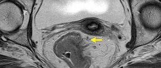
X-ray of the large intestine: irrigoscopy and irrigography
X-ray examination of the intestine is widely used as a method for diagnosing its condition. Since the organ being examined is hollow, the procedure performed can only be informative using a contrast agent.
Performing the irrigography procedure involves fixing the image obtained by X-ray irradiation on a special film, while with irrigoscopy the image is displayed on the monitor screen and examination of the large intestine in real time. There are no differences in terms of technique or methods of preparation.
Irrigoscopy itself can be of two types - it depends on the contrasting technique:
- classical irrigoscopy involves the introduction of a contrast agent in a liquid state;
- irrigoscopy with double contrast: in this case, the patient is first injected with liquid contrast that envelops the intestinal walls, after which gas or air is gradually introduced into the intestinal cavity.
Who reviews the research results and where can they be obtained?
The images are analyzed by a radiologist: a doctor who specializes in performing x-ray examinations and interpreting their results. After examining the images, the radiologist draws up and signs a report, which is sent to the attending physician. In some cases, the report can be collected from the radiology department itself. The results of the study should be discussed with your doctor.
A follow-up x-ray examination is often required, the exact reason for which will be explained to the patient by the attending physician. In some cases, additional examination is carried out when doubtful results are obtained that require clarification during repeated images or the use of special imaging techniques. Dynamic observation allows timely identification of any pathological abnormalities that arise over time. In some situations, repeated examination allows us to talk about the effectiveness of treatment or stabilization of tissue condition over time.
Up
Indications and contraindications for irrigoscopy
The list of reasons why a doctor may send a patient for examination of the condition of the large intestine includes characteristic pathologies affecting this section of the gastrointestinal tract. Typically, patients visit a doctor with typical symptoms:
- bowel dysfunction (disorder or constipation, if long-term);
- flatulence;
- bleeding from the anus;
- pain, a feeling of heaviness in the abdomen;
- the presence of blood in the stool.
However, the symptoms themselves are not grounds for prescribing irrigoscopy. The doctor interviews and examines the patient, prescribes the necessary tests and ultrasound of the abdominal organs. Based on the results obtained, a decision is made on the need for contrast radiography of the large intestine.
The indication for prescribing a procedure, most often, is a suspicion of the presence of a pathology or condition, if the results of preliminary tests and examinations do not allow an accurate diagnosis and determination of the etiology of the process. The doctor refers the patient for radiography, suspecting:
- the presence of tumors and polyps;
- gastric and intestinal bleeding with a tendency to recur;
- Crohn's disease;
- intestinal obstruction of any nature;
- diverticulitis;
- dolichosigma;
- Hirschsprung's disease (congenital anomaly);
- presence of foreign bodies;
- ulcerative, ischemic, pseudomembranous colitis;
- various inflammatory processes in the intestines.
Pain syndromes in the anus, anus, abdominal cavity, excessive organ mobility, prolonged and painful constipation, and systematic intestinal disorders are also indications for irrigography.
In what cases should a patient not undergo the procedure? Doctors list the following factors as contraindications to irrigography:
- rapidly progressing ulcerative colitis;
- presence of pregnancy;
- toxic megacolon;
- severe colitis;
- presence of allergies or intolerance to contrast agents;
- cardiovascular diseases, hypertension;
- early postoperative period, if the intervention took place in the abdominal cavity.
In what areas is radiography of the lower gastrointestinal tract used?
X-ray of the lower gastrointestinal tract is used in the diagnosis of the following conditions:
- Benign tumors such as polyps.
- Malignant tumors.
- Other intestinal diseases.
The test is often used to discover the causes of the following symptoms:
- Chronic diarrhea
- Blood in the stool
- Constipation
- Irritable bowel syndrome
- Unexplained weight loss
- Changing the stool
- Suspicion of chronic blood loss
- Stomach ache
X-rays of the small and large intestines are also used to diagnose inflammatory bowel disease, a group of diseases that includes Crohn's disease and ulcerative colitis.
Up
How to properly prepare for a colon x-ray
The procedure requires special preparation to obtain diagnostically significant results, which are influenced by the presence of feces in the intestine, when the contrast agent cannot be fully distributed throughout the organ.
Therefore, before the procedure, the patient should take care of cleansing the large intestine.
Preparation includes compliance with dietary restrictions, mechanical and medicinal cleansing measures. Two to three days before the scheduled date of the x-ray, you are prohibited from consuming:
- raw vegetables: beets, carrots, cabbage;
- millet, pearl barley and oatmeal;
- apples;
- apricots;
- black bread;
- peas and other legumes;
- alcohol and carbonated drinks;
- fried or grilled foods.
On the eve of the procedure, before going to bed, you should do a cleansing enema and take a laxative recommended by your doctor. Be sure to do an enema on the morning of the procedure.
When is an intestinal x-ray prescribed?
An x-ray is done to look at the large intestine or appendix.
An X-ray with a barium solution is prescribed when there is a need to conduct a detailed examination of the large intestine and the part of the small intestine that communicates with it, as well as the appendix.
It is this procedure that allows you to make the most accurate diagnosis and evaluate the effectiveness of pre-prescribed treatment.
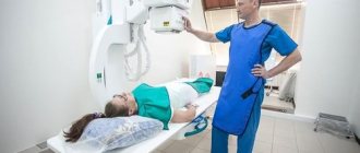
Irrigoscopy for adults and children: technique
The diagnostic procedure can only be carried out in a medical institution, in some cases with the patient admitted to a hospital. The doctor makes every effort to ensure that the examination is carried out as quickly and safely as possible for the patient.
Initially, the radiologist needs to take several survey images, while the patient must alternately take a lying and standing position.
The patient is placed on the couch in the left lateral position. A contrast agent is injected through the anus and rectum using a special contrast enema.
For this purpose, two types of contrast agents are used:
- a preparation with barium, in which the main substance is barium sulfate, and the auxiliary substances are tannin, sodium citrate, gelatin or cellulose;
- sodium amidotrizoate is part of Urografin or Verografin.
The second type of drugs is mainly used to examine newborns, or for patients of any age when perforation of the intestinal wall is suspected.
The procedure is performed in several phases. First, a phase of weak filling, when the contrast gradually envelops all the folds of the mucous membrane, visualizing them well. This is followed by a tight filling phase - during which the intestine is completely filled with contrast, which makes it possible to assess the thickness of the lumen, shape, location, contours, the presence of foreign bodies, places of narrowing and expansion. Based on how quickly the contrast is eliminated by the intestines, the doctor can draw conclusions about the elasticity of the walls and intestinal motility.
For the study you will need about 500 milliliters of solution. If you are having a double contrast procedure, slightly less fluid will be needed. Gas or air is introduced into the cavity to more clearly visualize the thickness of the walls and folds of the mucous membrane, and the organ as a whole. This procedure is called the double contrast phase.
After the injection of the substance, when the rectum and sigmoid colon are filled with contrast, the patient is placed on his back and then on his right side and a series of photographs are taken at different phases of filling. Next, if necessary, air is introduced into the intestine and a number of images are taken, this time with double contrast.
The patient then has a bowel movement and after all the contrast has left the bowel, the doctor takes a final image of the abdomen without contrast.
Best materials of the month
- Coronaviruses: SARS-CoV-2 (COVID-19)
- Antibiotics for the prevention and treatment of COVID-19: how effective are they?
- The most common "office" diseases
- Does vodka kill coronavirus?
- How to stay alive on our roads?
The irrigoscopy procedure for children is no different from that for adult patients. Whenever possible, they try to prescribe alternative methods of examination for children, since preparation requires significant time and effort, and the procedure itself is not pleasant, although it is painless. Children of primary school age may be prescribed irrigography under general anesthesia.
When performing irrigography, the patient may experience some difficulties. If preparation is poor, there may be stool in the intestines, visible in the photographs. If the sphincter is weak, the patient will experience contrast incontinence. In addition, the contrast agent may be unevenly distributed throughout the large intestine, making it difficult to analyze the resulting images.
The danger for the patient is the situation of perforation of the pathologically altered intestinal wall due to the administration of contrast. In each specific case, the radiologist decides whether or not to continue the procedure. If necessary, the doctor stops the examination at any stage and begins providing emergency medical care to the subject.
When is fluoroscopy prescribed?
The decision to perform fluoroscopy is made by the doctor. Typically, the procedure is prescribed when a person experiences the following signs of gastrointestinal distress:
- chronic constipation,
- persistent diarrhea,
- significant weight loss over a relatively short time with the usual diet,
- change in color of stool to black in the same situation,
- vomiting blood,
- constant pain after eating or defecating.
Indications for fluoroscopy include a number of gastrointestinal diseases, including cancer.
Human digestive system
The use of x-rays allows us to evaluate the anatomical changes in the intestine caused by pathologies or age (x-ray anatomy).
Benefits and risks of lower gastrointestinal radiography
Advantages:
- X-ray examination of the lower gastrointestinal tract is a minimally invasive procedure with rare complications.
- X-rays of the lower gastrointestinal tract often avoid more invasive procedures such as colonoscopy.
- Allergic reactions accompany the study extremely rarely, since barium is not absorbed into the blood.
- After completion of the examination, no radiation remains in the patient’s body.
- When used for diagnostic purposes, X-rays do not cause any side effects.
Risks:
- With excessive exposure to X-ray radiation on the body, there is always an extremely small risk of developing malignant tumors. However, the benefits of accurate diagnosis significantly outweigh this risk.
- The effective dose of radiation is different for all patients.
- In rare cases, barium can leak through undetected holes in the intestinal wall, causing inflammation in the surrounding tissue.
- Even more rarely, barium can cause gastrointestinal obstruction.
- With rectal administration of iodine-containing contrast material, allergic reactions are possible, but extremely rare.
- A woman should always tell her doctor or radiologist about the possibility of pregnancy.
A few words about reducing the effects of radiation on the body
During an x-ray examination, the doctor takes special measures to minimize radiation exposure to the body while trying to obtain the best quality image. Experts from international radiological safety councils regularly review radiology standards and produce new technical recommendations for radiologists.
State-of-the-art X-ray machines allow you to control the dose of X-ray radiation and provide filtration, which minimizes beam scattering. In this case, the patient’s organs and systems that are not examined receive a minimal dose of radiation.
Up