Duration
180 minutes
On average, a contrast study takes 2-3 hours.
Soreness
No pain
X-ray of barium passage through the small intestine is a non-invasive, painless procedure.
Anesthesia
No anesthesia required
Anesthesia is not needed, because
the procedure is not accompanied by painful sensations. Additional factors
Use of contrast agent
The contrast agent allows the radiologist to evaluate the functioning of the intestine, its motor-evacuation function, and the rate of removal of the contrast.
Preparation
Preparation required
Preparation consists of a diet that helps reduce gas formation and cleansing the intestines.
Rehabilitation
Not required
Because
The procedure is non-invasive and does not require a recovery period. Work ability
Saved
To restore energy balance after the procedure, you need to eat immediately.
However, the ability to work remains. Sports activities
No restrictions
You can maintain your usual rhythm of life and exercise if you do not feel weak.
The small intestine is located immediately after the stomach and is connected to organs such as the liver, gallbladder and pancreas. The small intestine produces a large number of enzymes, so the process of digesting food is completed here. It is also responsible for such an important process as the absorption of digested food products into the lymphatic and blood capillaries.
Contact a specialist if you are concerned about the characteristic symptoms of intestinal diseases: vomiting blood, blood in the stool, diarrhea or constipation, causeless weight loss; difficulty swallowing, constant heartburn, incessant pain in the epigastric region. A referral for an x-ray of barium passage through the small intestine is usually issued by a general practitioner, oncologist or gastroenterologist.
The small intestine is difficult to access for endoscopic examination methods, and therefore X-ray examination with contrast plays a leading role in the diagnosis of diseases of this organ.
In order to assess the condition of the small intestine, its motility, the health of its walls, the presence of narrowings or dilations, pathologies, tumors, inflammatory processes and various diseases, the attending physician prescribes special diagnostic measures to the patient, one of which is an x-ray of the small intestine .
The word "passage" means the passage of contrast. The contrast agent allows the radiologist to evaluate the functioning of the intestine, its motor-evacuation function, and the rate of removal of the contrast.
Indications and contraindications for the study
Using intestinal radiography, the following are diagnosed:
- polyps;
- ulcers;
- erosion;
- scarring;
- diverticula (protrusion of the walls of hollow organs);
- tumors (both benign and malignant);
- Crohn's disease;
- interintestinal abscess;
- IBS (irritable bowel syndrome);
- enteritis;
- internal abdominal hernia (a rare pathology that can cause acute intestinal obstruction).
As you can see, the study helps to identify various pathologies.
Contraindications to radiography:
- rapidly developing ulcerative colitis;
- the period immediately after taking intestinal tissue for biopsy - until the walls heal;
- intestinal obstruction;
- heart, kidney and liver failure;
- intense and severe abdominal pain.
Radiography of internal organs without contrast is not characterized by high information content, because parenchymal (almost all abdominal organs are composed of functional tissue called parenchyma) and air tissues do not reflect radiation. Because hollow organs are examined, it is necessary to use a contrast agent to identify pathologies.
Intestinal obstruction is characterized by a violation of the passage (patency) of intestinal contents. The study allows you to use the example of a passage of contrast agent to see if you have such a pathology.
The barium passage technique is highly informative for studying the small intestine and allows you to evaluate its structure and peristalsis (motor activity). This part of the gastrointestinal tract has many anatomical formations, bends, and narrowings. Therefore, it is almost impossible to do without using contrast here.
Indications for the procedure
A referral for a barium X-ray of the intestines is issued by an oncologist or gastroenterologist; in some cases, a general practitioner can refer you for the procedure. An examination is prescribed in cases where the doctor suspects a disease that poses a threat. Therefore, a referral for an X-ray of the intestines with barium should be taken with particular seriousness.
There is a whole range of signs of dangerous diseases of the digestive tract, in which a specialist prescribes an examination of the intestine with a barium-based contrast agent using radiography.
Serious weight loss for no significant reason
Severe weight loss without lifestyle or diet changes clearly indicates pathology. The patient often experiences lethargy and apathy. The source of the symptom may be a disruption of the intestines. To find out the cause, your doctor may order a barium X-ray of your small or large intestine.
The occurrence of loose stools for no reason
Loose stools for no apparent reason indicate a disruption in the digestive process. With long-term problems, it can cause intestinal damage. Therefore, the symptom requires prompt diagnosis and proper treatment.
Change in stool color
Loose, black stools are also called “tarry” stools. The discharge resembles a black paste with a pungent, unpleasant odor. The symptom indicates bleeding in the initial part of the duodenum or in the small intestine. If the bleeding is heavy, the patient may show signs of blood loss.
These conditions are extremely dangerous. Therefore, at the first appearance of tarry stools, it is better to immediately contact a specialist and undergo an intestinal examination with barium as prescribed; X-rays will show the source of bleeding and help the doctor make a diagnosis.
The appearance of severe pain in the abdominal area
Pain in the abdominal area can be caused by intestinal deformation or dysfunction. Often pain in the abdominal area indicates an ulcer. To determine the source of pain, various procedures are prescribed to examine the intestines, including the use of barium during x-rays.
Serious pathologies (emerging tumor processes)
Various neoplasms can interfere with normal intestinal motility, leading to pain, impaired defecation, loss of appetite, etc. To localize the neoplasm and establish its nature, the patient is prescribed an intestinal X-ray; the use of barium in an X-ray helps to obtain a contrast and informative image.
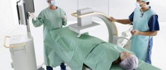
How do you do a barium x-ray of your intestines? Preparation and indications for the procedure
An intestinal X-ray is a diagnostic procedure that allows you to identify the pathology of this organ by irradiating it with X-rays. An X-ray image can be displayed either on a screen, which allows you to monitor the procedure immediately at the time of its implementation, or printed on a special film and then studied by a radiologist.
- Why is radiographic examination performed?
- Preparation for the procedure
- Performing fluoroscopy
- What does an intestinal x-ray show?
- Contraindications
- Price
Why is radiographic examination performed?
X-rays may be taken with or without contrast. Barium is used as a contrast agent for X-rays. It is not harmful to the body, is not absorbed in the intestines and is easy to remove from the body. An X-ray of the large intestine with a contrast agent is called irrigoscopy.
An X-ray can only be prescribed by a doctor, most often a highly specialized specialist - a gastroenterologist, oncologist.
X-ray diagnostics are performed for:
- frequent constipation or diarrhea of unknown etiology;
- suspected Crohn's disease, diverticulitis, peptic ulcer;
- suspected adhesions, formation of fistulas, polyps;
- chronic inflammatory process in the intestines;
- ingestion of solid non-edible objects into the gastrointestinal tract (stomach, and then intestines) - a frequent indication for x-rays for children;
- suspected oncological tumors;
- patient complaints of abdominal pain, bloating, indigestion, changes in the consistency and color of stool;
- examination of a patient with congenital intestinal anomalies.
Preparation for the procedure
X-ray of the intestines requires careful preparation for the examination, and if everyone is accustomed to the fact that an X-ray of an arm or leg can be done at any time, then with the organs of the gastrointestinal tract things are more complicated. The intestine is a hollow organ; in order to examine it, it is necessary that there is nothing in the small and large intestines, not even food. Preparation for x-rays begins two to three days before the expected examination.
What does an x-ray show?
Radiography, which uses a contrast agent, is considered one of the most informative methods for studying internal organs.
Intestinal shape
Using radiography, a specialist visualizes the shape of the intestine, its appearance and motor functions. Also, barium X-rays of the small intestine and colon are often used because they show areas of narrowing and obstruction.
Location and size of the lumen
Based on how barium fills the lumen of the colon during an examination of the intestines and how it looks on an x-ray, a specialist can diagnose the pathology and prescribe treatment. For example, if the contrast is deposited in flakes, the absorption function of the intestine is impaired; if barium is unevenly distributed on the x-ray, this is a clear sign of a neoplasm.
Elasticity degree
When filling the intestines with a barium-based contrast agent, specialists can use x-rays to evaluate the elasticity of the tissues and their ability to stretch.
How is an X-ray of the small intestine done?
X-rays of the small intestine will help diagnose diseases.
The x-ray will take about half an hour. The patient will need to drink half a liter of solution, which will resemble milk in appearance and taste like lime.
When it is necessary to carry out double contrast, barium must be drunk using a tube that goes from the machine, which supplies air a little at a time.
After this, after 5 hours, you can start x-raying, since during this time the contrast will enter the small intestine. While the solution passes through the small intestine, it is necessary to take about 8 x-rays in different positions. This happens every 45 minutes.
If the solution is not distributed evenly throughout the intestines, then the doctor, by palpating the abdominal wall, will be able to distribute the substance evenly. The procedure will come to an end when the substance completely fills the space between the small and large intestines - this is the ileocecal angle, and the solution fills the cecum.
Contraindications
Methods for examining the intestines using barium, including x-rays, have a number of contraindications. One part of them is dictated by the impact of ionizing radiation, the other – by the contrast agent.
Pregnancy
Ionizing radiation and barium can have a negative effect on the development of the fetus. Therefore, during the period of bearing a child, it is impossible to conduct an examination of the intestines using barium, especially using X-rays.
Intestinal perforation
Perforation (through damage) of the intestine makes it impossible to use barium, as it can enter the abdominal cavity. If there is a suspicion of such damage to the large or small intestine, contrast is not used during the examination.
The patient is in a serious or even unconscious state
When the patient is in serious condition, especially with acute inflammatory processes, the contrast can be harmful to his health. Therefore, examination of the intestines using barium using radiography in a pre-fainting state or in acute inflammatory processes is not carried out.
Ulcerative colitis
With ulcerative colitis, inflamed areas appear on the intestinal mucosa, which makes the use of barium in X-rays impossible. In this case, conventional radiography is prescribed.
Tachycardia
If barium is used to examine the intestines, various complications are possible that will lead to stress on the heart, so the use of contrast when X-raying a patient with tachycardia is not recommended.
X-ray of the stomach and intestines with barium
The X-ray method allows you to see the condition of human organs without surgical intervention. The essence of the method is simple - a beam of X-rays passes through the tissues of the body and leaves a negative image on a light-sensitive screen or special film. The quality of the image depends on the ability of the area being examined to transmit x-rays. Examination of hollow organs, such as the stomach and intestines, is difficult due to the fact that X-rays pass through them unimpeded, leaving no visual imprint. An element such as barium, or rather one of its compounds, came to the rescue.
Barium sulfate is a contrast agent that absorbs X-rays well. It is not dangerous for the human body, since it does not dissolve in the acidic environment of the stomach and, accordingly, is not absorbed into the blood. The contrast agent is taken orally and examined as it passes through the gastrointestinal tract.
Important!
The reliability of the results depends on the correct preparation for the procedure. To ensure a comprehensive examination, the digestive tract is cleared of large volumes of feces, strictly following the prescribed diet.
You should follow a slag-free diet for two days before the test. Fatty and fried foods, coffee, fresh vegetables and fruits, as well as foods that promote gas formation (rye bread, legumes, baked goods, pearl barley and oatmeal porridge, milk, etc.) are excluded from the diet. Instead of fresh bread, you can eat Lenten cookies and crackers made from wheat buns.
It is recommended to drink plenty of fluids (up to 2 liters per day, in the absence of contraindications). Drinks such as fermented milk drinks, dried fruit compote, and weak green or herbal tea are allowed.
The day before the test, breakfast and lunch should be light. The last meal is the day before at 19:00, after which you are allowed to drink weak tea and water.
Radiography of the barium passage must be carried out! on an empty stomach, in the morning before the procedure it is forbidden to have breakfast.
To conduct a study, the doctor must first study existing information about your disease.
We ask you to bring your medical history or outpatient card with you to the study.
If there is no data on the course of the disease in the medical history or outpatient card, this information can be provided in any form - in extracts, research forms, copies of medical documents. The more the doctor knows about the patient's situation, the more effective the diagnosis will be.
You must also bring with you dry and wet wipes (for hygiene after bowel movements) and a clean sheet. You can ask for a sheet in advance from the nurses in the department where you are being treated.
What is barium passage
Barium passage (barium passage x-ray) is an x-ray examination of the small intestine, which allows you to see the general condition and motor-evacuation function of the small intestine using x-rays. This part of the abdominal cavity (small intestine) has many anatomical formations, bends, anatomical narrowings, areas of impaired motility and secretory activity, which can be visualized precisely with this study. Most often used to identify pathologies such as intestinal narrowing, obstruction or tumors.
Indications for barium passage radiography
- prolonged constipation or diarrhea
- suspicion of intestinal obstruction
Contraindications to barium passage radiography
- pregnancy
- massive intestinal bleeding
Features of colonoscopy
The procedure is carried out using an endoscopic device - a colonoscope. The main feature is the need for careful preparation, including a special diet and taking medications that help cleanse the intestinal mucosa. Preparation lasts at least 2 days.
The study reveals:
- Oncological diseases.
- Sources of intestinal bleeding.
- Assess intestinal motility.
- Diagnose inflammatory processes in the intestine.
Which technique is preferable?
What's better
: X-ray examination of the intestines or colonoscopy - the doctor decides depending on the individual characteristics of the patient. Irrigoscopy with barium is often preferable, as it does not create discomfort for the patient.
But often it is impossible to do without endoscopic examination. This technique is the “gold standard” for diagnosing colon cancer; it allows detection of tumors at the earliest stages of development. X-ray methods do not provide such accuracy; their resolution is much lower.