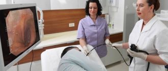
MRI of the abdominal organs in our clinic is performed using a modern tomograph. The procedure is performed under the supervision of qualified doctors. At the same time, many patients are interested in whether it is possible to do an MRI of the intestine instead of a colonoscopy. Non-invasive scanning in such cases is quite informative. Modern medical fiberscopes contain optical fiber, which gives the device the required flexibility. Moving forward, the probe captures and transmits the image to a computer screen. Using a fiberscope, you can obtain a piece of tissue for subsequent examination.
Intestinal anatomy
The hollow organ of the digestive system is an elastic tube, the walls of which have a complex structure.
There are 4 intestinal linings:
- mucous;
- muscular;
- serous;
- submucosa.
The structure of the inner layer depends on the department and the functions performed. The mucous membrane consists of:
- epithelium;
- own record;
- muscle part.
In the small intestine, there are villi on the inner surface of the wall that increase the area for absorption of nutrients. The underlying sections have epithelial folds (crypts).
The submucosal layer contains:
- blood vessels;
- nerves;
- digestive glands.
The muscular layer consists of circular and longitudinal fibers. Part of the peritoneum forms a serous layer, where processes with a high content of fatty tissue (omentums) are located.
The section of the intestine that opens in the initial part with the pylorus of the stomach and ends with the appendix is called thin. The main function is the digestion of food bolus with the participation of specific enzymes and the absorption of nutrients. Intestines are distinguished:
- duodenum;
- skinny;
- ileum.
Due to the structural features, active transport of nutrients occurs here through the penetration of molecules through the membranes of mucosal cells.
The part of the digestive tube that begins at the ileocecal angle and ends at the anus is called the large intestine. The inner layer is responsible for the absorption of water and dissolved minerals, the muscular layer ensures the formation and movement of feces towards the anus. The mucous membrane is populated by beneficial microorganisms involved in digestion.
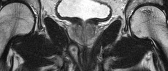
Malignant neoplasm of the rectum (indicated by an arrow) on an MRI image
The digestive system is represented by the following sections:
- cecum;
- ascending, transverse, descending and sigmoid colons;
- rectal section.
The rectum forms the internal and external muscular sphincters of the anus.
The length of the thin and thick sections in a tonic state is about 4 meters. The intestinal loops are quite mobile, which is associated with peristalsis of the muscle layer.
Do they do MRI of the rectum?
Resonance imaging is not a priority type for studying the pathologies of any organs filled with gas. More often, doctors prescribe a CT scan, but if there are contraindications to the use of x-rays, an MRI of the rectum can be done.
Using tomographic images, the condition of the walls of the distal part of the large intestine (mucosal and muscular layers), lumen, and patency of the anal canal are visualized.
Distal intestine on tomograms
Enhanced MRI shows a clearer picture of the location of blood vessels. Malignant tumors have a developed capillary network with a large number of anastomoses (connections), so they accumulate contrast more slowly.
MRI is characterized by a high degree of visualization of rectal tissue and surrounding structures. The method allows you to assess the condition of the peritoneum, fiber, and pelvic organs. Based on the results of MRI, the doctor draws conclusions about the extent of damage to the rectal intestine and the spread of the pathological process.
Intestinal diseases
Pathological conditions of the gastrointestinal tract are accompanied by a deficiency of nutrients, which affects the vital organs and systems of the human body.
Intestinal diseases are classified depending on the etiology, location and nature of the lesion. The most common are:
- inflammatory changes in the mucous membrane (enteritis, colitis, Crohn's disease, etc.);
- oncological processes (benign and malignant neoplasms);
- erosions, gastric and duodenal ulcers;
- parasitic diseases (helminthiasis, amoebiasis, etc.);
- irritable bowel syndrome (IBS);
- congenital pathologies (diverticula, stenoses);
- innervation disorders (Hirschsprung's disease, idiopathic megacolon);
- intestinal obstruction (mechanical and dynamic);
- vascular pathologies (hemorrhoids, Behcet's disease, mesenteric ischemia, etc.);
- traumatic injuries.
One of the common complications of intestinal diseases is the development of adhesions. Pathological changes are localized at the site of damage to the peritoneal epithelium. Adhesions are overgrown connective tissue strands that disrupt the functioning of the digestive system.
Crohn's disease on magnetic resonance imaging (affected area highlighted)
The following symptoms are reasons to check your intestines using MRI:
- pain in the epigastric and iliac regions of a pulling, cutting, spastic nature;
- long-term changes in stool (diarrhea, constipation);
- frequent nausea and vomiting for no apparent reason;
- pathological impurities in stool (pus, mucus, blood);
- loss of appetite, sudden weight loss.
Intestinal infections are accompanied by fever, weakness, chills, and deterioration in general condition. IBS is characterized by flatulence, corky stools, and severe pain in the iliac region.
When identifying intestinal diseases, MRI, CT, ultrasound and other types of hardware diagnostics, endoscopic and laboratory tests are used. Treatment of gastrointestinal pathologies requires an integrated approach, using both conservative and surgical methods.
results
After computer processing of the received signals, digital images appear on the screen in 3 projections. After the procedure is completed, the doctor carefully analyzes the images of the intestines and formulates a conclusion. Some clinics provide MRI results the next day.
MRI, given its safety, can be performed repeatedly without any restrictions. Such a need may arise after surgical (or other) treatment in order to monitor the dynamics of the process and the effectiveness of treatment.
Is it possible to do an MRI of the intestine?
Magnetic resonance imaging is prescribed:
- In case of insufficient information content of other types of hardware diagnostics.
- If there are contraindications to alternative methods of examining the digestive system.
The doctor may recommend a scan at the patient's initial visit. The gastroenterologist conducts an examination, listens to complaints, studies the results of previous examinations and decides whether there is a need to prescribe an MRI of the intestines.
The method is one of the safest types of radiation diagnostics. Existing limitations are associated with the effect of a magnetic field on some metals. MRI is not prescribed if patients have:
- pacemakers, insulin pumps and other electronic devices;
- non-removable metal medical structures in the scanning area;
- breast implants (for women) with magnetic guides;
- tattoos made with metal-containing inks.
Tomography is contraindicated for women in the first trimester of pregnancy; starting from the 13th week, native scanning can be done. Children over 5 years of age are examined in the presence of their parents. Whether it is possible to do an MRI of the intestine for a younger child is decided by the attending physician.
Contrast enhancement is not prescribed to pregnant women; lactation is not a contraindication to the use of gadolinium solution. For children under 12 years of age, intravenous injections are given in a hospital setting.
Colon tumor (indicated by arrow) on magnetic resonance imaging images
End-stage liver and kidney diseases are considered a limitation for enhanced MRI. The drug is eliminated with the participation of the filtration organs and can cause decompensation of existing pathologies.
Relative contraindications to MRI diagnosis of intestinal diseases in men and women are:
- claustrophobia;
- overweight (body weight more than 150 kg, chest and abdominal circumference more than 120 cm);
- unstable psycho-emotional background;
- severe pain syndrome.
In the presence of these factors, MRI of the intestine requires additional preparation. In some cases, the procedure is carried out with certain restrictions.
Preparing for a procedure with contrast
Special preparation is required to perform an MRI of the abdominal organs with contrast. This format of the procedure is recommended for inflammation, suspected tumor formation, and vascular examination. In addition to the measures indicated above, the following actions are necessary:
- In case of renal failure, determine the content of keratinin in the blood by passing the appropriate test;
- In case of severe anemia, take a hemoglobin test, based on which the doctor will determine whether tomography with contrast is possible;
- During lactation, express milk in advance and do not breastfeed for two days - during this time the contrast agent will be removed from the body. To speed up this process, it is recommended to drink more fluids;
- In all other cases, make sure there is no allergy to the drug used as a contrast.
The body's response to the injection of a contrast agent may include a stinging sensation, dizziness, a metallic taste in the mouth, or nausea. Therefore, before the examination, a small snack is allowed about an hour before the start of the procedure. For this purpose, products included in the list of permitted products before an MRI of the abdominal cavity are suitable.
How to prepare for an MRI of the intestine?
Examination of the digestive system requires compliance with preliminary measures. Increased gas formation, the presence of contents, and accumulation of feces lead to the appearance of defects in layer-by-layer photos. Increased peristalsis prevents a consistent response, reducing the effectiveness of MRI. The reliability of the examination largely depends on proper preparation for diagnosis.
To increase the information content of the scan results, doctors recommend eliminating foods that cause flatulence from the diet 2-3 days before the procedure:
- beans, beans, peas;
- milk;
- confectionery;
- baked goods made from yeast dough, bread;
- fresh fruits, vegetables;
- cabbage in any form.
You should limit the consumption of fatty, fried, smoked foods, and give up alcohol, carbonated drinks, and juices.
If you have problems with bowel movements or a tendency to constipation, you should consult your doctor about taking laxatives. MRI of the rectal intestine is performed after cleansing, both natural and with an enema.
To improve the quality of the examination, scanning is done on an empty stomach, 4-6 hours after eating. The question of prescribing sorbents, antispasmodics and other medications is decided by the attending physician.
Premedication may be required for patients suffering from claustrophobia. In this case, specialists prescribe sedatives. In case of severe pain, the doctor will select analgesic medications.
Rectal cancer in a man (MRI images in transverse and lateral projections)
Before scanning, you need to change into a comfortable suit (pajamas), remove jewelry and metal accessories.
Preparing for an MRI of the intestine with contrast
The use of a “staining” solution helps to diagnose neoplasms and vascular pathologies in the early stages of development. The use of the drug may cause weakness and nausea. To prevent side effects, the patient is advised to have a snack one hour before the procedure.
Breastfeeding women need to prepare milk or formula to feed the baby. After scanning with contrast enhancement, the breast is expressed for 6-12 hours.
Preparation for an MRI of the intestine using a gadolinium solution also includes diet and cleansing.
When is MRI contraindicated?
It is strictly forbidden to do an MRI for people who have metal particles in their bodies (metal plates after fractures, etc.), hearing aids, devices for regulating the heart (pacemakers), neurostimulators and other medical devices. MRI is also contraindicated for patients suffering from claustrophobia.
Children under 7 years of age are examined only in exceptional cases.
For more information about MRI, watch the video:
How is an MRI of the intestine done?
Before the procedure, the patient must warn the specialist about existing contraindications and concomitant diseases. The scan is carried out in a lying position, the session lasts from 15 to 45 minutes. Hydro-MRI begins with the introduction of a contrast agent into the intestinal lumen, for which the patient is asked to drink a special solution half an hour before the scan.
Intravenous injection is carried out using a catheter and an automatic device that ensures a constant rate of injection of the “coloring” substance.
The patient is positioned face up on the tomograph table, his body is fixed with bolsters and holding devices. Remaining still is necessary to obtain clear images of the scanned area. To protect against the noise of a working device, use headphones.
Medical personnel are located behind a partition; communication with the patient is carried out through a microphone. This measure prevents the interaction of external electromagnetic pulses with the force field generated by the tomograph.
The table with the patient is rolled into a wide tunnel. Scanning is carried out in transverse, direct and lateral projections.
The clinic staff will tell you how to do an MRI of the intestines before the procedure.
After a series of native images, a gadolinium solution is injected through a previously installed catheter. The procedure is continued after the drug fills the vascular bed in the area of interest (after 10-15 minutes).
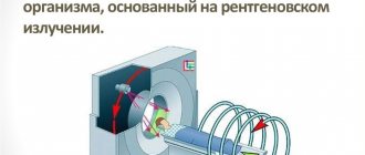
MRI of the intestine is performed both on open tomographs and on closed-loop devices; the image quality in the second case is much higher than in the first. Good resolution of layer-by-layer photos is possible with a magnetic field strength of 1.5 Tesla. Closed-type devices have the required power.
Preparation and conduct of the study
On the day of the procedure, you must arrive at the clinic in advance: paperwork will take some time. You need to take with you your passport, the results of previous studies (if available), certificates that may be important for the study.
The patient removes all jewelry and other objects made of metal, and then lies on the tomograph platform, lying on his back. Legs and arms are fixed to prevent involuntary movements. When conducting a contrast study, a special substance is injected into a vein.
The platform slides into a device that forms a magnetic field, takes pictures, and transfers them to a computer. After processing the information, three-dimensional images are obtained. After studying them, the doctor writes a conclusion and transfers it to the patient along with the images and recording on electronic media.
Based on the specifics of the procedure, a necessary part of preparation is a psychological attitude. To begin with, it is advisable to study information about how the study is carried out. It is important to remember that the device makes noise, but to reduce its level, the patient is given headphones or earplugs.
The reliability of the results largely depends on the quality of preparation for the study.
They are informative, so this issue should be taken seriously. Rate this article: (1 rated 5 out of 5)
Will an MRI show colon cancer?
Malignant neoplasms of the digestive system occur equally often in men and women. An asymptomatic course in the initial stages makes timely diagnosis difficult, so suspicion of the development of oncology is one of the main indications for intestinal MRI.
Magnetic resonance imaging makes it possible to study the pathologically changed area in detail. If a tumor is present, the doctor evaluates the size, localization of the lesion, and the interaction of the tumor with surrounding structures. The malignant process is characterized by the absence of a clear contour; atypical cells “grow” into healthy tissues.
A cancerous tumor accumulates gadolinium chelates more slowly on contrast-enhanced MRI of the intestine. This occurs due to the pathological tortuosity of its own blood vessels. Magnetic resonance scanning shows formations with a diameter of 3 mm, suggesting the nature of the process. The final diagnosis is made based on the results of laboratory tests.
The advantages of this technique include:
- Spatial reconstruction, clarifying the location of healthy tissue areas and those in which there are pathological changes.
- No invasive interventions.
- No pain.
- Possibility of reliable assessment of the condition of the intestinal walls and adjacent structures.
This procedure has only one drawback - intestinal peristalsis negatively affects the quality of images. This can make diagnosis difficult when the pathological focus is small.
Is an MRI of the intestines informative?
The method well visualizes the condition of the walls of the thin and thick sections, shows other organs of the abdominal cavity and small pelvis. MRI of the intestine is informative in identifying:
- adhesions;
- ulcerative processes;
- intestinal endometriosis (not found in men);
- oncological diseases;
- pathologies of the blood vessels of the abdominal cavity and pelvis;
- foreign bodies
Vascular pathologies, ulcers of the mucous membrane, malignant neoplasms can cause bleeding. In this case, treatment largely depends on what the intestinal MRI shows.
The method helps determine the etiology of the pathological process by visualizing the features of innervation, blood supply, structure of the digestive system and surrounding organs. Based on layer-by-layer photographs taken in three projections, the doctor can reconstruct a 3D model of the department in question. A three-dimensional MRI image shows how the intestinal loops are located in the abdominal cavity and pelvic area.
Peutz-Jeghers syndrome (arrows indicate multiple polyps)
Allowed foods and drinks
During preparation for an abdominal MRI, foods are baked in the oven, boiled, or steamed. The diet may include:
- Veal or beef, turkey or rabbit (optimally in the form of minced meat);
- Low-fat fish;
- Buckwheat or rice, oatmeal (it is better to exclude semolina);
- Fruits, vegetables, heat-treated and pureed;
- Vegetable oils;
- Omelettes;
- Dry types of cookies.
Honey is allowed, but in small quantities. Wholemeal bran bread, watermelon, and parsley are acceptable. Rosehip tincture helps reduce gas formation.
In addition to diet, it is important to maintain a drinking regime. Drinking plenty of fluids helps prevent constipation, but you need to choose the right drinks. These can be unsweetened compotes made from a small amount of berries or fruits, water, herbal tea. Of the juices, varieties that do not contain pulp are allowed; it is better to dilute them with water in a 1:1 ratio.
Indications
MRI of the intestine is a procedure during which the middle-lower gastrointestinal tract is examined layer by layer. Indications for such diagnostics are as follows:
- Frequent bowel movements: both constipation and diarrhea;
- Systematic bleeding from the upper or lower parts of the digestive tract;
- Pain in the lower abdomen;
- Weight loss;
- Controlled vomiting;
- Pain and feeling of fullness in the anus;
- Partial or complete intestinal obstruction;
- Flatulence.
An MRI of the intestine is also performed to evaluate the effectiveness of treatment or as preparation for surgery. The procedure allows for differential diagnosis of tumors and traumatic injuries.
MR imaging of the intestine for diagnosing Crohn's disease
Crohn's disease is a fairly common, severe disease that damages the small intestine. It is extremely difficult to reach it with an endoscope without worsening the patient’s condition. Ultrasound also does not provide an understanding of the complete clinical picture. Therefore, MRI is more often used for diagnosis. The sensitivity of this method is more than 95%, the procedure is well tolerated by patients and does not cause complications.
In case of Crohn's disease, it is necessary to conduct regular examinations to monitor the patient's current condition and adjust the treatment regimen. The most effective and safest method is MRI, which allows you to obtain detailed visualization of the entire intestine, detect the inflammatory process, thickening or dissection of the walls, abscesses and fistulas
As we said earlier, colonoscopy is strictly contraindicated for many patients with this disease, and CT gives a high dose of radiation and cannot be performed frequently.