Diseases of the gastrointestinal tract are a very common problem even in progressive developed countries. This is due to the accelerated pace of life, poor nutrition, and poor quality of food. One of the indirect causes of serious illnesses is considered to be failure to seek medical help in a timely manner. Most pathologies in the digestive tract are accompanied by unpleasant symptoms, pain, weight loss or obesity. To establish an accurate diagnosis and treatment, doctors use many examination methods. The main one remains radiography of the duodenum and stomach.
What does a gastrointestinal x-ray show?
An examination of the digestive organs is necessary to assess their condition, functioning, detect problems and eliminate them. Today there are several technologies for this: endoscopy, MRI, computed tomography, angiography.
The very first research technique was x-ray (x-ray) - radiation diagnostics of the internal structure of the body. With its help, one-time images of the stomach and duodenum are obtained.
The result obtained is recorded on film, from which the doctor will evaluate the condition of the internal organs. This procedure is also called gastrography.
Content:
- What does a gastrointestinal x-ray show?
- Indications for examination
- Contraindications to the procedure
- How to prepare for an x-ray
- How does this happen
- Where to get an X-ray of the gastrointestinal tract
- Possible complications
- Alternative examination methods
Often, images alone are not enough to fully study the anatomy of the digestive tract. In such cases, radiography is combined with fluoroscopy.
The latter is carried out using a special amplifier - a contrast agent. In laboratory conditions, a special barium-based solution is prepared, which does not transmit x-rays.
The patient drinks this cocktail during the procedure, as a result the doctor can observe in real time the patency of the esophagus, the peristalsis of the stomach, and have a good look at its internal walls and folds.
As a rule, these two procedures are carried out in combination. Radiography provides images, and fluoroscopy allows you to see the movement of internal organs and record data on electronic media. This makes it possible to study in detail the structure of the walls of the esophagus, duodenum, and stomach without invasive intervention.
Radiation diagnostics remains the most accessible method of examining a patient; it can be carried out in every hospital. The success of this process depends on the experience and skill of the radiologist.
But this technique also has a significant drawback: during the examination, the patient receives a certain dose of radiation, especially during fluoroscopy. Therefore, doctors resort to it only in extreme cases. The doctor must first carry out all non-radiation examination methods and only if they do not give the desired result, prescribe an x-ray.
X-ray of the gastrointestinal tract shows:
- diseases of the esophagus: diverticulum (protrusion of the mucous membrane), tumors, narrowing, varicose veins;
- foreign bodies in the digestive tract;
- pathologies of the duodenum: ulcers, cancer, spastic narrowing;
- stomach diseases: gastritis, cancer, ulcers, impaired absorption and weak peristalsis, etc.;
- other abdominal organs are not the main target of diagnosis, but their contours are still visible on the image. Based on the image, the doctor can notice problems in other organs.
In some cases, the patient has to undergo several procedures at once, since each type of diagnosis shows different areas. For example, endoscopy reflects the condition of the internal mucous membrane and allows you to take material for a biopsy. And on the x-ray you can see the outer part of the organs, a little internal, tumors and neoplasms, narrowing of the esophagus are visible.
What is fluoroscopy indicated for?
During an X-ray of the stomach, the specialist receives a detailed image of the internal structure of the organ. This allows us to draw conclusions about the shape and size of the stomach and the condition of the mucous membrane.
Definitions of stomach shape
The shape of the stomach is clearly visible on the x-ray. Thanks to the image, the specialist can diagnose local narrowing of the lumen. Pathology may indicate the presence of a neoplasm in the wall of the stomach or in neighboring organs. If fluoroscopy shows widening of the lumen, there may be a bulge in the stomach wall (diverticulum).
Condition of the mucous membrane
In some cases, when performing an X-ray of the stomach, the use of a contrast agent is required; the procedure is good because it shows changes in the structure of the mucous membrane. The study is prescribed if an ulcer, chronic atrophic gastritis or other anomalies is suspected.
Organ size
An increase in the size of the stomach occurs due to overeating, stress or inflammation, as a result of complications after surgery or for other reasons. The condition is characterized by pain, nausea, bloating, tachycardia and low blood pressure. To diagnose the pathology, a fluoroscopy of the stomach is done with contrast.
Relief
To determine the relief of the mucous membrane, an x-ray of the stomach is performed with double contrast. In normal condition, the mucous membrane has a relief in the form of twisting grooves. If fluoroscopy of the stomach reveals a change in the direction of the grooves, their increase or decrease, pathologies are possible.
Structural structure
Changes in tissue structure and disruption of the integrity of the stomach walls indicate a serious illness. The pathology will be visible immediately after specialists take an X-ray of the stomach.
Indications for examination
A therapist, surgeon, or gastroenterologist refers you to gastrography. Other specialists may also suspect problems in the area of digestion; the procedure is carried out directly by a radiologist, who also deciphers the results. Based on the deciphered data, the treating doctor determines the treatment tactics. It is also sometimes necessary to undergo the procedure again to monitor the progress of therapy.
Radiography is prescribed to determine various pathologies. Among them:
- irritable stomach syndrome;
- hernia of the diaphragm and esophagus;
- inflammation of the digestive tract;
- foreign bodies in the gastrointestinal tract;
- malignant and benign tumors;
- ulcers and gastritis;
- deformation of the duodenum and its parts.
At the same time, during the examination, problems with the liver, spleen, and kidneys may be identified. Gastrointestinal pathologies make themselves felt by deteriorating well-being; often such diagnostics are prescribed based on the patient’s complaints.
But, as already noted, the doctor first conducts other types of examinations to reduce the radiation dose to the patient.
Suspicious symptoms include:
- constant nausea and vomiting after eating;
- frequent attacks of heartburn, hiccups, belching;
- abdominal pain;
- cutting hunger pain in the stomach area, which goes away after eating;
- frequent bowel movements;
- unexplained weight loss;
- obesity with normal nutrition.
If one or more symptoms are detected, it is recommended to consult a gastroenterologist. Problems with the digestive system can progress into more serious pathologies, and timely seeking help will be the key to successful treatment. Also, people who may have a genetic predisposition to developing tumors of the stomach and intestines are also recommended to undergo preventive examinations.
When is the procedure indicated?
The examination is carried out if diseases and pathologies are suspected that may pose a serious danger to the patient. In most cases, barium X-ray of the stomach allows you to confirm or clarify the diagnosis and prescribe the correct treatment.
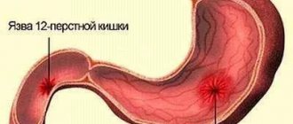
Suspicion of the formation of gastric and duodenal ulcers
Peptic ulcer of the stomach and duodenum (DPC) leads to the periodic appearance of ulcers. It can take years to develop and cause serious harm to health. Using a barium X-ray of the stomach, the specialist finds the ulcer and evaluates its size, shape and other parameters, as well as establishes the stage of the disease and detects accompanying symptoms.
Detection of a malignant process in an organ
If the development of a malignant process is suspected, an X-ray of the stomach with barium is recommended without fail, since the consequences of the disease can be deadly for the patient. In the photographs, the cancerous process looks like a defect in the mucous membrane. Often, after a barium X-ray of the stomach, an endoscopic examination is performed and a sample of the tumor is taken for laboratory testing.
Diverticulosis and other deformities of the gastric walls
To diagnose wall protrusion (diverticulosis), a barium X-ray of the stomach requires additional preparation. After administration of the contrast agent, the patient alternately lifts the upper and lower parts of the body so that the contrast fills the entire organ cavity. On a barium X-ray of the stomach, small diverticulosis looks like an ulcer, but has a horizontal level and a neck.
Any inflammation of the stomach
Foci of inflammation in the stomach can appear as a result of various processes. Including peptic ulcers or oncology. An X-ray of the stomach with barium allows you to find the source of inflammation and, based on indirect signs, determine its cause.
Swallowing dysfunctions
Dysphagia, or swallowing dysfunction, is often expressed by the patient complaining of the inability to swallow food and its accumulation in the esophagus. In this case, the patient cannot always accurately indicate the location of the obstacle. Therefore, barium radiography of the stomach is used to accurately localize the pathology.
Pain in the abdominal area or navel area
Pain in the navel and abdominal area may indicate the development of malignant processes in the stomach and duodenum. Therefore, with such symptoms, the specialist often refers the patient to an X-ray of the stomach and duodenum with barium.
Contraindications to the procedure
Due to the increased radiation dose, x-rays are not prescribed during pregnancy, since radiation rays can unpredictably affect the development of the fetus. For the same reason, this procedure is prescribed to children only in severe cases.
A categorical contraindication is a through formation in the stomach or intestines - perforation. The contrast agent may enter the abdominal cavity, causing further complications.
Gastrography is performed with caution in cases of acute obstruction and inflammatory processes. Such research is prohibited for people with allergies to iodine and barium.
Carrying out the procedure
To properly prepare for a stomach x-ray, you need to understand what it is and how the examination is carried out. Depending on the purpose of the procedure, the technique may vary. Therefore, preparing a patient for an X-ray of the stomach, in addition to general requirements, may include specific steps that the doctor warns about.
In most cases, the specialist prescribes an x-ray of the stomach without contrast. In this case, the patient takes a horizontal position on the couch, after which a series of photographs is taken.
If the examination is carried out with contrast, preparation for an x-ray of the stomach includes the use of a barium-based drug. The substance does not cause harm to health and is eliminated naturally. X-ray data of the stomach with contrast are more complete than with a conventional examination.
In cases where a detailed examination of the walls of the stomach is required, their natural folds must be straightened. Then the specialist prescribes a fluoroscopy of the stomach with double contrast.
After taking the contrast agent, air is pumped into the stomach through a tube. Under slight pressure, the folds of the walls are straightened, which allows you to obtain more detailed images of the surface.
How to prepare for an x-ray
The procedure takes place on an empty stomach. The abundance of gases or the presence of food in the gastrointestinal tract makes diagnosis difficult and can distort the data, so it is recommended to prepare 2 days before the scheduled session. To do this, you need to follow a diet or artificially cleanse the intestines.
At the time of issuing a referral for examination, you must consult with your doctor about detoxification methods. The easiest way is a cleansing enema; it is done the day before and in the morning before the procedure. An enema can be replaced with special medications that cleanse the gastrointestinal tract. In this case, Fortrans is recommended - a soluble powder that removes gases and food residues well without enemas.
Preparatory measures also include an allergy test for iodine and barium. You can take medications only with the permission of your doctor. Before the x-ray, you need to warn the specialist if you have taken any medications.
A couple of days before the session, you need to remove gas-forming products from the menu. These include:
- beans and grains (except rice);
- dairy products;
- Rye bread;
- cabbage, radish, radish;
- potato;
- carbonated drinks.
On the day of diagnosis, it is important to stop smoking; alcohol is excluded 2-3 days before the session. You are prohibited from eating 8 hours before the examination.
What can be detected with radiography?
If a barium stomach x-ray is performed correctly and with proper patient preparation, the examination can provide enough comprehensive information to make a diagnosis in most cases. At the same time, to detect a particular disease, a specialist evaluates a number of parameters.
Gastric lumen disorders
Disturbances in the lumen of the stomach may indicate the development of a malignant process or diverticulosis. If the lumen of the stomach is narrowed on a barium X-ray, diffuse fibroplastic cancer is possible. With diverticulosis, on a barium X-ray of the stomach, the lumen appears larger than it should be normally.
Abnormal placement of the stomach
Gastroptosis (prolapse of the stomach) is often diagnosed with an abdominal wall hernia or diaphragmatic hernia. The disease is clearly visible using barium X-ray of the stomach.
Niche symptom
A niche is a shadow of a contrasting mass that fills a defect during an ulcer or the development of a malignant process. Depending on the location of the defect, an X-ray of the stomach with barium distinguishes between a contour and a relief niche. In the first case, the silhouette of the defect is visible from the full face, in the second - in profile.
The lack of filling is visible in the image as a dark area
A dark area on a barium x-ray of the stomach indicates that there is a place where the contrast could not reach. As a rule, this happens with atrophic gastritis or tumor.
How does this happen
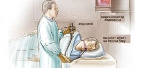
The study is carried out only on an empty stomach, after preliminary preparation of the patient. In the X-ray room, the patient takes off his clothes and puts on a special gown. Be sure to remove all jewelry and notify your doctor if there are metal inserts in your body. Afterwards, an overview photograph is taken in a standing position; further process is possible only after the first photographs. This is necessary to eliminate the possibility of internal bleeding, organ ruptures, and acute obstruction. The resulting images are examined by a doctor; if there are no contraindications, contrast is performed.
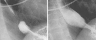
A mixture is prepared in the laboratory: a solution of barium sulfate in water. The subject takes a few sips of the drink. It tastes and smells like chalk and does not cause any unpleasant sensations. After entering the esophagus, the barium mixture covers the mucous membrane of the gastrointestinal tract, and the esophageal tube is clearly visualized on the screen. For subsequent examination of the stomach and duodenum, the patient drinks the remainder of the solution - 200-250 ml. The fluid fills the organs and they are better visible in the image; ulcers, neoplasms, deformations, and narrowings are easily visible.
To get a complete picture, the patient is placed on a table and a series of photographs are taken. Diagnosis is carried out in direct, lateral and oblique projections. To do this, the patient is asked to change his body position several times.
After receiving the data, the radiography procedure is considered complete. In some cases, an x-ray of the lower intestine is needed, then the process is repeated a few hours later, when the barium enters the large intestine.
Once the radiologist has enough information, he will write a report and describe the images. Sometimes this takes several days, sometimes the patient can receive results after 30-60 minutes. With this data, he goes to the doctor, who referred him for examination. The attending doctor will be able to establish a diagnosis and determine treatment, or, at best, determine that there are no diseases or complications.
X-ray of the stomach, esophagus and duodenum
Duration
60 minutes
We make an appointment for an hour.
The study itself lasts 30-40 minutes. Within half an hour after the fluoroscopy, you will receive the images on disk or by email. Soreness
No pain
X-ray of the stomach is a non-invasive technique.
Anesthesia
No anesthesia required
The examination is a non-invasive, painless procedure and no anesthesia is required.
Additional factors
Use of contrast agent
To perform an objective x-ray examination, additional contrast with an aqueous solution of barium sulfate in an amount of 250-300 ml is used.
Preparation
Preparation required
Preparation for fluoroscopy of the stomach is simple.
The study is carried out on an empty stomach. It is advisable to make an appointment for the first half of the day. For fluoroscopy of the esophagus (when the esophagus is examined separately), almost no preparation is required. Rehabilitation
Not required
After your exploration, you will be offered sweet tea or coffee.
You will feel good. Work ability
Saved
You can safely go to work after the procedure.
Sports activities
No restrictions
The procedure is non-invasive.
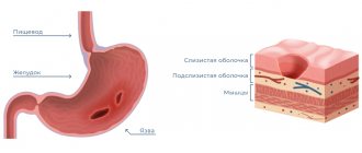
You can exercise even on the day of the procedure. X-ray of the stomach is a diagnostic study, which is a non-invasive technique that makes it possible to examine the shape, size, position of the esophagus, stomach and duodenum, as well as to identify their anatomical features and pathological changes.
The study helps gastroenterologists identify ulcers, gastritis and other diseases and pathologies. Don’t worry and remember: the main thing is to start treatment at an early stage!
X-ray examination of the esophagus, stomach and duodenum allows us to detect and differentiate the following pathological processes:
- impaired movement of the contrast mass through the esophagus, stomach and duodenum (impaired gastrointestinal motility);
- inflammatory changes in the esophagus, stomach and duodenum: esophagitis, gastritis, duodenitis;
- peptic ulcer of the stomach and duodenum;
- diseases of the esophagus: achalasia of the esophagus, varicose veins of the esophagus, hiatal hernia, diverticula of the esophagus and others;
- deformation of the duodenum and its parts due to scarring, stenosis, external compression;
- neoplasms in the walls of the stomach and duodenum, etc.
About stomach examination
The stomach is an abdominal organ with a hollow structure. To perform an objective x-ray examination, additional contrast is used with an aqueous solution of barium sulfate in an amount of 250-300 ml, which is harmless to the patient’s body, is not absorbed in the gastrointestinal tract and is excreted unchanged.
Sometimes, if it is necessary to examine the inner surface of the stomach and antrum, double contrast is used.
When conducting a study that lasts 30-40 minutes, the doctor must evaluate the passage of the contrast mass through the esophagus, stomach and duodenum, note the changes and take x-rays. They are processed at the end of the study and enable the radiologist to correctly assess the condition of the organs being examined and make a diagnosis.
The examination is usually carried out in an upright position. We also perform fluoroscopy in the Trendelenburg position. This is the name for the position of the patient on the operating table when the pelvic organs are slightly above the head.
Indications for the study:
- difficulty swallowing, pain in the chest when swallowing;
- acute or chronic pain in the abdominal cavity;
- vomit;
- heartburn and reflux (backflow of partially digested food or digestive juices);
- diarrhea or constipation;
- sudden weight loss;
- pathological change in the color and consistency of stool, blood in the stool.
Preparing for the study is simple. The main thing is that fluoroscopy of the stomach is performed on an empty stomach. You can read more in this section.
X-ray of the esophagus + radiography (as a separate study):
To correctly make a diagnosis, taking into account the patient’s complaints, the radiologist conducts an independent examination of the esophagus. To do this, the patient is given several sips of barium suspension and a series of polypositional (in different positions: sideways, obliquely to the right and left, lying and standing) images are taken under the scope of an RG-scope. The shape, position of the esophagus in the chest cavity, the speed of passage of the barium suspension, the location of the folds of the mucous membrane, additional narrowings or protrusions in the wall of the esophagus (diverticula) are assessed. When examining the esophagus, the doctor can detect a hiatal hernia, assess its size and position, which will tell cardiologists and gastroenterologists a plan for further treatment of the patient.
With diseases of the esophagus, patients often complain:
- for swallowing disorder - dysphagia;
- to “lump in throat”;
- for heartburn, chest pain;
- for heart rhythm disturbances;
- for pain in the heart.
preparation is required for fluoroscopy of the esophagus. It is only important that the patient comes to the clinic on an empty stomach.
Attention! After gastroscopy, X-ray examination is not performed.
Contraindications for carrying out
X-ray is a safe examination that has no absolute contraindications and allows you to quickly obtain clear images of internal organs.
However, pregnant women (especially in the first trimester) and children under 14 years of age are not recommended to undergo x-rays. Examinations using a contrast agent are contraindicated in those who have recently had an intestinal biopsy.
At MedikCity, fluoroscopy is performed using a digital X-ray machine GMM Opera T30csx (ITALRAY CLINODIGIT COMPACT). One of its important advantages is high image detail: the examination area can be enlarged on the screen without loss of quality. This allows you to see even small details.
Fluoroscopy results are not the only basis for diagnosis. Anamnesis, laboratory tests and other instrumental diagnostic methods are also taken into account.
Our X-ray room is open for you from 9.00 to 21.00. You can find out the cost of x-ray examination in a special section.
You can learn more about preparing for fluoroscopy from your attending physician or call center operators.
The material was prepared with the participation of a specialist:
Kostacheva Galina Aleksandrovna
Radiologist
Where to get an X-ray of the gastrointestinal tract
Every clinic provides a similar service. It does not require special equipment, although private clinics have digital devices that provide quick results on electronic media. An X-ray of the stomach and duodenum can be taken at a public clinic, hospital, or clinic.
Private medical institutions also conduct X-ray examinations of the gastrointestinal tract. The price for an x-ray will be 40-50 dollars, fluoroscopy is priced separately - from 45 to 65 dollars. The price includes contrast agent and diagnostics. In some private diagnostic centers you will have to pay separately for decoding the results.
Decoding the results
When deciphering the results of an X-ray of the stomach, the specialist evaluates a whole range of data: from the shape and size of the organ, the condition of the tissues and the relief of the inner surface of its walls to the speed of movement of the contrast agent along the digestive tract. Deviations from the norm can indicate many pathologies.
For example, if fluoroscopy of the stomach reveals slow progress of contrast through the esophagus, stomach and intestines, motor disturbances are possible. Abnormalities in the mucous membrane may indicate a peptic ulcer, and changes in the lumen of the stomach may indicate a neoplasm.
Possible complications
Immediately after the x-ray, it is recommended to drink a lot of clean water, this will help remove the barium mixture from the body faster. It does not dissolve in liquid, so it is not absorbed into the walls of the digestive tract and does not cause damage to the body. Among mild complications, patient reviews note attacks of nausea and vomiting, but this is only possible with increased sensitivity. Constipation may occur for 2-3 days after the session; this is a common reaction that goes away with time. The stool also turns whitish or gray. If bowel problems do not go away after three days, you should contact your doctor.
Such side effects do not cause significant discomfort, so they are not called complications. Rare and serious consequences include an allergic reaction. This is possible if the preparation rules were violated and the patient did not undergo a barium test.
Sometimes the barium suspension is replaced with an iodine solution, so the reaction to it must also be checked. In case of allergies, the patient may experience swelling of internal organs, difficulty breathing, and Quincke's edema. If the patient is well prepared for the procedure, gastrography will take place without negative consequences.
Operating principle
The essence of this diagnostic method is based on exposure of the abdominal cavity to a permissible dose of radiation. When X-rays pass through a cavity filled with contrast, a fairly clear picture is obtained, on the basis of which a radiologist can identify many pathological changes and establish the correct diagnosis.
Despite the fact that the patient is exposed to radiation during the study, barium radiography of the abdominal cavity is actively used in modern medicine. Firstly, the radiation dose is insignificant and does not exceed the established daily norm. Secondly, the procedure does not take much time and does not cause discomfort to the patient. And thirdly, x-rays with a contrast agent are an inexpensive and fairly informative diagnostic method.
Content:
- Operating principle
- What will a barium x-ray of the abdomen show?
- Indications for radiography
- Preparing for the study
- Features of the procedure
- Contraindications and restrictions
Modern equipment used for radiography procedures makes it possible to strictly regulate radiation activity. Thanks to this, neighboring organs are practically not exposed to radiation.
Causes of operated stomach syndrome
Gastric resection itself is the cause of dumping syndrome. The provoking factor is considered to be the rapid transfer of undigested food from the stomach to the intestines. Irritation of the small intestinal mucosa by poorly processed food leads to increased blood circulation in the intestines. Insufficient blood supply to the brain occurs, the effect on the parasympathetic nervous system increases, hormonally active substances - catecholamines - are released into the blood, which in turn aggravate hypovolemia in the human body.