Good day!
My name is Khalisat Suleymanova - I am a herbalist. At the age of 28, I cured myself of uterine cancer with herbs (read more about my experience of recovery and why I became a herbalist here: My story). Before being treated using traditional methods described on the Internet, please consult with a specialist and your doctor! This will save your time and money, since the diseases are different, the herbs and treatment methods are different, and there are also concomitant diseases, contraindications, complications, and so on. There is nothing to add yet, but if you need help in selecting herbs and treatment methods, you can find me at my contacts: Khalisat SuleymanovaInstagram page: instagram.com/fitoterapevt1
Telephone: 8
Email: [email protected]
I consult for free.
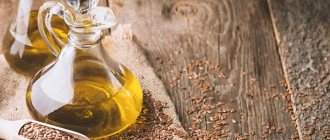
There are a large number of different diseases in the world to which humans are susceptible. And many people dream of such a miracle cure that would help cure them. Most often, diseases in our body arise as a result of nervous stress and poor nutrition. All this can lead to such a serious disease as stomach and duodenal ulcers. Doctors recommend the use of fraction ASD 2 for humans with gastric ulcers.
Properties of the drug
In the middle of the last century, scientists developed a drug that many people began to call the “elixir of life”, and in science - ASD, which meant antiseptic Dorogov stimulant. Patients who were disappointed in well-known medications resorted to its use. They noted that they can cure diseases such as:
- damage to the nervous system,
- skin diseases,
- oncology,
- tuberculosis,
- cardiovascular problems,
- bronchial asthma.
There is also a positive effect of using ASD fraction 2 for stomach and duodenal ulcers.
3 types of this drug were invented. The first fraction did not contain any beneficial properties, but the next 2 had a healing effect.
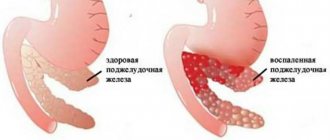
INTERESTING fact: ASD 2 for the pancreas in cancer and pancreatitis
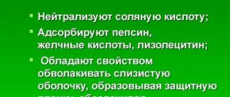
ASD 2 - water soluble. It can be used for both internal and external applications. Particularly positive effects were noted with its complex use. Most often it is used to treat vomiting of various organs and systems.
Among the diseases are:
- nervous system dysfunction,
- immune system problems,
- phlebeurysm,
- loss of skin elasticity,
- diseases of the endocrine system,
- mastopathy,
- trichomoniasis,
- breast cancer,
- chlamydia,
- uterine cancer,
- fibroma,
- myoma,
- respiratory system diseases,
- skin problems,
- gynecological troubles,
- gastrointestinal diseases,
- oncology.
ASD 3 is used for external use only. It is only soluble in alcohol or fat. It is used to disinfect wounds and treat:
Simple ways to treat complex diseases:
Did you know that salt dressings work wonders? This recipe was published in Healthy Lifestyle in 2002. Unfortunately, it is not well forgotten, but deliberately erased from memory and discredited by pharmaceutical companies in pursuit of ... Read more
Honey and flax - strong health! Honey with flax seeds is the best remedy for blood vessels and immunity! Detoxify with flax seeds! Colon detoxification is possible using natural remedies. … Read more
MIRACLE MIXTURE FOR THE BODY I always determined the age plus or minus 1-2 years. Once I was visiting and there was a woman there, everyone thought that she was about 35 years old, but from the conversation we realized that we were mistaken. They asked how many… Read more
8 natural products that destroy parasites in the body Most of the world's population suffers from parasitic diseases. Common natural remedies will help your body fight parasites. Parasitic… Read more
Even 1 date causes an irreversible process in the body! Dates are an amazing fruit, which in many countries is credited with the properties of improving health and prolonging life. They say that in China there are long-livers, the basis... Read more
- skin parasites,
- skin pathologies (trophic ulcers, eczema, psoriasis, dermatitis, acne, trophic ulcer, neurodermatitis),
- various fungi.
All types of Dorogov’s drug are considered not only a powerful antiseptic, but also a biogenic stimulant. It not only takes part in metabolic processes, but also exhibits the qualities of an immunomodulator.
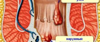
Complex treatment of trophic ulcers
This work is devoted to studying the possibility of increasing the effectiveness of complex therapy for trophic ulcers and postoperative wounds of the lower extremities using a topical immunomodulator.
The study involved 44 patients aged 30 to 81 years (30 women, 14 men) with chronic trophic ulcers of the lower extremities. Trophic ulcers developed due to circulatory disorders in the lower extremities due to thrombophlebitis or diabetic angiopathy. In most patients, thrombophlebitis occurred as a complication of varicose veins. The diagnosis of “varicose veins, postthrombophlebitic syndrome (PTPS)” was made to 41 patients. At the time of the study, 9 people had signs of acute thrombophlebitis against the background of PTFS, and 5 people had signs of erysipelas of the skin. In one patient the trophic ulcer was combined with osteomyelitis of the tibia, in two patients it was combined with incipient gangrene of the toes. In the anamnesis, three patients underwent phlebectomy, 1 patient underwent sclerotherapy for varicose veins.
The vast majority of the patients studied had 1 trophic ulcer, and only two patients had 2 ulcers. In most cases, the ulcers were located on the anterior, inner or outer surface of the leg; in 2 patients with diabetic angiopathy, the ulcerative process was localized on the foot. The sizes of trophic ulcers varied from 0.5x0.5 cm to 3x4.5 cm. The ulcer crater was filled with fibrinous masses in 30 patients, fibrinous-purulent - in 6 patients, purulent-necrotic tissue fragments - in 5 patients. In three cases, upon admission of the patient, the ulcer looked clean, without fibrinous, purulent or necrotic masses.
In 5 patients with necrotic-bullous form of erysipelas, the skin of the lower leg had signs of acute inflammation and blisters with serous-purulent contents. In 2 other patients there were signs of incipient gangrene: respectively, the first and third toes of the right foot were bluish-black.
Treatment
When treating all 44 patients, the following treatment principles were observed:
- bed rest with an elevated position of the sore limb to eliminate stagnation of blood and lymph;
- careful cleaning of the skin around the ulcer;
- creating a flow of tissue fluid from the ulcer into the bandage at the beginning of treatment. For this purpose, dressings with a hypertonic solution of NaCl were used in combination with an alcohol solution of chlorophyllipt, which ensured cleansing of the ulcer and improved nutrition of the living tissues of the bottom and walls of the ulcer;
- activation of the body's regenerative abilities after cleansing the ulcer crater.
All patients with PTFS received general and local treatment. The general components of treatment were infusion (reopolyglucin 200 ml + trental 5 ml + ascorbic acid 5 ml intravenously, every other day No. 5), aescusan 15 drops 3 times a day, aspirin 0.5 g - 1 time a day, troxevasin 2 capsules 3 times a day for 15 days or diovenor 600 mg, 1 tablet. 1 time per day for 30 days. In addition to the treatment described above, patients with signs of acute thrombophlebitis received injections of heparin solution 5000 IU subcutaneously 4 times a day for 6 days.
Local treatment in the first days (from 1 to 4 days) - alcohol chlorophyllipt in combination with a hypertonic solution, dressings were done every day. After cleansing the ulcer, bandages with Gepon ointment or solcoseryl were applied (control group, 10 people). As noted above, in 3 patients the ulcer did not contain fibrinous, purulent or necrotic elements. These patients immediately began treatment with Gepon ointment, an immunomodulator that has the ability to increase the effectiveness of immune defense, as well as a direct antiviral effect.
The ointment was prepared directly in the pharmacy of City Clinical Hospital No. 1 and had the following composition: Gepona 0.006; lanolin 10.0; olive oil 10.0; water for injection 10.0. The finished ointment was stored at +4°C and used for 10 days.
The ointment was applied in a thin layer to the surface of the trophic ulcer, the bandages with Gepon ointment were changed every other day. Treatment was carried out for 10 days (5 dressings).
Treatment of uncomplicated trophic ulcers
In all patients, already on the 3rd day of treatment with Gepon, rapid growth of granulation tissue was observed in the ulcer crater. After 8-10 days of treatment with Gepon, a scar formed from connective tissue.
In the control group, 10 patients received the same general therapy, but local treatment after cleansing the ulcer was carried out with solcoseryl ointment. Healing of ulcers in patients in this group took 5–15 days longer than when using Gepon ointment. One patient in the control group worsened during treatment with solcoseryl ointment and developed erysipelas of the skin (necrotic-bullous form). This patient was prescribed adequate surgical treatment; in addition to general treatment, antibiotics and biseptol were used; for local treatment, Gepon ointment was used instead of solcoseryl.
Treatment of ulcerative skin defects after necrectomy for necrotic-bullous erysipelas
Patients with necrotic-bullous erysipelas, in addition to infusion therapy, received injections of cefazolin 1 g intramuscularly 3 times a day for 7 days, as well as biseptol 480 mg, 1 tablet. 3 times a day for 10 days. Against the background of conservative treatment, surgical intervention was performed - necrectomy. The blisters were opened, necrotic tissue was removed, and the open wound was treated with a solution of potassium permanganate. Further, large skin defects that opened after necrectomy were treated with Gepon, like trophic ulcers. All patients had good treatment results. 3–4 days after the start of using Gepon ointment, a pronounced growth of granulation tissue was observed, followed by the formation of a connective tissue scar in the shortest possible time.
Treatment of postoperative wounds of the lower extremities in patients with diabetic angiopathy
When treating patients with diabetic angiopathy, conservative treatment was supplemented with adequate doses of insulin (s.c.). Lincomycin 600 mg IM 2 times a day was used as an antibiotic for 14 days. In case of incipient gangrene of the finger, against the background of conservative treatment, adequate surgical intervention was performed - amputation or limited excision of necrotic elements. In the postoperative period, the wound and fistula tracts were washed with Gepon solution (0.002 g in 10 ml of saline), and bandages with Gepon ointment were applied, as described above. The results of treatment indicate significant activation of the growth of granulation tissue and accelerated healing of the postoperative wound under the influence of Gepon.
It is obvious that the use of Gepon in the clinical cases described above stimulated the active growth of granulation tissue. Typically, in patients with diabetic angiopathy, the patency of the capillary bed is minimal; during surgical procedures, blood is released, as a rule, only from the subcutaneous vessels, the internal tissues are practically bloodless and have a pale pink color. The growth of granulation tissue in such patients is either not observed at all or is very sluggish, postoperative wounds do not chronically heal, and trophic ulcers remain. The use of Gepon made it possible to achieve accelerated healing of postoperative wounds and non-healing ulcers in patients with diabetic angiopathy.
Literature
- Ataullakhanov R. I., Holmes R. D., Narovlyansky A. N., Katlinsky A. V., Mezentseva M. V., Shcherbenko V. E., Farfarovsky V. S., Ershov F. I. Mechanisms of antiviral action drug "Gepon": changes in the transcription of cytokine genes in transplanted human cells // Immunology. 2002.
- Bibicheva T.V., Silina L.V. Gepon immunomodulator for local therapy of herpes viral infection. In the book: Abstracts of the IX Russian National Congress “Man and Medicine”. M., 2002. 55 p.
- Bibicheva T.V., Silina L.V. Treatment of recurrent genital herpes with the immunomodulator Gepon. In the book: Abstracts of the IX Russian National Congress “Man and Medicine”. M., 2002, p. 56.
- Dudchenko M. A., Parasotsky V. I., Lysenko B. F. Effective treatment of gastric and duodenal ulcers with the immunomodulator “Gepon”. In the book: Abstracts of the IX Russian National Congress “Man and Medicine”. M., 2002, p. 141.
- Kladova O.V., Kharlamova F.S., Shcherbakova A.A., Legkova T.P., Fildfiks L.I., Znamenskaya A.A., Ovchinnikova G.S., Uchaikin V.F. First experience of intranasal use of Gepon in children with respiratory diseases // Pediatrics. 2002. No. 2. P. 86-88.
- Kladova O. V., Kharlamova F. S., Shcherbakova A. A., Legkova T. P., Fildfiks L. I., Uchaikin V. F. Effective treatment of croup syndrome using the immunomodulator Gepon // Russian Medical Journal. 2002. T. 10. No. 3. P. 138-141.
- Polyakova T. S., Magomedov M. M., Artemyev M. E., Surikov E. V., Palchun V. T. A new approach to the treatment of chronic diseases of the pharynx // Attending Physician. 2002. No. 4. P. 64-65.
- Tishchenko A.L. A new approach to the treatment of recurrent urogenital candidiasis // Gynecology. 2001. T. 3. No. 6. P. 210-212.
- Khaitov R. M., Ataullakhanov R. I., Holmes R. D., Katlinsky A. V., Pichugin A. V., Papuashvili M. N., Shishkova N. M. Increasing the efficiency of immune control of opportunistic infections in the treatment of patients HIV infection with the immunomodulator “Gepon” // Immunology. 2002.
- Khaitov R. M., Holmes R. D., Ataullakhanov R. I., Katlinsky A. V., Papuashvili M. N., Pichugin A. V. Activation of the formation of antibodies to HIV antigens during the treatment of patients with HIV infection with the immunomodulator "Gepon" » // Immunology. 2002.
Clinical example
Patient O. L. O., 52 years old (IB No. 5039).
Diagnosis upon admission: varicose veins, postthrombophlebitic syndrome, trophic ulcer of the right leg.
The diagnosis is final: varicose veins, postthrombophlebitic syndrome, trophic ulcer of the right leg. Erysipelas of the right leg (necrotic-bullous form).
Complaints upon admission: pain in the area of the right shin, aggravated by walking, the presence of a trophic ulcer on the anterior surface of the lower third of the right shin.
Anamnesis morbi: considers himself sick for 20 years, when varicose veins of the right leg first appeared. She was repeatedly treated for this disease by an angiosurgeon at her place of residence, but refused surgical treatment. A trophic ulcer appeared about a month ago, attempts to self-treat did not bring relief, so she went to the surgical department of the 1st City Clinical Hospital.
Anamnesis vitae: does not remember childhood illnesses, Botkin's disease, tuberculosis, the presence of sexually transmitted diseases in himself and his immediate family. There is no allergic history.
Status praesens objectivus: the patient’s general condition is satisfactory, consciousness is clear, position in bed is active. The patient has high nutrition, the musculoskeletal system is without pathology. The skin and visible mucous membranes are of normal color. Regional lymph nodes are not enlarged, mobile, painless. In the lungs, breathing is vesicular, respiratory rate is 16 per 1 min. Heart sounds are rhythmic, pulse 68 beats. in 1 min, blood pressure 130/80 mm Hg. Art. The tongue is moist, pink, the abdomen is soft, painless, the liver is at the edge of the costal arch, the spleen is not palpable, the “tapping” symptom is negative on both sides. Physiological functions are normal.
Locus morbi: the right lower limb is swollen, the lower leg is bluish in color, painful on palpation. On the anterior surface of the lower third of the leg there is a trophic ulcer 2x2 cm, the edges are hyperemic, there is fibrinous discharge in the crater.
Tests: blood RW - negative; biochemical blood test - protein 54 g/l, creatinine 76 µmol/l, urea 5.5 mmol/l, residual nitrogen 25 mmol/l, diastase 20 g/(h/l), bilirubin 16 - 4 - 12 µmol/l , glucose 3.2 mmol/l; coagulogram - prothrombin 85%, fibrinogen 3.2 µmol/l, recalcification time 90 s; general blood test: E - 5.5 billion/ml, L - 6.4 million/ml, Hb - 115 g/l, color index - 0.92, ESR - 25 mm/h; General urine test is normal.
Treatment: heparin solution 5000 units subcutaneously every 6 hours, aspirin 0.25 g, 1 tablet. 1 per day; The trophic ulcer was treated locally with an alcohol solution of chlorophyllipt, the surface of the ulcer was smeared with Troxevasin ointment 2 times a day, and at night with solcoseryl ointment. After 5 days of treatment, the patient’s general condition deteriorated significantly, body temperature increased to 39.5°C. The skin of the right lower limb is sharply hyperemic, hypertrophied, and painful. A diagnosis was made: erysipelas of the right lower limb.
Correction of treatment: cefazolin 1 g 2 times a day, biseptol 480 mg 1 tablet. 3 times a day. Two days later, blisters with serous fluid appeared in the area of the affected limb, under which areas of dermal necrosis subsequently formed (necrotic-bullous form of erysipelas).
Due to the lack of a positive effect from previous therapy, the patient was treated with Gepon.
Locally, the blisters were dissected and necrotic tissue elements were removed. Baths with potassium permanganate, infusion therapy (reopolyglucin 200 ml + trental 5 ml + ascorbic acid 5 ml intravenously, every other day No. 5), aescusan 15 drops 3 times a day, aspirin 0.5 g - 1 table 1 time per day, troxevasin 2 capsules 3 times a day for 15 days or diovenor 600 mg 1 tablet. 1 time per day for 30 days.
Locus morbi at the start of Gepon therapy: on the anterior surface of the right leg there are 3 ulcerative skin defects 10x10 cm, the wounds are filled with fibrous-purulent discharge. After sanitation of the wound surface with a solution of rivanol, bandages with Gepon were applied. The dressings were changed every other day. Already after the second dressing, a significant growth of granulation tissue appeared, and by the end of treatment (a total of 5 dressings over 10 days), the wounds had cleared. An autodermoplasty operation was performed using the brand method (15 brands). Gepon in the form of an ointment continued to be used on the entire postoperative surface. With the use of Gepon, all 15 brands took root, and a scar formed in the shortest possible time.
Clinical example
Patient K.L.N., 78 years old (IB No. 6784).
Diagnosis upon admission: obliterating atherosclerosis of the vessels of the lower extremities. Diabetes.
The final diagnosis is stage III diabetes mellitus. Diabetic angiopathy of the lower extremities. Beginning gangrene of the third toe (ungual phalanx) of the right foot.
Upon admission, complaints of constant pain in the lower extremities, especially in the area of the third toe of the right foot, general weakness, and malaise.
Anamnesis morbi: considers himself sick for about 20 years, when diabetes mellitus was first discovered. She was repeatedly treated in endocrinological and surgical hospitals. The last exacerbation began 3 weeks ago, when the complaints listed above appeared. Attempts at self-treatment were unsuccessful, so she went to the surgical department of the 1st City Clinical Hospital.
Anamnesis vitae: appendectomy in 1950. He denies Botkin's disease, tuberculosis, and sexually transmitted diseases in himself and his immediate family. There is no allergic history. Notes long-term purulent processes in any minor injuries.
Status praesens objectivus: general condition of moderate severity, clear consciousness, active position in bed. The patient has a normal diet, the musculoskeletal system is without pathology. Regional lymph nodes are not enlarged, mobile, painless. In the lungs - vesicular breathing, respiratory rate 16 per 1 min. Heart sounds are rhythmic, slightly muffled, Ps 68 beats. in 1 min, blood pressure 130/90 mm Hg. Art. The tongue is slightly dry, the abdomen is of regular shape, participates in the act of breathing, and is painless on palpation. Symptoms of peritoneal irritation are negative. The liver is along the edge of the costal arch, the spleen is not palpable. The “tapping” symptom is negative on both sides. Physiological functions are normal.
Locus morbi: the skin of both lower extremities is pale and dry. The skin on the feet is cool to the touch. The skin of the third toe of the right foot in the area of the nail phalanx is bluish-black. Movements in the finger are preserved.
Tests: RW - negative. Complete blood count: E - 4.2 billion/ml, L - 9.2 million/ml, Hb - 105 g/l, color index - 0.95, ESR - 17 mm/h. Blood biochemistry: glucose (on admission) 18.5 mmol/l, glucose (after correction) 5.4 mmol/l; bilirubin 20.3-5.8-14.5 µmol/l, ALT - 0.43 mmol/(h/l), AST - 0.3 mmol/(h/l). General urine analysis is normal. Coagulogram: prothrombin index 90%, fibrinogen 8.8 µmol/l, recalcification time 100 s.
Treatment: insulin injections (s.c.) 28 units in the morning, 16 units in the evening, lincomycin solution 600 mg intramuscularly 3 times a day for 14 days. Infusion therapy (reopoliglucin 200 ml + trental 5 ml + Actovegin 5 ml intravenously, every other day No. 5).
Locally, lincomycin was injected into the third toe of the right foot under a tourniquet. At night after the injection, “jerking” pain appeared in the area of the third finger. In the morning, an oval incision about 2.5 cm long was made in the area of skin necrosis, necrotic elements were excised in the area of the lysed area of the nail phalanx, sequesters were removed, a rubber release was placed, and an aseptic bandage was applied. From the next day, they began to apply bandages with Gepon ointment, dressings were carried out every other day No. 5. During dressings, necrotic elements were removed down to the “living” tissue. Amputation of the finger was avoided. Subsequent treatment was successful according to the type of treatment for bone panaritium. Rapid wound cleansing, vigorous growth of granulation tissue, and formation of a connective tissue scar were noted.
Clinical example
Patient B. L. A., 65 years old (IB No. 4571).
Diagnosis upon admission: diabetic angiopathy of the vessels of the lower extremities, beginning gangrene of the first toe of the right foot.
The final diagnosis: diabetes mellitus type II of moderate severity in the decompensation stage. Diabetic angiopathy of the lower extremities. Beginning gangrene of the first toe of the right foot. Diabetic nephropathy stage I.
Complaints upon admission: constant pain in the area of the right foot, black color of the skin of the first toe of the right foot, general weakness and malaise.
Anamnesis morbi: considers himself sick for about 20 years, when diabetes mellitus was first diagnosed. She was repeatedly treated in the endocrinology department. The complaints listed above appeared about 2 weeks ago. I tried to treat myself - without results. I turned for help to the surgical department of the 1st City Clinical Hospital.
Anamnesis vitae: does not remember childhood illnesses. He denies Botkin's disease, tuberculosis, and sexually transmitted diseases in himself and his immediate family. There is no allergic history. Periodically notes pain in the heart area and increased blood pressure.
Status praesens objectivus: general condition is satisfactory, consciousness is clear, position in bed is active. A patient with increased nutrition. The skin is pale, the musculoskeletal system is without pathology. Regional lymph nodes are not enlarged, mobile, painless. In the lungs - vesicular breathing, respiratory rate 16 per 1 min. Heart sounds are muffled, rhythmic, Ps 82 beats per minute, blood pressure 140/80 mm Hg. Art. The abdomen is soft and painless on palpation. The liver is along the edge of the costal arch, the spleen is not palpable. The symptom of “tapping” on both sides is negative. Physiological functions are normal.
Locus morbi: both legs and feet are cool to the touch. The pulsation on A.dorsalis pedis is significantly weakened. The first toe of the right foot in the area of the nail and middle phalanges is bluish-black, movements in the toe are preserved.
Tests: RW - negative; E - 3.2 billion/ml, L - 13.5 million/ml; Hb - 104 g/l, color index - 0.97; ESR - 56 mm/h; prothrombin - 100%, fibrinogen 4.8 µmol/l, recalcification time 90 s: blood glucose 12.5 mmol/l; general urine test - L for the entire field of view.
Rheovasography - the general blood flow of the right leg is reduced, the left leg is sufficient. Vascular tone is increased. Venous outflow is obstructed, more on the right.
Treatment: surgery - amputation of the first toe of the right foot with the head of the first metatarsal bone.
Mode II, diet 9. Insulin injections (s.c.) 26 units in the morning, 16 units in the evening. Injections of lincomycin solution 600 mg IM 3 times a day for 14 days. Locally - dressings with an alcohol solution of chlorophyllipt, then with levomikol ointment.
Locus morbi at the start of treatment with Gepon: a postoperative wound in the area of the first toe of the right foot up to 3 cm in diameter, there is a fistulous tract to the stump of the metatarsal bone with purulent discharge. Treatment with Gepon included washing the fistula tract and postoperative wound with Gepon solution (0.002 g in 10 ml of saline). Procedures using Gepon were repeated every other day, a total of 5 procedures over 10 days. On the 6th day of treatment with Gepon, a sequester was removed from the fistula tract. On the 10th day, the wound cleared and decreased in diameter to 1.5 cm. The edges of the wound were connected with an adhesive plaster like guiding sutures. Treatment with Gepon ointment was continued. By the 14th day, granulation tissue appeared, but sequesters of the metatarsal stump continued to emerge from the fistula tract. By the 30th day the wound had healed by secondary intention.
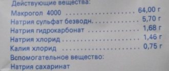
Is it possible to take ASD if you have a stomach ulcer and how to drink the fraction correctly
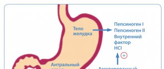
When taken orally, this drug activates the motility of the digestive system, stimulates the central and autonomic nervous system, promotes the secretion of digestive enzymes and glands, normalizes digestion, and increases the body's resistance to external negative factors. When used externally, it promotes rapid regeneration of damaged skin, has an anti-inflammatory and antiseptic effect.
INTERESTING fact: Kidney treatment ASD 2
How to drink
There is often a question about how to take the medicine correctly. It all depends on what disease needs to be cured. Each has its own specific dosage regimen. But regardless of the disease, there are certain requirements for admission:
- The water must be boiled. If the unpleasant smell cannot be tolerated, then milk can be used.
- 1 ml contains 30-40 drops of ASD 2.
- When applying compresses, you should use parchment paper. It will prevent evaporation. You need to put a thick layer of cotton wool on the top and bandage it.
- The drug must be stored in a dark, cool place protected from light.
- While taking the drug, it is prohibited to drink alcohol.
For stomach or duodenal ulcers, you should drink 20 drops per ½ glass of water. Take twice a day half an hour before meals.
Before taking ASD 2 for stomach ulcers, you should consult your doctor. He will take into account the presence of concomitant diseases, as well as the nature of the complexity, so as not to harm the body.
Be healthy!