When is the procedure indicated?
The examination is carried out if diseases and pathologies are suspected that may pose a serious danger to the patient. In most cases, barium X-ray of the stomach allows you to confirm or clarify the diagnosis and prescribe the correct treatment.
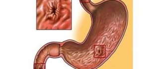
Suspicion of the formation of gastric and duodenal ulcers
Peptic ulcer of the stomach and duodenum (DPC) leads to the periodic appearance of ulcers. It can take years to develop and cause serious harm to health. Using a barium X-ray of the stomach, the specialist finds the ulcer and evaluates its size, shape and other parameters, as well as establishes the stage of the disease and detects accompanying symptoms.
Detection of a malignant process in an organ
If the development of a malignant process is suspected, an X-ray of the stomach with barium is recommended without fail, since the consequences of the disease can be deadly for the patient. In the photographs, the cancerous process looks like a defect in the mucous membrane. Often, after a barium X-ray of the stomach, an endoscopic examination is performed and a sample of the tumor is taken for laboratory testing.
Diverticulosis and other deformities of the gastric walls
To diagnose wall protrusion (diverticulosis), a barium X-ray of the stomach requires additional preparation. After administration of the contrast agent, the patient alternately lifts the upper and lower parts of the body so that the contrast fills the entire organ cavity. On a barium X-ray of the stomach, small diverticulosis looks like an ulcer, but has a horizontal level and a neck.
Any inflammation of the stomach
Foci of inflammation in the stomach can appear as a result of various processes. Including peptic ulcers or oncology. An X-ray of the stomach with barium allows you to find the source of inflammation and, based on indirect signs, determine its cause.
Swallowing dysfunctions
Dysphagia, or swallowing dysfunction, is often expressed by the patient complaining of the inability to swallow food and its accumulation in the esophagus. In this case, the patient cannot always accurately indicate the location of the obstacle. Therefore, barium radiography of the stomach is used to accurately localize the pathology.
Pain in the abdominal area or navel area
Pain in the navel and abdominal area may indicate the development of malignant processes in the stomach and duodenum. Therefore, with such symptoms, the specialist often refers the patient to an X-ray of the stomach and duodenum with barium.
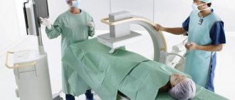
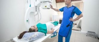
Carrying out
An X-ray of the esophagus and stomach begins with a general image of the abdominal cavity. After this, the patient is asked to drink prepared barium sulfate. The initial targeted photograph is taken after two sips of the drug. At this moment, the relief of the walls of the esophagus with barium is determined. Then the patient is allowed to finish the rest of the drug. During the examination, the doctor can press on the patient's abdomen to promote better distribution of the contrast. During the process, various pictures are taken in different positions - lying on the back as standard or with the pelvis elevated at an angle of 45°, lying on the side, standing. At the same time, at the command of the radiologist, the patient must hold his breath. Modern X-ray rooms are equipped with a special table that rotates while taking pictures. As a rule, to examine the upper part of the gastrointestinal tract, it is enough for an adult to take 250–300 ml of dissolved barium sulfate (in some cases, the dose may be increased). If radiography is prescribed for a child, then the required amount of suspension is calculated based on the age category. For children, as a rule, 100 ml of barium paste is enough.
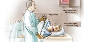
Bowel examination
X-ray of the small intestine with barium is performed in stages. The procedure begins with the patient drinking 0.5 liters of barium suspension. If the study is carried out with double contrast, the drug enters the body through a special tube that is inserted into the patient’s mouth. Along with the contrast, air or inert gas is supplied. After this, wait at least 2 hours - during this time the barium has time to reach the small intestine. As contrast fills the small intestine, the radiologist takes a series of pictures, asking the patient to assume different body positions. And after relieving themselves, they take the last control shot. After filling them with contrast, the diagnostician examines different segments of the small intestine in detail on the monitor for half an hour. The contrast procedure allows you to evaluate intestinal motility and its mucous membrane. While the contrast agent is still present in small quantities, the relief of the inner wall of the intestines is examined, and when there is a lot of contrast, the shape, size, contours and functionality of the intestines are assessed. With maximum filling of barium, it is possible to identify inflamed segments, ulcerative processes and identify neoplasms. If the passage of barium is disrupted, then the radiologist gently presses on the anterior wall of the peritoneum to distribute it evenly. During a procedure with contrast, as the substance is distributed in the intestinal lumen, an experienced radiologist can draw conclusions about the presence of pathological processes. If the barium suspension is distributed in the form of flakes, then this is a clear sign of impaired absorption. And if the contrast fills the lumen unevenly, this may indicate oncopathology.
What can be detected with radiography?
If a barium stomach x-ray is performed correctly and with proper patient preparation, the examination can provide enough comprehensive information to make a diagnosis in most cases. At the same time, to detect a particular disease, a specialist evaluates a number of parameters.
Gastric lumen disorders
Disturbances in the lumen of the stomach may indicate the development of a malignant process or diverticulosis. If the lumen of the stomach is narrowed on a barium X-ray, diffuse fibroplastic cancer is possible. With diverticulosis, on a barium X-ray of the stomach, the lumen appears larger than it should be normally.
Abnormal placement of the stomach
Gastroptosis (prolapse of the stomach) is often diagnosed with an abdominal wall hernia or diaphragmatic hernia. The disease is clearly visible using barium X-ray of the stomach.
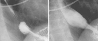
Niche symptom
A niche is a shadow of a contrasting mass that fills a defect during an ulcer or the development of a malignant process. Depending on the location of the defect, an X-ray of the stomach with barium distinguishes between a contour and a relief niche. In the first case, the silhouette of the defect is visible from the full face, in the second - in profile.
The lack of filling is visible in the image as a dark area
A dark area on a barium x-ray of the stomach indicates that there is a place where the contrast could not reach. As a rule, this happens with atrophic gastritis or tumor.
The essence of the examination and its purpose
An X-ray of the stomach should be prescribed by a gastroenterologist.
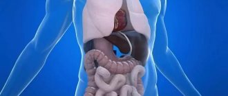
X-ray of the stomach is a technique for examining the human gastrointestinal tract.
The essence of diagnosis is to assess the condition of a given part of the body and make a specific diagnosis for the patient or state that he is completely healthy.
X-ray of the gastrointestinal tract is based on the use of ionizing radiation, which, unevenly passing through different tissues of the body, ultimately allows special equipment to form a more or less clear image of the insides of a person.
This image displays all the structures of the human body in two forms: dark and light shades. The first way is to form all the sections and components of the body that allow air to pass through, the second way is to form the bone structures.
Thanks to the fact that in medicine it is clearly known about the form of the normal human gastrointestinal tract, specialists are able to check the health status of the examined patient and give him the correct diagnosis.
Since the stomach and intestines are cavitary organs, in modern gastroenterology it is customary to conduct a contrast examination of them. The essence of such an X-ray is that before radiation begins to pass through the patient’s body, a special substance is introduced into his body, which improves the quality of the final image of the human insides.
How should a patient prepare for the procedure?
Since the patient needs time to prepare for a barium stomach x-ray, it is best to start a few days before the examination. The first step is to exclude from the diet foods that cause increased peristalsis and gas formation. 8 hours before the examination you should completely abstain from food. To relax the stomach before the x-ray and prepare it for filling with barium, the doctor may recommend taking a special drug half an hour before the examination. Contrast is administered orally a few minutes before the procedure. When a double contrast examination is required, after the patient has prepared for a stomach x-ray and taken barium, he will need to drink a gas-forming mixture.
Preparation
It is necessary to prepare for the study in advance, since its results and their reliability depend on the quality of the preparation.
How to prepare:
- limit food and water intake (the procedure is carried out on an empty stomach);
- follow a diet;
- take medications (if necessary);
- to refuse from bad habits.
More information about this and some other features of the preparatory stage can be found in a separate article.
When is x-ray prohibited?
An X-ray of the stomach with barium is a safe examination, but since it is done using ionizing radiation and a contrast agent, the method has a number of contraindications.
Hematopoietic disorders caused by pathological conditions of the bone marrow
Ionizing radiation can worsen the disease. Therefore, if hematopoiesis is disrupted, an X-ray of the stomach with barium is not performed.
Cataract
When X-raying the stomach with barium, the radiation dose does not exceed permissible limits. However, with cataracts, even a limited amount of ionizing radiation may be enough to have a negative effect on the condition of the lens.
Malignant neoplasms of the bronchi and lungs
The examination is contraindicated if the patient is undergoing radiation or chemotherapy treatment.
Pregnancy
During pregnancy, even minor radiation can have a detrimental effect on the development of the fetus. Therefore, X-rays of the stomach with barium are contraindicated for expectant mothers.
Childhood (before the onset of puberty)
In childhood, the body reacts poorly to exposure to even small doses of ionizing radiation. Therefore, doctors refer children for an X-ray of the stomach with barium only if absolutely necessary.
Possible complications
Immediately after the x-ray, it is recommended to drink a lot of clean water, this will help remove the barium mixture from the body faster. It does not dissolve in liquid, so it is not absorbed into the walls of the digestive tract and does not cause damage to the body. Among mild complications, patient reviews note attacks of nausea and vomiting, but this is only possible with increased sensitivity. Constipation may occur for 2-3 days after the session; this is a common reaction that goes away with time. The stool also turns whitish or gray. If bowel problems do not go away after three days, you should contact your doctor.
Such side effects do not cause significant discomfort, so they are not called complications. Rare and serious consequences include an allergic reaction. This is possible if the preparation rules were violated and the patient did not undergo a barium test.
Sometimes the barium suspension is replaced with an iodine solution, so the reaction to it must also be checked. In case of allergies, the patient may experience swelling of internal organs, difficulty breathing, and Quincke's edema. If the patient is well prepared for the procedure, gastrography will take place without negative consequences.
Price
Pricing policies in different regions of the country have some differences. It is determined by the prestige of the clinic, its location, equipment, and qualifications of the staff.
- The highest price for an x-ray of the esophagus in Moscow and St. Petersburg is from 4,500 rubles.
- In the regions, prices are slightly lower: in Nizhny Novgorod and Novosibirsk - from 3000, in Voronezh - from 2000, in Krasnodar and Ufa - from 1000 rubles.
The official websites of clinics do not always have accurate data on the cost of the study. Often, when a client contacts him personally, it increases due to various reasons (for example, overestimation, outdated data). Moreover, the price may not be indicated in full for the entire procedure, but only for fluoroscopy or 1 x-ray. But it is also possible for the price to decrease during promotions or discounts. Therefore, when choosing a clinic, you should remain vigilant and clarify financial issues before the procedure begins.