Villous tumor of the rectum is a benign epithelial formation that is prone to malignancy. The insidiousness of the disease is that it may not manifest itself for a long time, while treatment in the initial stages is the least traumatic and very effective.
- Features of villous tumor
- Causes of development and risk factors
- Classification
- Clinical picture
- Diagnostics
- Treatment
- Complications
- Forecast
- Prevention
Features of villous tumor
The neoplasm has a round or oval shape, pinkish color and a peculiar papillary or velvety surface. The main element of the tumor is the villus. It is an elongated connective tissue formation that contains many vessels and is covered on the outside with columnar epithelium. Numerous villi merge with each other and form lobules, which are visible to the naked eye on the surface of the formation.
The villous tumor grows mainly in the lumen of the rectum. The size of the tumor can vary: from a few millimeters to 6-8 cm or even more.
Causes
Rectal cancer can occur due to diseases - Crohn's disease, Ulcerative colitis.
The main cause of rectal cancer is considered to be a complication of chronic intestinal diseases. These include:
- Familial multiple intestinal polyposis. This disease has a genetic predisposition and is manifested by the formation of a large number of polyps.
- Crohn's disease - the risk is 26%.
- Ulcerative colitis – the risk is 50% if the disease lasts for 30 years or more.
- Chronic constipation.
- Fistulas of the rectum and large intestine, proctitis.
- Hereditary intestinal diseases - Gardner's syndrome and Turcot's syndrome (a combination of intestinal polyposis with brain tumors and epidermoid cysts).
Additional factors influencing the development of this type of cancer are work at a chemical plant (asbestos, hydrocarbons, nitro compounds), anal sex, HPV (human papillomavirus), sedentary lifestyle, obesity. Separately, it is necessary to note such an important factor as an unbalanced diet, the predominance of red meat, fatty, fried and smoked foods in the diet, a small proportion of vegetables and fruits in the menu.
Causes of development and risk factors
The exact causes of benign neoplasms of the rectum, including villous tumors, are not completely clear. Factors that, according to a number of researchers, may influence the increase in incidence of this type are:
- Decreased level of physical activity.
- Dietary features (addiction to high-calorie foods with high fat content, insufficient fiber intake).
- Deterioration of the environmental situation.
Villous tumors usually form in older people. Men suffer from this disease somewhat more often than women.
Prevention of adenocarcinoma of the large intestine
- A diet in which “healthy” foods will prevail: fruits, vegetables, vegetable fats, cereals, etc.
- Timely treatment of infections and diseases of the gastrointestinal tract.
- Avoiding frequent stressful situations and exposure to harmful factors (for example, hazardous working conditions). This may include strictly limiting the consumption of alcohol and tobacco products or completely abstaining from them.
- Moderate use of medications, especially those that can irritate the walls of the stomach and intestines.
- Do not abuse anal sex, but rather avoid it.
- Studying your pedigree to see if any of your relatives have malignant neoplasms. You can consult a specialist about this.
As a rule, more than one risk factor influences the appearance of a dangerous disease, so you should definitely listen to the above advice. Thus, the less harmful the effect on the body, the less likely it is to get cancer.
Be smart and pay more attention to your health!
Classification
There are two forms of villous tumor:
- Nodular, more common, has the appearance of a single tumor node on a wide base.
- Creeping - lobular growths line the inner surface of the rectum. Damage to the mucous membrane can have a significant extent.
Based on the nature of the surface, these new formations are:
- Fringed, which have pronounced villi.
- Lobed, resembling cauliflower in appearance.
Solitary villous tumors are more common. Multiple formations of this type are diagnosed extremely rarely.
Modern methods of treating colorectal cancer
Modern methods of treating cancer include surgery, chemotherapy and radiation therapy. Also, at the present stage, biological and immune drugs are used.
- Radiation therapy is used if, before surgery, the doctor discovered that all three layers of the intestinal wall are involved in the process, and there are metastases in the lymph nodes. Radiotherapy is indicated before surgery, as one of the stages of preparation for surgery.
- A combination of chemotherapy and radiation is used for local spread of cancer, damage to the lymph nodes, and tumor invasion of the intestinal fascia (when the tumor is motionless relative to the pelvis).
The specific type and sequence of treatment is determined on the basis of clinical, histological, and instrumental studies. Cancer treatment must be comprehensive. It involves supporting the body during chemotherapy and radiation exposure, and high-quality postoperative treatment.
Operational tactics
- 1. If the size of the tumor allows, the intervention is performed endoscopically. This type of intervention is possible if tumor growth affects only the mucosal and submucosal layers.
- 2. The modern method of treatment is transanal endomicrosurgery. It allows you to preserve the intestinal walls as much as possible and is characterized by the most comfortable postoperative period.
- 3. If the tumor has reached the muscle layer, the entire section of the intestine that is involved in the process is removed. The surrounding tissue and lymph nodes are also removed. An open or laparoscopic technique can be used.
Most often, the location of the tumor makes it difficult to preserve the natural anus and requires the formation of a colostomy. Modern surgeons are maximally focused on preserving the functionality of the organ, so they can form a preventive colostomy, which reduces the risk of infectious complications and inflammatory processes in the suture area. With a favorable prognosis, the colostomy is closed within several months after surgery.
Combined surgical interventions are required if several organs are involved in the process. Distant metastases require sequential removal.
Prevention and life prognosis
Even the best treatment does not exclude the possibility that cancer cells will remain in the body. They are so small that they are difficult to detect using diagnostics. However, they are fraught with danger and can result in a relapse.
The earlier a relapse is diagnosed, the easier it is to treat. Therefore, even after surgery and a course of therapy, the patient should undergo diagnostics. After rectal cancer, polyps may reappear and become malignant over time. It is important to diagnose this process and provide treatment before the process becomes uncontrollable.
The first two years after treatment are the time of greatest risk for recurrence of the disease. Monitoring of new cases of the disease should be especially intensive at this time.
It is advisable to be examined by a doctor every six months for the first 2 years and once a year for the 3rd, 4th and 5th year after surgery. According to studies, after 5 years from the moment of treatment, the risk of relapse sharply decreases.
The consultation includes a physical examination, analysis to measure tumor markers, colonoscopy, chest x-ray, CT scan, ultrasound.
Prevention of colorectal cancer includes the following measures:
- balanced diet high in fiber;
- preventive colonoscopy for early detection of polyps and chronic inflammation;
- avoidance of stress, adequate sleep and normal physical activity;
- maintaining normal metabolism;
- prevention of HPV infection;
- it is necessary to minimize exposure to carcinogens;
- maintaining and strengthening the immune system.
Clinical picture
The disease may be asymptomatic for a long time. Therefore, often a neoplasm is discovered only when a person consults a proctologist as planned or for another reason.
Patients with rectal villous tumor may complain of:
- Constipation and difficulty in defecation, feeling of incomplete bowel movement.
- Discharge of a mucous nature, which can lead to maceration and itching of the perianal area.
- An admixture of blood in stool and mucus. With regular bleeding, the patient may experience a clinical picture of anemia: pale skin, weakness, dizziness.
- Pain in the rectum.
If located low, the villous tumor of the rectum may fall out during defecation, and the patient has to straighten it with his fingers. With large tumors, partial or complete intestinal obstruction may form.
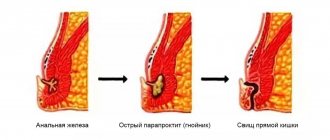
What do they do in particularly difficult cases?
Then, when the malignant process has caused complications, surgical intervention is performed urgently.
First, the tumor is removed and complications are eliminated, then the formation of a colostomy begins; it can be made single-barrel or double-barrel. In the case of the first option, the movement of feces passes directly through the colostomy.
With the second option, feces can be eliminated naturally. After the appropriate operation, the intestines begin to function normally after 2-5-7 months, this depends on the success of the operation and the individual characteristics of the body of each individual patient.
Diagnostics
Instrumental methods for diagnosing a neoplasm include:
- Total colonoscopy or sigmoidoscopy with biopsy is an informative study that allows you to detect a tumor, identify its size and location, and also obtain biological material for histological analysis. This requires 3-5 grips with standard endoscopic forceps. The specificity and effectiveness of the method increases with the use of modern technologies (magnifying and narrow-spectrum endoscopy, fluorescent diagnostics).
- Ultrasound using a rectal probe makes it possible to assess the degree of invasion of the walls of the rectum by the tumor.
- MRI of the pelvis, which is preferably performed in the presence of large tumors (more than 3 cm) and if malignancy is suspected. The method helps to clarify the main characteristics of the tumor and assess the condition of regional lymph nodes.
If it is impossible to perform an endoscopic examination, irrigoscopy or CT colonography is recommended.
Diagnosis of villous polyp of the rectum
The classic clinical symptom of rectal villous polyp is the discharge of mucus from the anus during bowel movements. The amount, color and consistency of mucus varies from patient to patient. Most often, mucus is released with feces without pain, but sometimes tenesmus is observed. Mucus from villous polyps of the rectum does not irritate the skin and does not cause itching in the anal area.
Another important symptom of villous tumors is the discharge of scarlet blood from the rectum. Most often, a mucous-bloody fluid is released. It can be mixed with feces or excreted on its own.
Doctors at the Yusupov Hospital carry out early diagnosis of villous polyps of the rectum using modern screening programs. Proctologists widely use a non-invasive research method - a stool occult blood test (hemoccult test). The prerequisite for its implementation is that the villous polyp of the rectum bleeds to varying degrees. For the test to be reliably positive, a daily blood loss of at least 20 ml is required. For a positive test result, hemoglobin released from red blood cells must also be present in the stool.
To obtain reliable results of the hemoccult test, the patient is recommended to follow the following rules:
- Eliminate beef and vegetables with high peroxidase activity from the diet;
- Do not take medications that contain iron;
- Avoid taking high doses of ascorbic acid.
At least two smears are carried out over three days.
If there are specific complaints, the patient is examined by a proctologist at the appointment. The doctor collects anamnesis, studies clinical manifestations and additionally prescribes examinations. If the villous polyp is located at the bottom of the rectum, the proctologist can detect it during a digital examination. In cases where the tumor is located high, doctors at the proctology department perform sigmoidoscopy or colonoscopy using the latest endoscopic equipment from the world's leading manufacturers. During the examination, a functional diagnostics doctor may see the following changes:
- The intestinal lumen is deformed;
- The bends of the intestine have lost mobility;
- Increased bleeding;
- The villous polyp contains ulcers, fibrous and necrotic masses.
Such symptoms indicate that the villous polyp has transformed into a malignant tumor.
If endoscopic methods are not available, radiologists perform irrigoscopy. This diagnostic method is not as accurate and may give a false negative result. If there are doubts, proctologists at the Yusupov Hospital repeat the examination after 6-8 weeks. The final diagnosis is made based on histological studies, for which doctors take biological material during endoscopic examinations.
With a digital examination, the proctologist is able to examine a section of the rectum up to 10 cm from the edge of the anus. This primary diagnostic method precedes sigmoidoscopy, since it is a fairly informative way to identify both villous polyps and other diseases of the rectum (hemorrhoids, fistulas, fissures), surrounding tissue (cysts and tumors), and the prostate gland in men (adenoma, prostatitis, cancer ). The method allows you to evaluate the shape, consistency, mobility, presence or absence of a stalk of the villous polyp. Low-lying polyps are always detected by digital examination. Small adenomatous polyps, which are localized above 5–6 cm from the anus, are difficult to determine using the finger method.
Treatment
All villous tumors of the rectum must be surgically removed due to the precancerous nature of these tumors. Depending on the size of the lesion and its location, the following types of surgical treatment can be used:
- Endoscopic removal using a diathermic loop. The method is preferable for tumors less than two cm, with the exception of signs of malignancy. The operation is performed in an endoscopy room without anesthesia. The patient can be operated on using one of the technical methods: one-step excision of the tumor, or fragmentation by removal in one stage or during several planned visits.
- Transanal excision of a tumor using mirrors. This method is used for tumors whose lower edge is no higher than 7 cm of the skin of the perianal area. Surgery is performed under general anesthesia or epidural anesthesia.
- Posterior proctotomy - removal of highly located tumors the size of the rectal wall.
- Resection of the rectum is a full-fledged abdominal operation under endotracheal anesthesia with removal of the tumor along with the affected area of the intestine. The use of this method is preferable when detecting large tumors located in the rectosigmoid region.
If at the diagnostic stage signs of malignant degeneration were identified, the scope of surgical intervention is significantly expanded. In this case, the rectum can be partially or completely removed, and pararectal tissue and regional lymph nodes are also excised.
Neoplastic polyps and villous adenomas of the colon
A tumor is a pathological process represented by newly formed tissue, in which changes in the genetic apparatus of cells lead to disruption of the regulation of their growth and differentiation (M. A. Paltsev, N. M. Anichkov, 2001). Based on the nature of their growth, tumors are divided into benign and malignant.
Definition.
The definition of a polyp is not clear. Currently, a true polyp is considered to be a proliferation of glandular epithelium, forming an elevation above the level of the mucous membrane in the form of a wide mushroom-shaped, often branched formation, sitting on a narrower stalk, and sometimes on a wide base.
Etiology and pathogenesis.
It is very difficult to establish the true incidence of benign colon polyps, since they are practically asymptomatic, and they are most often discovered by chance in patients who complain of discomfort, pain in the anus, intestinal dysfunction, pathological discharge from the anus, etc. , which are signs of other diseases (hemorrhoids, paraproctitis, anal fissure, colitis, rectal cancer, etc.). In this regard, a close to true frequency of polyps can only be established as a result of targeted preventive examinations of the population or autopsies. As a result of the work of Russian and foreign scientists, it was established that the frequency of detection of colon adenomas (using only sigmoidoscopy) ranges from 2.5 to 7.5% of the total number of patients examined. However, the true incidence of their occurrence is certainly higher, since during the examination the authors did not examine other parts of the colon, in which about 50% of all colon adenomas are located.
The most accurate method for assessing the condition of the mucous membrane of the rectum and colon can be considered their study during pathological and anatomical autopsies. According to the literature, the frequency of detection of colon polyps during autopsies for economically developed countries averages about 30%. According to the State Scientific Research Center (1987), when studying the results of preventive examinations (digital examination and sigmoidoscopy) of two groups of patients (15,000 people) - practically healthy and complaining of discomfort in the anorectal area - it was found that in the structure of diseases of the colon, polyps accounted for only 16 %, while in the group of practically healthy individuals this figure is significantly higher - 40.6%. This difference is due to the fact that some patients whose polyps are asymptomatic do not come to the attention of doctors.
The etiology of polyps of the rectum and colon is not clear. Works that study the viral nature of these diseases are theoretical in nature, as is the creation of an animal model of colon polyposis.
The increase in the incidence of benign tumors of the colon is associated with environmental influences (metropolises, the presence of large industries), and a decrease in physical activity. Many researchers believe that an important factor influencing the increase in the incidence of colon disease is the change in the nutritional pattern of the population in the context of industrialization.
It has been established that the main feature of the diet of residents of economically developed countries is the predominance in the diet of high-calorie foods with a high content of animal fats and a small amount of fiber. All this leads to the fact that chyme containing little fiber enters the large intestine, which affects the decrease in motor activity of the intestine, and a large amount of bile acids, which, as has been established, during the digestion process are converted into substances that have a carcinogenic effect on the mucous membrane. A decrease in the rate of passage of chyme through the intestine creates longer contact between carcinogens and mucous membranes. All this causes disruption of the microbial landscape, which in turn changes the composition of enzymes of microbial origin. Some researchers have established a certain relationship between the frequency of detection of adenomas and the male gender of the deceased, as well as diseases such as atherosclerosis, malignant tumors, diverticulosis and other diseases of the gastrointestinal tract, and chronic nonspecific lung diseases.
Pathological anatomy.
According to the International Histological Classification of Tumors, benign neoplasms of the colon are presented as follows.
1. Adenoma: a) tubular (adenomatous polyp), b) villous, c) tubular-villous.
2. Adenomatosis (adenomatous intestinal polyposis).
Tumor-like lesions.
1. Hamartomas: a) Peutz-Jeghers polyp and polyposis; b) juvenile polyp and polyposis.
Heterotopias.
Hyperplastic (metaplastic) polyp.
Benign lymphoid polyp and polyposis.
Inflammatory polyp.
Deep cystic colitis.
Endometriosis.
Hyperplastic polyps look like small ones (up to 0.5 cm in diameter), slightly rising above the level of the mucous membrane, formations of soft consistency and normal color. They are characterized by elongation and cystic expansion of the crypts. The epithelium in such polyps is sawtooth-shaped, with a reduced number of goblet cells.
Glandular and glandular-villous (tubular adenomas) are larger formations (up to 2-3 cm in diameter), usually having a pronounced pedicle or a wide base. In color they are close to the surrounding mucous membrane, but have a denser consistency, move along with the mucous membrane, and rarely bleed or ulcerate. According to the degree of morphological differentiation of the epithelium, three groups of tubular adenomas are distinguished: with mild, moderate and significant dysplasia. With a weak degree, the architectonics of the glands and villi are preserved; the number of goblet cells decreases, their nuclei are elongated, somewhat enlarged, but arranged in one row; the number of mitoses increased slightly. With severe dysplasia, the structure of the glands and villi is disrupted, the nuclei can be located in all parts of the cell, their increase is noted, many mitoses appear, including pathological ones; goblet cells disappear. Moderate dysplasia is characterized by intermediate changes.
Villous adenomas have a slightly lobulated surface, resembling a raspberry. As a rule, they are larger in size than tubular adenoma.
Juvenile polyps cannot be classified as adenomas, because they do not have glandular hyperplasia and atypical changes in the glandular epithelium. This formation is quite large, often hanging into the intestinal lumen on a long stalk, smooth, more intensely colored (bright red, cherry color). Under microscopy, it appears as a cystic granulating polyp, the dilated glands of which are lined with typical intestinal epithelium and contain a mucous secretion.
Classification.
According to the clinical picture, all benign tumors of the colon can be divided into two main groups: epithelial tumors, which are most common (92%) and pose the greatest risk of growth and malignancy, and rare neoplasms, the frequency of individual forms of which ranges from 0.2-3. 5% (overall 8%), the likelihood of their malignancy is low, except for melanoma and carcinoid. The division of epithelial tumors according to histological structure, size and multiplicity factor is of great clinical importance.
Based on their histological structure, polyps are divided into:
— hyperplastic (2%);
— glandular (51.6%);
— glandular-villous (21.5%);
- villous (14.7%).
The probability of its malignancy depends on the size of the benign tumor: the larger the size of the benign tumor, the higher the probability of its malignancy.
Based on the multiplicity factor, epithelial tumors are divided into:
1. single;
2. multiple:
- group;
- absent-minded.
3. diffuse (familial) polyposis.
The multiplicity factor is important in the prognosis of the disease - single polyps rarely become malignant (1-4%) and have a more favorable prognosis. Multiple polyps can be compactly located in one of the sections of the colon or can be found in 1-2 or more in each section (scattered), malignizing up to 20%. Scattered multiple polyps are difficult to differentiate from diffuse polyposis of the colon. The latter is usually characterized by the massiveness of the lesion (there are hundreds and thousands of polyps, and sometimes there are no areas of unaffected mucous membrane left at all), and most importantly, it is inherited, i.e., it is familial, genetically determined and has a significant tendency to malignancy (80-100 %).
Among the epithelial polypoid formations of the colon, there are peculiar, exophytically growing, spreading along the intestinal wall, soft to the touch formations of a small lobular structure. Histologically, these are villous adenomas, and the clinical term villous tumor can be applied to them.
There are two forms of villous adenomas based on their microscopic appearance: creeping and nodular. The nodular form is more common and is located on one of the intestinal walls in the form of a compact exophytic node with a wide and short base or stalk. In the creeping form, villous growths are located flat on the surface of the mucous membrane, almost circularly covering the intestinal wall.
Macroscopically, villous tumors are reddish in color due to the abundance of blood vessels in their stroma. Thin and delicate villi are easily injured and bleed, so bleeding in itself is not evidence of malignancy of these formations.
Malignant transformation of a large colon adenoma can be diagnosed with a high degree of probability if two or more of the following endoscopic signs of malignancy are present: dense consistency of the villous formation, the presence of areas of compaction, surface tuberosity, fibrin overlay, surface ulceration and contact bleeding. It is advisable to distinguish villous tumor of the colon as an independent nosological unit.
Most epithelial neoplasms (polyps) go through successive stages of development from small to large, from low to high proliferative activity, up to the transition to an invasive cancer process.
The appearance of hyperplastic polyps precedes the appearance of glandular (adenomatous) polyps, which, as they grow, can undergo villous transformation, and signs of invasive growth can be detected in the villi. The development of polyps occurs slowly from the simplest structure to sharp degrees of atypia and dysplasia of the mucous membrane, up to the development of cancer, and this process takes at least 5 years, and on average lasts 10-15 years.
Clinical picture.
In most patients, benign neoplasms of the colon are asymptomatic and are detected mainly during endoscopic examination. However, when villous tumors reach large sizes (2-3 cm), bloody-mucous discharge, pain in the abdomen and anus, constipation, diarrhea, and anal itching may occur. In giant villous tumors, loss of protein and electrolytes due to hyperproduction of mucus can sometimes lead to significant disturbances of homeostasis (dysproteinemia, water-electrolyte imbalance, anemia). With them, symptoms of acute complete or partial obstruction (due to intussusception) may appear. The malignancy index of villous tumors is quite high and amounts to 40%.
Diagnostics.
If the symptoms listed above are present, it is necessary to conduct a digital examination of the rectum and sigmoidoscopy. With digital examination, it is possible to examine a section of the rectum up to 10 cm from the edge of the anus. This primary diagnostic method should always be used. It must necessarily precede sigmoidoscopy, since this is a fairly informative way to identify other diseases of the rectum (hemorrhoids, fistulas, fissures, etc.), surrounding tissue (cysts and tumors) and the prostate gland in men (adenoma, prostatitis, cancer).
Sigmoidoscopy requires special preparation using cleansing enemas or oral laxatives (fortrans, etc.). This research method is more informative and makes it possible to detect the majority of colon polyps, since more than 50% of them are localized in the rectum and sigmoid colon, i.e., within the reach of a proctoscope (25-30 cm from the edge of the anus). If polyps are detected in the rectum or sigmoid colon, a thorough examination of the overlying sections of the colon and stomach is necessary, since polyps often affect different parts of the gastrointestinal tract in combination. For these purposes, X-ray and endoscopic examinations of the colon and stomach are used.
Irrigoscopy has important clinical significance; it allows you to diagnose most polyps more than 1 cm in diameter; smaller formations can be detected much less frequently. Therefore, during preventive examinations, it is better to use a colonoscope, which can detect almost any formation (less than 0.5 cm in size).
During an endoscopic examination of the colon, hyperplastic polyps appear as small (less than 0.5 cm in diameter), slightly rising above the level of the mucous membrane, formations of soft consistency and normal color. Often hypertrophied lymphatic follicles simulate hyperplastic polyps (this is confirmed by histological examination). Adenomatous polyps are more than 0.5 cm in size and can reach 2-3 cm in diameter, have a stalk or are located on a broad base, are close in color to the surrounding mucous membrane, but have a denser consistency, are displaced with the mucous membrane, ulcerate and rarely bleed .
Adenopapillomatous polyps (glandular-villous) usually exceed 1 cm in diameter, have a velvety surface, which gives the impression of dullness in color, sometimes appear finely lobulated due to the uneven surface, can be eroded, and the bottom of the ulcers is covered with fibrin, from under which a small amount is released blood.
Villous polyps are large in size (from 2 cm or more), may have a thick stalk (polyps) or spread across the mucous membrane (tumors), sometimes taking on a creeping character. They occupy a large area, only slightly rise above the surrounding mucous membrane and do not have clear boundaries. The color of such formations differs little from the color of the mucous membrane, their surface is characteristically velvety and dull, the presence of ulcerations allows us to suspect the onset of malignancy. Negative biopsy results cannot serve as evidence of the absence of malignant growth, and the final conclusion is made after removal of all villous tumor.
Treatment.
Conservative methods for the treatment of polyps and villous adenomas of the colon currently do not exist. The method of treating polyposis with the juice of the celandine herb proposed by A. M. Aminev (1965) has not found widespread use due to its dubious effectiveness. Its use is inappropriate, since attempts at conservative treatment only lead to postponing the operation and progression of the disease until the polyp becomes malignant.
A biopsy is not essential in determining the treatment strategy for colon polyps. Small areas of the polyp taken for biopsy cannot characterize the essence of the pathological process in the entire tumor. Information about a polyp based on a biopsy is incomplete and may be erroneous. A completely excised polyp is the best material for histological examination.
In modern conditions, only removal of polyps endoscopically and surgically guarantees the success of treatment. The most common methods of surgical treatment of polyps and villous adenomas of the colon are:
• polypectomy using a rectoscope or colonoscope with electrocoagulation of the stalk or bed of the polyp;
• transanal excision of the tumor;
• colotomy or bowel resection with tumor;
• transanal resection of the rectum with the formation of a rectoanal anastomosis for circular or almost circular villous tumors of the lower ampullary rectum;
• transanal endomicrosurgical excision of the tumor.
All methods of removing polyps are used after special preparation of the colon using laxatives and cleansing enemas. This preparation also serves to prevent complications.
One of the main complications is bleeding, which can occur up to 10 days after the intervention. The appearance of blood from the anus on the 1st day after removal of the polyp is associated with insufficient coagulation of the vessels of the polyp leg. Later bleeding develops as a result of scab rejection, which is most often observed 5-12 days after surgery. Both early and late bleeding can be insignificant, or they can be massive, posing a danger to the patient’s life. To eliminate this complication, a repeat endoscopic examination is required, during which electrocoagulation of the bleeding vessel is performed. Sometimes such measures do not help, and you have to resort to laparotomy and intestinal resection.
The second most common complication is perforation of the intestinal wall, which can also occur either during the intervention or some time, even several days, after it. The occurrence of a late complication is explained by a deep burn of the intestinal wall at the base of the removed tumor during electrocoagulation.
If this complication occurs on the intra-abdominal part of the colon, a laparotomy is performed and the defect in the intestinal wall is sutured, this section is disconnected from the stool passage by applying a colostomy to the overlying sections, or, if the perforation occurs high enough, the damaged area is removed in the form of a double-barreled colostomy. In the future, such patients are treated as patients with peritonitis, despite the fact that after preparation there is no content in the intestine and during perforation only gas enters the abdominal cavity. With the availability of modern antibacterial agents and anti-inflammatory therapy, this can be dealt with without complications. If the postoperative course is favorable, the question of closing the colostomy can be raised after 2-4 months.
After removal, all colon tumors must undergo histological examination so that the degree of epithelial dysplasia or the presence of malignancy can be judged.
If adenomatous and villous polyps are detected, the patient can be discharged from the hospital under mandatory follow-up.
If areas of transition to adenocarcinoma are detected, a repeat colonoscopy or rectoscopy is necessary, with material taken from the tumor bed for histological or cytological examination. In the absence of adenocarcinoma complexes, the patient can be discharged from the hospital with mandatory monthly endoscopic examination; if a tumor recurrence is suspected, re-hospitalization, a thorough examination and a decision on further treatment tactics are necessary.
If complexes of malignant cells are detected in the material from the tumor bed, a decision is made on radical surgery.
Long-term results of treatment and follow-up.
Considering the possibility of recurrence of benign neoplasms of the colon and the occurrence of cancer, especially in the first 2 years after surgery, patients should be under constant follow-up. After removal of benign polyps, the first examination is carried out after 1.5-2 months, then every six months, and for villous tumors - every 3 months. during the first year after removal. Further inspection is carried out once a year.
After removal of malignant polyps, a monthly examination is required in the 1st year after surgery, and every 3 months in the 2nd year of observation. And only after 2 years are regular examinations every 6 months possible.
In the first 2 years after removal of benign tumors, relapse was noted in 13% of patients, and new polyps in various parts of the colon - in 7%. Relapses after glandular polyps were observed in 8% of cases, glandular-villous polyps in 13%, and villous polyps in 25%. Considering that the malignancy index of a villous tumor is 40%, an increase in the incidence of malignancy is possible. The occurrence of relapse is an indication for urgent re-operative intervention.
Complications
Possible complications of the disease are:
- Tumor malignancy.
- Formation of partial or complete intestinal obstruction.
- Bleeding with the formation of iron deficiency anemia.
- Impaired water-electrolyte balance and dysproteinemia, which can be observed with giant tumors.
The most dangerous and at the same time very common complication of villous tumor of the rectum is its malignant degeneration. The malignancy index of this type of neoplasm is 30-70%.
How is treatment for adenocarcinoma of the large intestine carried out?
It is important to note that the treatment process directly depends on the form and stage of the disease. More often, doctors resort to complex therapy.
In particular, a course of radiation is prescribed, after which partial death of pathological (cancer) cells is observed, which leads to a decrease in tumor size. This type of treatment is also carried out after surgical removal of the formation.
This minimizes the likelihood of tumor cells being transferred and reduces possible inflammation of nearby tissues. With all this, an important place is given to compliance with a special diet and drug therapy.
Prevention
The main method of prevention is regular (once a year) endoscopic examination of the rectum by people over 40 years of age. Such measures make it possible to identify a tumor in the early stages of development and carry out the necessary treatment, preventing the possible development of cancer.
It is worth noting that currently there are various options for helping cancer patients suffering from villous tumor of the rectum, even with a widespread tumor process. Having qualified oncologists on staff allows us to achieve the best possible result in each specific case.
Book a consultation 24 hours a day
+7+7+78
Characteristic symptoms of the disease
Intestinal discomfort may be a symptom of cancer.
The main risk group for this disease is those over 50 years of age, and this form of cancer is diagnosed in men one and a half times more often than in women. The insidiousness of the tumor is that the first stages are practically asymptomatic, or with minor manifestations, which can be attributed to many other intestinal diseases.
As the disease progresses, the manifestations become stronger, pain appears, indicating the development of the process. Symptoms of cancer:
- Intestinal discomfort - alternating constipation and diarrhea, bloating, frequent bowel movements.
- The appearance of blood and mucus in the stool, in the last stages – bleeding.
- Constantly elevated temperature.
- Cramping pain in the abdomen, turning into continuous pain in the later stages of the disease.
- Itching in the perineum, skin irritation with discharge.
- Genital dysfunction.
- Manifestations of intoxication – vomiting, headaches.
- Exhaustion, weakness, anemia due to metabolic disorders.
- Painful tenesmus - the urge to defecate, which does not end with the release of feces.
As the disease progresses, intestinal obstruction occurs, leading to inflammation of the peritoneum. Due to the absence or limitation of the act of defecation, bloating develops in the abdomen, it increases in size, intoxication develops, and “pencil” or “ribbon” stools appear. Vomiting and lack of appetite accompany these complications.
Bleeding is a characteristic sign of this disease; blood loss can reach 150-200 ml per day, anemia develops, and there may be profuse bleeding. The last stage of the disease can be complicated by the appearance of urogenital fistulas, the urine becomes cloudy and takes on the smell of feces. Severe cystitis and pyelitis are the consequences of this complication.