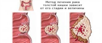
Colon tumors can be benign or malignant. Tubular adenoma of the colon and carcinoid of the appendix (worm-shaped appendix) are benign neoplasms. Colon adenocarcinoma is a malignant neoplasm that develops from glandular epithelial cells. This is one of the histological types of colon cancer.
At the Yusupov Hospital, the presence of colon tumors is determined using modern diagnostic methods. Histologists verify the type of tumor by examining tissue samples obtained during a biopsy under a microscope. In the presence of tubular adenoma of the colon, a description of the microslide is carried out.
Depending on the location and histological type of the tumor, the patient’s condition and the presence of concomitant diseases, oncologists develop an individual treatment plan for the patient. Severe cases of colon carcinoma are discussed at an expert council meeting. Candidates and doctors of medical sciences, doctors of the highest category take part in its work. Leading oncologists in Moscow make a collegial decision on patient management tactics.
Adenocarcinoma can develop from tubular-villous adenoma of the colon with dysplasia. The sequence “adenoma - cancer” has been confirmed by numerous scientific studies. The risk of developing malignant neoplasms of the rectum and colon in individuals with adenomatous polyps is 3–5 times higher than in the general population. Due to the high risk of malignant transformation of tubular adenoma of the colon with dysplasia, oncologists at the Yusupov Hospital carry out early diagnosis and take preventive measures to reduce the incidence of colon adenocarcinoma.
Histological types of colon adenomas
There are 3 histological types of colon adenomas:
- Tubular;
- Tubular villous;
- Villous.
The separation criterion is the ratio of villous and tubular structures. Tubular adenoma of the colon - what is it? Microscopically, tubular adenoma is represented by proliferating adenomatous epithelium. The tumor consists of branching and significantly convoluted glandular tubes, longer than in the normal intestinal mucosa. In tubular adenoma, no more than 25% villous tissue is present. Tubular adenoma of the colon has a mucous membrane-covered base. It is represented by connective tissue, smooth muscle cells and blood vessels. Tubular adenomas have a stalk and a smooth lobulated surface. Less often they are located on a wide base. Creeping tubular adenomas, which protrude slightly above the surface of the mucous membrane, are very rare.
In tubular-villous adenomas, the number of villi increases, which can be detected both on the surface of the polyp and inside large glands. The glands lengthen, acquire an irregular shape, and fit tightly to each other. The degree of epithelial dysplasia increases. In tubular-villous adenoma, the percentage of villous tissue varies from 25 to 75%. The tumor consists of pronounced lobules and has small areas with villi or very small lobules.
Villous adenoma consists of thin finger-like outgrowths of the connective tissue of the lamina propria, which are covered with epithelium. In villous adenomas, a small number of glands and 75% of the villous component can be found. Macroscopically, villous adenomas have a wide base and a “shaggy” surface. There is a special histological type of colon adenoma - serrated adenoma. The tumor is close in structure to a hyperplastic polyp, but has the potential for malignancy.
Adenomatous epithelium belongs to the category of neoplastic. For this reason, each adenoma has signs of dysplasia of varying severity. Histologists distinguish 3 degrees of dysplasia of tubular adenoma of the colon:
- 1st degree – weak;
- 2 degrees – moderate;
- Grade 3 – severe.
Tubular adenoma of the colon with low grade dysplasia is a poorly differentiated tumor. It can transform into adenocarcinoma.
Clinical manifestations
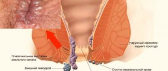
Most tubular adenomas do not cause any specific symptoms. They are detected incidentally during a full examination for another disease of the gastrointestinal tract. Relatively rarely, patients may complain of the appearance of mucous-bloody discharge from the anus, mainly after defecation. Nonspecific manifestations can also include subjective discomfort in the intestines, changes in stool such as diarrhea or constipation.
If there is a large polyp in the colon, obstructive intestinal obstruction may develop. Its possible symptoms are paroxysmal intense pain, which becomes constant as the disease progresses, retention of feces and gases, bloating, and vomiting. Without timely medical care, the patient's condition quickly deteriorates, which can lead to death.
Classification
Histologists distinguish the following types of malignant neoplasms of the large intestine:
- Well-differentiated colon adenocarcinoma;
- Moderately differentiated colon adenocarcinoma g2;
- Poorly differentiated adenoma.
Glandular cancer can generally be represented by the following types of colon carcinomas: tubular, mucinous, signet ring cell, squamous cell. Tubular adenocarcinomas consist of tubular structures. Tumors of this type occur in more than 50% of patients with glandular cancer. They have blurred contours and small sizes.
Mucinous adenocarcinoma consists of mucous components and epithelial structures and has no defined boundaries. Metastasis occurs through the lymphogenous route. The high risk of recurrence is due to insensitivity to radiotherapy.
Signet ring cell adenocarcinomas are characterized by a highly aggressive clinical course. Most patients with tumors of this type, who seek medical help at the Yusupov Hospital for the first time, already have metastases in the lymph nodes and liver. Oncological disease is most often observed in young patients.
Squamous cell adenocarcinomas form in the area of the anal canal. The tumor consists of flat epithelial cells. The clinical course of squamous cell adenocarcinomas is characterized by a high level of malignancy. They often recur, growing into the tissue of the vagina, ureters, bladder, and prostate gland. The five-year survival rate for squamous cell adenocarcinomas does not exceed 30%.
Diagnosis of villous tumor of PC
The diagnosis is based on examination by a proctologist and instrumental research methods. So, when examining the rectum using the finger method, you can palpate the tumor if it is located close to the anus. Characteristics of the tumor: mobile, soft, has a jelly-like structure. Of the instrumental methods, the most indicative are:
- sigmoidoscopy;
- colonoscopy.
These studies not only make it possible to reliably detect a tumor of the rectum, but also to see the structure of the intestine and examine in detail all parts of the colon. If there are contraindications, irrigoscopy can be used. Verification of the diagnosis involves histological examination of the tumor. The material is taken during instrumental examinations.
Reasons for education
The development of tubular adenoma of the colon is promoted by nutritional factors: high fat content and low dietary fiber. Changes in diet affect the likelihood of developing adenoma and adenocarcinoma. Refined fats can disrupt the proliferation of epithelial cells. Nutrient components found in fruits, vegetables and other foods can regulate colon carcinogenesis and influence the progression of adenoma to carcinoma.
The individual risk of developing colon adenoma increases in first-degree relatives of patients with colorectal cancer. The likelihood of developing colorectal carcinomas increases if a person has first-degree relatives diagnosed with colon cancer before the age of 50 years. The risk is especially great if these relatives are brothers or sisters. Environmental factors can interact with the genetic factors of the body, leading to the emergence or progression of “adenoma-carcinoma”.
An increase in the size of the polyp, the number of villi and severe dysplasia increase the risk of malignancy of colon adenoma. According to statistics, 4.8% of tubular, 22.5% of tubular villous and 40.7% of villous adenomas are converted into adenocarcinoma. The risk of transformation of benign neoplasms into malignant tumors increases with the degree of dysplasia. 5.7% of adenomas with mild dysplasia, 18% with moderate dysplasia and 34.5% with severe dysplasia transform into adenocarcinoma of the colon.
Villous, tubulovillous adenomas, and adenomas larger than 1 cm increase the risk of subsequent development of colon adenocarcinoma. This risk is higher in patients with multiple polyps.
Causes
There is still no consensus on the mechanism of development of benign intestinal neoplasms. Risk factors include:
- errors in nutrition - excessive consumption of fatty and meat foods and lack of plant fiber;
- tendency to constipation;
- the presence of chronic inflammatory processes in the gastrointestinal mucosa;
- diseases of the liver and biliary tract.
Non-modifiable risk factors for polyp formation also include age and genetic predisposition. A direct relationship has been proven between the aging of the body and the appearance of benign neoplasms. The peak incidence of tubular adenomas occurs at the age of 50-65 years.
The role of heredity also plays a major role in the development of intestinal polyps: there are many diseases associated with genetic predisposition. For example, diffuse familial polyposis and adenomatous polyposis syndrome are typical examples of genetically inherited forms of colon polyps. Some of these diseases are characterized by the formation of multiple benign tumors throughout the colon and a high risk of oncological transformation.
Symptoms and diagnosis
Most colon adenomas do not manifest themselves clinically. They are discovered incidentally during screening tests or examinations for complaints that are not related to them. Sometimes adenomas cause significant bleeding or lead to chronic anemia due to prolonged hidden blood loss. Large rectal adenomas may be accompanied by tenesmus and mucus secretion. The production of mucus in large quantities causes electrolyte imbalance. Distal rectal adenomas may prolapse through the anus.
Doctors at the Yusupov Hospital identify colon adenomas using sigmoidoscopy and colonoscopy. Adenoma of the large intestine most often takes the form of a polyp located on a broad base or connected to the intestinal wall with a pedicle. Its stalk length depends on the growth rate of the polyp localization. Rapidly growing adenomas have a wide base. Slowly growing ones are located on a stalk, which is formed as a result of peristalsis and stretching of the polyp by a peristaltic wave.
Some colon adenomas have a deepened or flat appearance. They do not rise above the surface of the mucous membrane. They can be visually identified by changes in color, structure of the mucous membrane, and the absence of a capillary network. The Yusupov Hospital uses a simple and effective method for their identification - chromoscopy with indigo carmine.
Diagnosis and treatment
Colon adenomas usually do not manifest any symptoms and are an incidental finding during screening or examination for other complaints.
In rare cases, a tubular adenoma of the colon may bleed, which becomes a reason to consult a doctor. In addition, patients may complain of abdominal pain, frequent constipation or diarrhea.
The gold standard for diagnosis is colonoscopy. In addition to it, irrigoscopy and sigmoidoscopy may be prescribed.
There is an opinion according to which endoscopic removal of only adenomas with a diameter of more than 5 mm is recommended, since smaller formations become malignant extremely rarely and dynamic observation is acceptable. However, a more common tactic is in which all adenomas are removed, regardless of the size of the tumor, since histological studies show that polyps even less than 5 mm in diameter in 60-70% of cases have tubular-type areas and, accordingly, have the potential to degenerate into malignant formations.
There is currently no effective conservative treatment for tubular adenomas, and patients are advised to undergo surgical treatment. The tactics of surgical treatment largely depend on the diagnostic method used to diagnose colon adenoma with dysplasia. If the patient undergoes a colonoscopy, then all detected polyps larger than 5 mm are removed. If polyps were identified during another type of intestinal examination (for example, irrigoscopy), then if a small tubular adenoma of less than 1 cm is detected, a biopsy must be performed, and after confirming the diagnosis, the patient is shown a colonoscopy to remove all visualized adenomas and their histological examination. If a colon adenoma larger than 1 cm in size has been identified, then there is no need for a biopsy - the patient must be immediately referred for a colonoscopy. Thus, colonoscopy for detected adenomas becomes both a diagnostic and therapeutic measure.
When adenomas are localized in the colon, removal is carried out endoscopically through a colonoscope. If the adenoma is located in the rectum, removal can be performed using an endoscope or by transanal endomicrosurgery. The peculiarity of this operation is that in this case the tubular adenoma is removed simultaneously with resection of the intestinal wall. This is explained by the fact that when an adenoma is localized in the rectum, in almost every third case, cancer cells are already detected at its base.
In the case of multiple polyps (so-called diffuse lesions), it is advisable to perform a colotomy or resection of the affected area of the intestine.
Unfortunately, tubular adenomas are prone to recurrence. The most common cause of relapse is incomplete removal of the base of the adenoma, if it is not located on a long stalk. In case of recurrent tubular adenoma, surgical removal of the affected area of the colon by laparotomy may be required, since postoperative changes may be an obstacle to complete endoscopic removal of the recurrent adenoma.
What is tubular adenoma of the rectum?
Tubular adenoma of the rectum is a type of polyp
Tubular adenoma of the rectum is a type of colorectal polyp. This type of benign neoplasm occurs in only 5% of patients.
Most polyps are considered harmless, but as such formations grow, malignant degeneration of the cells is possible. Also, in the later stages, rectal adenoma can cause unpleasant symptoms.
There are two main categories of intestinal polyps – neoplastic and benign. Benign polyps are usually of an inflammatory nature and are rarely characterized by oncological degeneration. Such tumors appear in patients of any age and most often have a long asymptomatic stage.
Screening tests, such as colonoscopy, can help detect polyps in the early stages and remove them to prevent colorectal cancer.
Tubular adenomatous polyps are by definition neoplastic, so they are more dangerous. Despite the general signs of a benign process, the cells of such adenoma are direct precursors of adenocarcinomas. The longer such polyps grow on the walls of the rectum, the higher the likelihood of cancerous growth.
Tubular adenoma can occur in other organs, but most often the tumor is found in the colon or rectum.
Treatment of serrated adenoma of the colon
As part of the treatment of serrated tumors, they are removed surgically. There are no other effective treatments.
The Euroonco clinic uses two techniques for this purpose:
- Electrocoagulation, also known as simultaneous loop excision.
- Complete excision - endoscopic mucosectomy.
Both techniques involve minimally invasive endoscopic intervention. All manipulations are performed under anesthesia, so the patient does not experience discomfort.
It is advisable to perform electrocoagulation for small polyps with a thin stalk. Otherwise, there is a high risk of developing serious complications - burns of the intestinal wall, intestinal perforation, bleeding. In this case, we use complete excision of the polyp. The first step is to remove the marginal zone of the polyp within the healthy mucosa. Then the mucous membrane along with the tumor is excised along the entire length of the polyp.
Over the past few years, colorectal cancer (CRC) has occupied one of the first places in the structure of cancer incidence. In 2010, in the Russian Federation, colorectal cancer was diagnosed in 53,763 patients, of whom 30% died during the first year. During this period, colon cancer was diagnosed in 29,922 patients, of whom 32% died within the first year. Cancer of the rectum, rectosigmoid and anus was detected in 23,841 patients in 2010, with a mortality rate during the first year of 28.6% [1]. At the same time, the number of patients with rectal adenomas, which are the main substrate for the development of adenocarcinomas of this localization, is increasing. Moreover, the size of benign epithelial tumors has a decisive influence on the rate of malignancy [2–4]. Thus, early diagnosis and radical removal of adenomas, especially large ones, can significantly reduce the risk of rectal cancer.
Currently, colorectal surgeons have several methods for treating benign rectal tumors. The most commonly used are loop endoscopic electroexcision and transanal excision of the tumor. However, the use of these techniques is significantly limited by such factors as size, growth pattern, and the height of the tumor from the dentate line [5–9]. Exceeding the indications for the use of endoscopic removal or transanal excision of rectal adenoma inevitably leads to an increase in the frequency of relapses of the disease [10-12]. Patients with large adenomas often undergo highly traumatic intra-abdominal interventions, which are accompanied by a significant incidence of postoperative complications, loss of the reservoir function of the rectum and, in some cases, dysfunction of the rectal obturator apparatus, which significantly reduces the quality of life of patients [13, 14]. Quite effective minimally invasive methods for the treatment of benign tumors of the rectum are currently transanal gas endomicrosurgery and its variant - open gasless transanal endosurgery. The use of an operating rectoscope and precision technology in these techniques allows one to clearly visualize and mark the boundaries of the tumor, radically remove it and, in most cases, restore the integrity of the rectal wall [15-22].
Thus, at present, standardized approaches to the surgical treatment of large rectal adenomas have not been fully developed. This makes this problem extremely urgent.
Material and methods
The present study included 408 patients with large primary benign epithelial tumors of the rectum, the size of which was 3 cm or more. All patients at the State Scientific Center from 1999 to 2010 underwent various minimally invasive treatment methods. Among the patients there were 180 (44.1%) men and 228 (55.9%) women aged from 18 to 89 years, the average age was 64.5±5.0 years.
All patients underwent a diagnostic algorithm, which included laboratory, endoscopic, X-ray examination methods, transrectal ultrasound and ultrasound examination of the abdominal and pelvic organs, physiological studies of the functional state of the rectum and its obturator apparatus. Morphological examination was used before surgery and after removal of rectal adenoma.
The size of rectal adenoma varied from 3.0 to 8.5 cm, the average tumor size was 3.8±1.5 cm. According to this indicator, patients were distributed as follows: with adenoma from 3 to 4 cm - 172 (42.1% ), from 4 to 5 cm - 161 (39.5%), from 5 cm or more - 75 (18.4%) patients.
The growth patterns of large rectal adenomas were different, most often the tumors had a nodular growth pattern. Multifocal adenomas were quite rare: nodular on a narrowed base - 123 (30.1%), nodular on a wide base - 137 (33.6%), creeping - 113 (27.7%), multifocal - 35 (8.6% ).
There were no significant differences in tumor location. Somewhat more often, the adenoma was located on the posterior wall - in 142 (34.8%) patients, on the anterior wall of the rectum in 102 (25.0%) patients, on the right side wall - in 77 (18.9%), on the left lateral wall - in 87 (21.3%) patients.
When examining the surgical specimen, tubular-villous adenoma was more often identified (49.5%), somewhat less often - villous (39.2%), and relatively rarely - tubular adenoma (11.3%). At the same time, the degree of tumor dysplasia varied from minimal (in 43; 10.5% of patients) to moderate (in 226; 55.4%) and significant (in 139; 34.1%).
The distance from the lower pole of the adenoma to the dentate line is one of the main factors determining the choice of one or another treatment tactic. According to the height of the location of the rectal tumor, patients were distributed as follows: lower ampullary section - in 134 (32.8%), middle ampullary section - in 184 (45.1%), upper ampullary section - in 90 (22.1%) patients.
In 408 patients with large rectal adenomas, three minimally invasive surgical treatment methods were used: transanal endosurgical operations in 158 (38.7%) patients, transanal excision in 65 (15.9%), and endoscopic removal in 185 (45. 4%). The study of the effectiveness of minimally invasive methods, as well as the determination of indications and contraindications for their use, was carried out on the basis of analysis and comparison of immediate and long-term results of treatment of patients.
Technique for performing minimally invasive surgical interventions
The main stages of transanal endosurgical operation are: installation of an operating proctoscope and revision; marking the boundaries of tumor removal (in recent years, a method has been used - preoperative endoscopic marking of the boundaries of tumor removal using argon plasma coagulation); dissection of the intestinal wall, mobilization and removal of the tumor; suturing a defect in the intestinal wall. In 99 (62.7%) patients, rectal tumors were removed en bloc, in 59 (37.3%) - using fragmentation. Fragmentary removal of rectal tumors was most often performed in patients with multifocal and creeping adenomas, and relatively rarely in cases of nodular growth of large adenomas.
In 61 (38.6%) patients, complete removal of the adenoma was performed, in 54 (34.2%) - within the muscular layer, in 43 (27.2%) - within the submucosal layer. Complete suturing of the rectal wall defect after tumor removal was performed in 114 (72.1%) patients, partial suturing was performed in 11 (7.0%) patients. In 33 (20.9%) cases, the intestinal wall defect was not sutured.
During transanal excision of an adenoma, the main stages of the operation were: installation of a rectal speculum; marking the boundaries of removal; dissection of the intestinal wall, mobilization of the tumor; tumor removal, wound defect treatment; suturing a postoperative defect of the intestinal wall. In 40 (61.5%) patients, large adenomas were removed en bloc, in 25 (38.5%) - using fragmentation. Complete excision of the tumor was performed in 13 (20.0%) patients, within the muscular layer - in 31 (47.7%), within the submucosal layer - in 21 (32.3%) cases. Complete restoration of the integrity of the intestinal wall was performed in 56 (86.2%) patients, partial suturing of the defect was performed in 6 (9.2%) patients, in 3 (4.6%) cases the postoperative wound was not sutured.
The main stages of endoscopic removal of large rectal adenomas were: application of a coagulation loop of the endoscope to the base of the tumor; tightening it at the base of the tumor; coagulation and drug removal. In 128 (69.2%) patients, the tumor was removed simultaneously as a single block, in 39 (21.1%) - using fragmentation during one procedure, in 18 (9.7%) the tumor was removed in several stages.
The distribution of patients by treatment method depending on the distance of the distal edge of the tumor to the dentate line is presented in Table. 1.
The size of benign rectal tumors did not influence the determination of treatment tactics in the patients studied (Table 2).
The growth pattern of large rectal adenomas also did not matter when determining the tactics of minimally invasive surgical treatment (Table 3).
Thus, the study material was homogeneous, and patients with large rectal adenomas had differences in the height of the tumor from the dentate line and in the method of treatment undertaken.
results
Minimally invasive surgical interventions in patients with large rectal adenomas were not accompanied by the development of intraoperative complications. In the early postoperative period, a complex of therapeutic measures was carried out, including microenemas with betadine, antibacterial therapy (tsiprobay + metronidazole), and drug retention of stool for up to 3-4 days. Postoperative complications after transanal endosurgical intervention developed in 4 (2.5%) patients (bleeding in 2 and urinary retention in 2). After transanal excision, complications occurred in 7 (10.8%) patients (4 had bleeding in the early postoperative period and 3 had urinary retention). Thus, when comparing the frequency of postoperative complications between these groups, it was found that their frequency was significantly higher after transanal excision (p = 0.03). After endoscopic electroexcision of large rectal adenomas, bleeding developed in 4 (2.2%) patients, which is practically no different from the same indicator after transanal endosurgery.
Dysfunction of the obturator apparatus of the rectum was noted in 1 (0.6%) patient after transanal endosurgery and in 3 (4.6%) patients who underwent transanal excision (p = 0.2). In all cases, sphincter function was restored 1 month after conservative measures.
The frequency of relapses of adenomas in patients who underwent minimally invasive surgical interventions was the main criterion for the effectiveness of the treatment.
The patients were followed up for periods from 6 to 118 months, the average follow-up time was 53.6±14.5 months. Most often, recurrent adenomas were diagnosed during the observation period from 6 to 12 months after surgery. Relapses of the disease occurred in 11 (7.0%) patients after transanal endosurgical surgery, in 13 (20.0%) patients who underwent transanal excision, and in 33 (17.8%) patients after endoscopic electroexcision (Table 4) .
Thus, it was found that the use of transanal endosurgery led to a statistically significant reduction in the relapse rate both when compared with transanal excision (p = 0.005) and endoscopic electroexcision (p = 0.007).
When studying the influence of various factors on the incidence of disease relapses, the following indicators were taken into account: the sex and age of the patients, the location of the tumor on one or another wall of the rectum, the distance of the distal pole of the adenoma from the dentate line, the size of the tumor, the growth pattern of the adenoma, the histological structure of the tumor and its degree dysplasia. We studied the significance of the features of performing surgical interventions - the depth of tumor excision and its removal en bloc or using fragmentation.
Factors such as the gender and age of the patients, the location of the adenoma on one or another wall of the rectum, as well as the histological structure and degree of dysplasia did not influence long-term results, regardless of the method of treatment undertaken.
In patients who underwent transanal endosurgical intervention, the main factors influencing the development of disease relapses were the size of the rectal tumor and the growth pattern of the adenoma. In large rectal adenomas measuring 5 cm or more, relapses of the disease were observed significantly more often than in patients whose tumor size varied from 3 to 4 cm (p = 0.001). When assessing the results of treatment in patients whose tumor size was from 4 to 5 cm, statistically significant differences were also obtained (p = 0.006). In patients operated on for creeping rectal adenomas, relapses occurred significantly more often than in nodular tumors (p = 0.001). After operations for tumors with multifocal growth, relapses were also diagnosed significantly more often than for nodular forms (p = 0.0005). No statistically significant differences were obtained between the relapse rate after removal of creeping and multifocal large adenomas (p = 0.69). When studying the effect on the frequency of relapses of the depth of excision of the intestinal wall and the method of tumor removal - en bloc or using fragmentation - it was found that none of these factors had an effect on the frequency of relapses of adenomas (p>0.05).
The frequency of relapses in patients who underwent transanal excision of large rectal adenomas was significantly influenced by factors such as tumor size, growth pattern of adenomas, distance from the distal pole of the adenoma to the dentate line, and method of tumor removal (en bloc or fragmentation).
Adenomas measuring 5 cm or more recurred significantly more often than tumors whose size varied from 3 to 4 cm and from 4 to 5 cm (p=0.0001 and p=0.001, respectively). When the tumor size was from 3 to 4 cm and from 4 to 5 cm, there were also statistically significant differences in the relapse rate (p = 0.03).
Transanal excision of creeping and multifocal adenomas was accompanied by a high recurrence rate - 42.1 and 42.9%, respectively. Compared with the recurrence rate after a similar removal of nodular neoplasms on a broad basis, there are statistically significant differences (p = 0.0015 and p = 0.0012). There were no statistically significant differences between the frequency of relapses after removal of creeping or multifocal large adenomas (p = 0.92).
A statistically significant relationship was revealed between the frequency of relapses and the distance from the distal edge of the adenoma to the dentate line. It was found that the more proximal the distal pole of the tumor was located, the more often relapses developed (p=0.001).
When the surgical specimen was removed en bloc, the relapse rate was significantly lower compared to tumor fragmentation, and the differences were statistically significant (p = 0.003).
Factors such as the depth of excision of the intestinal wall did not affect the long-term results of treatment. When the tumor was removed within the submucosal layer, relapses occurred in 4 (19.0%) of 21 patients. When the depth of adenoma removal was within the muscular layer, relapses developed in 6 (19.3%) of 31 patients. After full-thickness excision of the tumor, recurrence of the disease was recorded in 3 (23.1%) of 13 patients (p>0.05).
After endoscopic removal of large adenomas, the size of the tumor, the nature of its growth, as well as the method of removing the adenoma - en bloc or using fragmentation - had a significant influence on the incidence of disease relapses.
For large tumors 5 cm or larger, the recurrence rate increased sharply compared with smaller tumor sizes (79.1% versus 10.7% for adenomas 4 to 5 cm in size, and 6.5% for adenomas 3 to 4 cm in size). In patients with nodular adenomas on a narrowed base, relapses occurred in 5 (6.8%) patients; in patients with a wide base, in 6 (11.5%) patients. With creeping tumors, relapses developed in 19 (35.8%) patients, with multifocal tumors - in 3 (42.9%) patients. After removal of a large adenoma en bloc, relapses were recorded in 18 (14.1%) patients, and after fragmentation of the tumor - in 15 (26.3%) (p = 0.04).
Thus, the size of large adenomas and the nature of their growth played a statistically significant role in relation to the occurrence of disease relapses with all methods of treatment undertaken. The gender and age of the patients, the localization of adenomas on one or another wall of the rectum, the histological structure of the tumor and the degree of dysplasia did not affect the incidence of relapses. The height of the location of large adenomas, as well as the method of their removal (en bloc or using fragmentation) were important in patients who underwent transanal excision. During endoscopic removal, the rate of disease recurrence, in addition to the size and nature of tumor growth, was influenced by the method of tumor removal - en bloc or fragmentation.
Conclusion
The problem of treating patients with large rectal adenomas is very relevant, and standardized approaches have not yet been developed. Minimally invasive operations available to oncologists and coloproctologists allow in the vast majority of cases to radically remove a benign tumor of the rectum, but the indications and contraindications for the use of one or another treatment method are not clearly defined.
The indication for the use of transanal excision of benign tumors is their localization at the level of the dentate line and 1-2 cm proximal to it. With a higher location of large rectal adenomas, transanal excision is not advisable, as evidenced by the increase in the frequency of relapses. In addition, with this localization, transanal endosurgical removal can be successfully performed.
The study showed the high effectiveness of transanal endosurgical operations for large adenomas of any form of growth located in the mid-ampullary and lower ampullary sections of the rectum, when it is possible to install an operating proctoscope. In addition, the use of this method is also indicated when the tumor is localized along the posterior wall of the upper ampullary rectum.
Endoscopic removal of large adenomas is indicated when they are located in the upper ampullary region, since the use of transanal endosurgery in this situation can lead to perforation of the intra-abdominal part of the rectum, injury to internal organs and the development of purulent-inflammatory complications. Endoscopic electroexcision can be successfully used in patients with nodular tumors of the mid-ampullary rectum that have a narrowed base. In the presence of large adenomas of creeping or multifocal growth, preference should be given to transanal endosurgery, since the use of this method can significantly reduce the frequency of disease relapses compared to endoscopic electroexcision.
Complications in the presence of serrated adenoma
Complications of the disease are possible in 2 cases:
- In case of an incorrect diagnosis.
- If technology is not followed during surgery.
In the first case, the patient’s condition deteriorates due to an incorrectly selected treatment strategy. In the second case, the risk of bleeding increases. The development of internal bleeding is possible even if the surgeon performed the operation successfully. The danger of this remains in the first 10 days.
To prevent possible complications, after removal of tumors you should visit your doctor throughout the entire rehabilitation period. It is better if a systematic examination occurs in the first 2 years after surgery.
Types of disease
The type of polyps depends on a number of signs:
- Size.
- Architecture.
- External features.
- Quantity.
Traditional adenoma can be solitary, but can also be characterized by multiple neoplasms. There are types:
Tubular adenoma of the intestine in section
- Tubular type of adenoma with dysplasia. The most common variety. Localization is possible in the colon or sigmoid colon, also in the stomach. Characterized by the appearance of small tumors. Most often does not exceed a centimeter. The surface of the tumor is smooth with clearly defined boundaries.
- Villous. It is called so because the tumor is formed from the villi covering the mucous membrane of the gastrointestinal tract. A dangerous type of disease. This is due to the rapid progression of the disease, which activates oncological processes in the body. The new growths are soft in structure and have a velvety surface. Can form in the cecum.
- Tubular-villous. In medical practice it is observed quite rarely. The size of the tumor in the detailed type of adenoma reaches 2.5 cm.
The papillary type of adenoma can develop in organs with glandular epithelium. Characterized by papillary growths.
It is important to establish the type of disease during diagnosis. This will prevent possible complications.
Prevention
To prevent the development of tubular adenomas and other morphological variants of polyps, risk factors should be excluded. Patients need to adjust their lifestyle and diet, avoid constipation, give up bad habits and monitor their health. If you have diseases of the gastrointestinal tract, you must regularly undergo a comprehensive examination and follow the recommendations of your doctor.
Middle-aged and older patients are recommended to undergo diagnostic endoscopic examination of the large intestine annually. The modern option involves performing the procedure under intravenous anesthesia, which relieves the patient of psycho-emotional discomfort and any unpleasant sensations.
Book a consultation 24 hours a day
+7+7+78