It happens that women, for one reason or another, are prescribed an ultrasound of the pelvic organs. Sometimes during a diagnostic procedure, in addition to the problem that caused the examination, an endometriotic growth is discovered. The disease can be diagnosed in women of any age, but endometrial disease is more common after 35 years. Age does not affect the course of the disease. The pathology is benign. The absence of symptoms means that the growth is small in size. If the growth begins to rapidly increase, the patient must be operated on. The growth rate of the tumor growth depends on the individual characteristics of the patient and her state of health. A woman should know that this disease is dangerous not because of its size, but because of its metaplasia into cancer.
Polyp in the uterus - what is it?
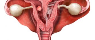
An endometrial polyp or polyp in the uterus is a neoplasm that forms on the inner layer of the reproductive organ (endometrium) and grows into its cavity.
Uterine polyps, for the most part, are benign neoplasms. Small polyps usually do not require treatment and do not manifest themselves in any way. Multiple large polyps are accompanied by a clinical picture and require treatment, since they greatly reduce a woman’s quality of life and can be dangerous. Some forms of endometrial polyps are prone to developing into cancer. Such neoplasms are easily identified during diagnosis. Recurrent polyps cause concern among doctors, therefore, when a uterine polyp reappears, a woman requires long-term therapy.
A uterine polyp is a cellular tumor growing from the functional layer - the endometrium. Polyps are classified depending on their structure. The treatment method is selected in accordance with the type of polyp, its location, the severity of clinical manifestations, the patient’s age and other health characteristics.
In ICD-10, a uterine polyp is designated by code N84.0 and stands for polyp of the uterine body. Code N84 contains subsections that specify the origin of the tumor: N84.1 – polyps of the cervical canal or cervix; N84.2 polyps in the vagina; N84.3 – vulvar polyps; N84.8 – polyps of the female genital organs (other parts); N84.9 – unspecified polyp. ICD-10 code N84 does not include adenomatous polyps (D28) and placental polyps (O90.8).
Symptoms of uterine polyps
Clinical manifestations of uterine polyps appear when the size of the neoplasm exceeds 2 cm. Small polyps up to 1 cm do not manifest themselves in any way. A woman finds out about such tumors unexpectedly during a routine ultrasound.
Throughout the menstrual cycle, the lining of the uterus undergoes hormonal changes. At the beginning of the cycle after menstruation, the thickness of the endometrium is minimal. By the time of ovulation, the endometrial layer undergoes proliferation and thickens. In the second half of the cycle, the secretory phase of the endometrium begins. Similar changes occur with the polyp of the uterine body. During the cycle, the tumor can increase and decrease.
Uterine polyps can cause different symptoms in women. As a rule, patients with uterine polyps complain of menstrual irregularities:
- heavy bleeding;
- scanty spotting;
- breakthrough bleeding not associated with menstruation;
- delays;
- premature start of the cycle;
- irregular periods;
- profuse leucorrhoea.
About 30% of patients experience pain due to uterine polyps. Discomfort occurs during physical activity and during intimacy. Large uterine polyps can cause infertility. Against the background of intense uterine bleeding with a polyp, a woman may develop anemia.
Complications after the formation of a polyp in the endometrium of the uterus
If a woman ignores the first symptoms of the disease and tries to cope with the problem on her own, in most cases serious complications cannot be avoided, including:
- severe forms of posthemorrhagic anemia;
- breakthrough prolonged bleeding, which poses the risk of large blood loss;
- physiological infringement of the growth as a result of a woman’s physical activity;
- uncontrolled pathological growth of the endometrial layer;
- uterine fibroids (in this case, the gynecologist recommends that the woman have a hormonal IUD installed);
- chronic endometritis;
- adenomyosis;
- ischemic changes in the polyp, leading to tissue necrosis;
- Oncology of the endometrium of the uterus.
If, after treatment for uterine polyposis, a woman’s menstrual cycle is disrupted, the gynecologist prescribes oral contraceptives to normalize the functioning of the ovaries.
Classification of uterine polyps
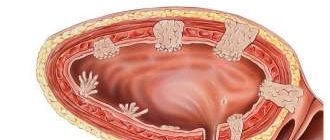
Uterine polyps can be multiple or single, small or large. Neoplasms are located in different segments of the uterine body. The polyp grows from the mucosa, having a wide base or stalk. Large pedunculated polyps can penetrate the cervix, then they speak of a “nascent” polyp.
Uterine polyps are classified according to their cellular structure:
- Glandular polyp is the most common type of neoplasm. Consists of endometrial glandular cells. It is detected in women of reproductive age. It is of benign origin and never degenerates into cancer.
- Fibrous polyp - consists of connective tissue. A fibrous polyp is detected in women in old age, postmenopause, and in women who have given birth. It is rare in young and nulliparous girls.
- A glandular fibrous polyp is a tumor based on connective tissue that is covered with a glandular structure. It can appear in women of different ages.
- Adenomatous polyp is rare and has a poor prognosis. This tumor is characterized by an altered cell structure. Adenomatous endometrial polyps are also called atypical or atypical. Such tumors have a high risk of malignancy - degeneration of the polyp into cancer.
- Placental polyp - detected in women after childbirth or surgical termination of pregnancy. A placental polyp is formed from the remains of the placenta.
- Cervical polyp is a separate type of polyp, since it is formed not from the mucous layer of the uterus, but in the cervical canal. Women can have a polyp of the cervical canal at different ages.
Make an appointment with a gynecologist
Signs of formations
Signs of uterine tumors in women can manifest themselves in different ways. Small tumors that do not affect the menstrual cycle exist asymptomatically. As the tumor grows, a clinical picture may appear.
As a rule, the main sign of uterine formation is changes in the menstrual cycle. Pain in the pelvis may also indicate the formation of the uterus. Other signs of tumors may include difficulty conceiving, endocrine disorders, and heavy discharge. Diffuse endometriosis and polyp are accompanied by infertility, heavy menstruation with clots, pain and infertility.
Polyps and pregnancy
A small endometrial polyp does not affect pregnancy in any way. A woman can become pregnant naturally and not even know that she has a polyp. Neoplasms up to 2 cm are considered harmless and cannot affect conception. If a woman has a large polyp, it may interfere with implantation. Also, polyps located at the mouth of the fallopian tubes can cause infertility in women.
During pregnancy, under the influence of new hormonal levels, the growth of an endometrial polyp may begin. In this case, there is a risk of complications such as infection, bleeding, and necrosis of the tumor. The polyp increases the risk of intrauterine infection, premature placental abruption, fetoplacental insufficiency and other problems.
There is no treatment for endometrial polyps during pregnancy. After childbirth, treatment tactics are determined individually and depend on the size, type, and location of the tumor.
Diagnosis of uterine polyps
Diagnosis of uterine polyps is crucial for choosing further tactics of action. During the examination, it is important to determine which endometrial polyp a woman has, what size of the neoplasm and make a prognosis. In the diagnosis of uterine polyps, various instrumental, hardware and laboratory techniques are used.
Ultrasound
Ultrasound examination of a uterine polyp is the primary diagnostic method. On ultrasound, the polyp looks like a neoplasm of the endometrium. If you have a good ultrasound machine and extensive experience of a specialist, you can determine the type of uterine polyp with a high degree of accuracy.
It is informative to do an ultrasound scan for a uterine polyp on days 5-8 of the menstrual cycle, after the end of menstruation. In the first half of the cycle, the size of the polyp will be minimal, since the endometrium has not yet grown. If a uterine polyp is accidentally detected, it is recommended to repeat the ultrasound after menstrual bleeding in the next cycle.
Histology
Histology of a uterine polyp is a laboratory study of part of a neoplasm. This type of diagnosis is the most reliable in determining the type of tumor. To conduct histology, it is necessary to obtain biological material. This can be done using curettage, hysteroresectoscopy or puncture. Histology of the polyp is done within 10 days.
Hysteroscopy
Hysteroscopy is a therapeutic and diagnostic procedure. It is carried out using an endoscopic device - a hysteroscope. Hysteroscopy allows the doctor to examine in detail the location and structure of the endometrial polyp. Performed under general anesthesia.
During hysteroscopy, a miniature device is inserted into the uterine cavity. A small camera transmits an enlarged image onto the screen. If necessary, a polyp can be punctured during hysteroscopy. Also, a survey hysteroscopy can turn into a therapeutic one, with the help of which a polyp of the uterine body is removed.
Hydrosonography
Hydrosonography is a diagnostic procedure that allows you to assess the condition of the uterus and fallopian tubes. It is performed without anesthesia; if necessary, the woman is given painkilling injections. During hydrosonography, a contrast agent is injected into the uterine cavity using a miniature catheter. At the same time, the behavior of the contrast is monitored using ultrasound. Under pressure, the contrast passes through the fallopian tubes and, if the oviducts are patent, enters the abdominal cavity. In addition, the contrast fills the uterine cavity, which allows you to examine the tumors in detail.
Make an appointment with a gynecologist
Causes of pathology
There is no consensus among doctors about the causes of endometrial polyp. Theoretical gynecology and practitioners agree that endometrial disorders are caused by an imbalance of hormones in the body, namely, an insufficient amount of progesterone. But this hormonal deviation provokes not only the formation of growths in the uterus, but also other pathologies in the female reproductive system.
- disturbances in the functioning of the endocrine system;
- ovarian dysfunction;
- mechanical damage to the endometrium of the uterus as a result of careless medical procedures;
- severe pregnancy or spontaneous termination;
- complicated childbirth, after which fragments of placental tissue remain in the uterine cavity;
- chronic inflammatory process in the genitourinary system;
- a woman being in a stressful state for a long time.
How to treat polyps in the uterus
The treatment method for endometrial polyp or cervical canal polyp is selected individually in each clinical case.
Removal operations are performed when the polyp is large or if the tumor reduces the patient’s quality of life. The best method of removal is selected for each patient separately, based on the results of clinical studies and in accordance with health indicators. Removal of uterine polyps is best performed in the first half of the cycle. The neoplasm is removed 3-5 days from the start of menstrual bleeding, but after its completion. Before removal, tests are performed in order to exclude complications and obtain maximum information about the patient’s health.
Drug treatment of uterine polyps is carried out for small polyps. As a rule, therapy is performed with hormonal drugs. Non-hormonal treatment involves taking anti-inflammatory and antibacterial agents, which is required for inflammation and infection.
Operative methods
Surgical treatment of uterine polyps requires hospitalization. How long to stay in the hospital depends on the chosen treatment method. Minimally invasive endoscopic operations are easy and without complications, and the woman can go home after 1-3 days. Open and radical operations to remove polyps require longer medical supervision, so the patient remains in the hospital for up to two weeks. The method of polyp removal depends on the type of tumor, size and location, as well as concomitant diseases.
A uterine polyp is removed in the following ways:
- surgical curettage;
- polypectomy;
- hysterectomy.
Indications for surgery
Surgery to remove a uterine polyp is performed when the tumor is large. Surgical intervention is also required for multiple tumors. A polyp on the fundus of the uterus, located on a stalk, with a high risk of torsion, also requires surgical intervention.
Indications for polyp removal:
- heavy, prolonged menstruation;
- breakthrough bleeding not associated with the menstrual cycle;
- contact bleeding;
- chronic pelvic pain;
- anemia caused by uterine bleeding;
- infertility caused by a polyp.
Hysteroscopy
Medical hysteroscopy allows you to remove a uterine polyp without complications or unpleasant consequences. The procedure is performed under anesthesia. The endometrial polyp is removed using a hysteroresectoscope, an endoscopic device that is inserted into the uterine cavity and allows the doctor to control the cauterization process. Removal of a polyp during hysteroscopy can be performed with various instruments - curette, laser, radio waves and others.
After hysteroscopy, the resulting biomaterial is sent for histology. The woman can go home the same day if there are no complications.
Hysteroresection
Hysteroresection of a uterine polyp allows you to remove the polyp within healthy tissue. The operation is performed for different types and sizes of tumors. Menstruation usually does not go astray after removal of a uterine polyp. But after surgery, a woman may experience bleeding due to a violation of the integrity of the vascular tissue.
Curettage of the uterine cavity
Curettage of a uterine polyp is one of the outdated methods of surgical treatment of a polyp. The procedure is performed under anesthesia. During the operation, the cervix is dilated, and a curettage instrument is inserted into the cavity. Subsequently, the material is sent for histology, and the woman is prescribed antibacterial therapy.
The regularity of menstrual bleeding after curettage of a uterine polyp is usually disrupted. The first day of the cycle can be considered the day of surgery. Curettage of a uterine polyp for a woman is associated with various risks: infection, perforation, bleeding.
Laser removal of uterine polyps
Removal of uterine polyps using a laser is one of the modern methods of treating uterine tumors. The procedure requires pain relief, but unlike other surgical treatments, it has a short recovery period.
In gynecological surgery, the method of laser removal of cervical canal polyps and cervical polyps is widely used. Laser treatment virtually eliminates the possibility of tissue infection, completely eliminates bleeding and leaves no scars.
Hysterectomy
Hysterectomy or hysterectomy is a radical surgical procedure that is rarely prescribed to women due to polyps. Removal of the uterus for a polyp may be necessary if a woman’s tumor has been confirmed to have transformed into cancer. Also, hysterectomy can be offered to elderly patients with multiple polyps, which are accompanied by frequent bleeding and greatly reduce the quality of life.
Hysterectomy for a polyp is performed using open surgery or laparoscopy. You will have to stay in the hospital for at least two weeks after such an operation. The rehabilitation period after a hysterectomy is long, and the consequence is always absolute infertility.
Polypectomy
Uterine polypectomy is an organ-sparing, minimally invasive and quick operation. The goal of the procedure is to remove the polyp while preserving healthy tissue and the reproductive organ as a whole. Polypectomy is performed in different ways. The most progressive method for removing uterine polyps is hysteroscopy. Polypectomy can also be performed using curettage.
Conservative treatment
Conservative treatment of polyps involves taking progestin drugs. Hormonal medications are prescribed individually (capsules, injections, patches or vaginal tablets). The dosage is also selected separately for each woman.
The goal of hormonal treatment is to normalize hormonal levels and reduce estrogen levels in a woman’s body. An auxiliary treatment method can be a diet for polyps. A woman is recommended to exclude from her diet foods that increase estrogens in the body (beer, pineapples, legumes, nuts, citrus fruits and others).
Drug treatment
Treatment of glandular, adenomatous and other polyps may require auxiliary drug therapy. Under certain circumstances, endometrial polyps can become inflamed, infected, and undergo necrotic changes. Such processes require antibacterial and anti-inflammatory therapy. The woman is also prescribed proteolytics - drugs that resolve hematomas, suppuration, and adhesions.
Drug treatment of a polyp cannot be an alternative to surgical and hormonal treatment. Symptomatic and etiotropic drugs are prescribed to a woman individually, based on the results of diagnostic procedures.
Treatment of recurrent uterine polyps
Recurrence of a polyp is usually not a good indicator. The number of relapses of endometrial tumors after hormonal conservative treatment reaches 40%. After surgical removal, polyp recurrence is much less common. For recurrent polyps, a woman needs an extensive examination that will help find the causes of tumors and choose the right treatment tactics.
Diagnosis and treatment of pathology
Modern medicine has all the necessary tools and technologies for diagnosing and treating uterine polyposis.
Diagnosis of the disease
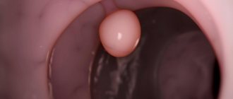
Depending on the location of the lesion, it is detected by a specific diagnostic method. Additionally, the patient undergoes tests. So, if a parietal polyp has grown on the walls of the cervical canal, the doctor will see these round pink lumps during a gynecological examination. This location of tumors is easier to detect than the presence of pathology in other parts of the female reproductive system. Diagnosis using ultrasound of the pelvic organs makes it possible to detect fibrous and glandular-fibrous tumors. The pathology is indicated by the enlarged cavity of the reproductive organ and echo signs of endometrial hyperplasia, reserve cell activity. Histological examination and biopsy are considered an accurate diagnostic method. During the procedure, the gynecologist, while examining the woman on the chair, takes a sample of tumor tissue and sends it to the laboratory. Hysteroscopy is widely used in the diagnostic examination of endometrial polyposis. The technology involves inserting a special probe into the uterus, equipped with a video camera and LEDs. It allows you to determine with high reliability the type and size of pathological growths in the uterus. The peculiarity of hysteroscopy is that it can be both diagnostic and operative. In the first case, the doctor collects information about the pathology, and in the second, he removes the pathogenic tumor.
- the doctor suspects uterine cancer;
- the mucous membrane grows rapidly, which is the initial stage of endometriosis;
- the doctor has reason to suspect the presence of fibroids in the uterine cavity;
- An ultrasound revealed remnants of membranes and bloody clots in the uterine cavity;
- the patient complained of very heavy or irregular menstruation;
- the presence of 2 or more miscarriages in the woman’s history;
- a woman has infertility and an unsuccessful attempt to give birth to a child through IVF;
- the appearance of spotting in a girl in the middle of her cycle, which may cause stomach pain;
- pain a week after menstruation.
Treatment of the disease
Postmenopause signals the end of a woman's reproductive function. During menopause, after menopause, symptoms disappear. With the onset of menopause, uterine polyps resolve on their own. There are no relapses during menopause or postmenopause. If a woman is still of childbearing age, a neoplasm of a polypous nature must be removed.
- Monitoring the progression of the anomaly.
- Drug therapy in combination with hormonal drugs.
- Surgical removal of the growth.
The choice of treatment regimen is determined by the doctor, based on the patient’s age, the size of the tumors, and the severity of the symptoms of the pathology. During treatment, the woman’s desire to become a mother in the future and safely bear a child is also taken into account.
Surgery to remove the polyp is recommended only if the patient has fibrous and atypical uterine growths. In other cases, a disease without atypia is treated without surgery.
The main goal of conservative therapy is to stop the growth of the uterine mucosa, create conditions for the disappearance of the polyp, prevent relapse and the adverse consequences of the pathology. Conservative therapy is used for polyps in nulliparous women. These technologies are also applicable if the patient has contraindications for surgery or if the girl has a categorical personal protest against the operation. Drug therapy takes a lot of time. It includes hormones and homeopathic medicines. As part of this approach, it is prescribed to take “Zhannine”, “Nafarelin”, “Duphaston”. “Zhannine” and “Duphaston” normalize the menstrual cycle, normalize the functioning of the endocrine system, stimulate the production of progesterone in the female body and restore homeostasis. “Nafarelin” reduces the level of estrogen and prevents the growth of the endometrium. If a pregnant woman has polyps localized in the cervix, she is prescribed antimicrobial drugs. For recovery, she must follow clinical recommendations. Folk remedies are acceptable as a way to combat placental and glandular type polyps. Infusions of celandine are effective. Viburnum fruits are also effective in combating endometrial growths. A woman should definitely see a gynecologist at the antenatal clinic to prevent relapse of the disease.
Polypectomy
Removing a polyp through surgery today is more effective than conservative treatment methods and folk remedies. The operation is performed under general anesthesia using a hysteroscopy machine.
If the endometrial polyp is large and forms on a stalk, the doctor removes it by rotating the head in a circle. After removal of the tumor, curettage of the uterine cavity is mandatory. Fragments of the pathological growth are sent for histological diagnosis. As an anti-inflammatory measure, after removal of the tumor, the site of its localization is treated with liquid nitrogen or the endometrium is exposed to high-frequency electric current. Also, in some cases, a woman is prescribed a course of antibiotics. After hysteroscopy, a repeat diagnostic hysteroscopy is done and maintenance medications are taken in order to facilitate the body’s recovery process. Maintenance therapy drugs are prescribed by the attending physician, based on the specific course of the pathology, the patient’s health status and the presence of concomitant anomalies.
Is it dangerous to remove polyps in the uterus?
Removing uterine polyps is not dangerous. It is much more dangerous to ignore the doctor’s recommendations and, when an operation is prescribed, to refuse surgery. When prescribing surgical treatment, the doctor takes into account all the pros and cons, benefits and possible risks. An experienced gynecologist, after a comprehensive examination, will determine whether the uterine polyp needs to be removed or not, and how dangerous the operation can be.
Polyp removal is prescribed during a period that is as safe and comfortable for the body as possible. If necessary, hormonal correction is carried out before surgery, which helps to reduce the size of the tumor.
Is it possible not to remove the tumor?
Whether the polyp needs to be removed is determined by the results of the examination. If the tumor does not tend to grow, its size does not exceed 1 cm, and the woman does not have any symptoms, then you can choose a wait-and-see approach. In such a situation, the polyp is monitored - an ultrasound is done every 3 months. If the polyp begins to grow, it is removed.
The need to remove cervical polyps is also discussed individually. Cervical polyps can be dangerous. Any neoplasm of the cervix requires an extensive examination and exclusion of atypical changes in the cells.
When the examination confirms the high tendency of the polyp to become malignant, the woman is prescribed surgery. In this case, you don’t have to choose whether to remove the polyp or not. The tumor must be removed, as it can develop into cancer. Often such tumors have to be removed along with the uterus.
Surgitron
Surgitron is a modern radiosurgical device. It was developed by American. The device is used in gynecology to remove cervical polyps. Removal of a polyp of the cervical canal using the radio wave method brings extremely positive results. Removal of a polyp on the cervix with surgitron has the following advantages:
- No contact impact;
- Minimal damage to tissues located near the polypous formation;
- Elimination of blood loss when exposed to radio waves;
- Painlessness during the procedure;
- Clear visual control of all stages of the operation;
- Minimal postoperative period and rapid recovery.
Radiosurgical removal of polyps of the cervix and cervical canal with surgitron is carried out in the presence of the following indications:
- The size of the polyps is more than 10mm;
- The patient's age is over 45 years;
- Problems with conception and pregnancy;
- Lack of positive effect from conservative treatment;
- Presence of adenomatous polyps.
Reviews from women who had a cervical polyp removed using the radio wave method are positive.
Bloody discharge after surgery
Discharge after a polyp and surgical treatment is normal. Bleeding may last from several days to two weeks. Bleeding after polyp removal is caused by vascular damage. In the first days after surgery, the discharge may be quite heavy. Gradually, the intensity of bleeding decreases, after which it is replaced by ichor. Bleeding after removal of a polyp by curettage can be mistaken for a new menstruation. After such operations the cycle changes.
Make an appointment with a gynecologist
How does a polyp affect menstruation?
Small polyps do not affect the menstrual cycle in any way. If the polyp is large, it can cause anovulatory cycles. In such a situation, a woman experiences various menstrual irregularities: delays are replaced by premature menstruation, and scanty discharge is replaced by heavy uterine bleeding.
The vast majority of patients with uterine polyps experience heavy and long periods, which are accompanied by painful sensations. In the first days of the menstrual cycle, women are forced to take painkillers. With the development of iron deficiency anemia in a woman during menstruation, performance decreases.
Pregnancy with a polyp can occur. Despite the fact that in some situations a polyp causes infertility, if the tumor is small, it cannot affect conception. Therefore, if a woman has a delay in menstruation due to a polyp, pregnancy should first be excluded. Only if the result is negative can hormonal or surgical treatment begin. If a woman becomes pregnant with a polyp, treatment is postponed until after childbirth.
Diagnostics
A professional gynecologist will begin collecting anamnesis at the initial appointment. The doctor will be interested in information regarding the symptoms, age of the patient, past pathologies, regularity of the menstrual cycle, its quality, number of pregnancies and their course.
Next, the specialist proceeds to a gynecological examination on a chair, during which he will immediately take a smear for microflora and cytology. For greater visualization of the pathology, the gynecologist performs a colposcopy, using a hysteroscope if the object of study is the uterine cavity. Using these optical instruments, it is possible to determine the location, number, and size of growths.
If the polyp is located in easily accessible places, you can immediately take a fragment of it for a biopsy to determine the type of cells and rule out oncology.
An ultrasound and x-ray with a contrast agent are required to exclude a malignant process and other pathologies of the genital organs.
Additionally, laboratory tests of blood, urine, and secretions are performed. The doctor must know about existing infectious diseases and the specifics of hormonal balance.
Restrictions after the procedure
After removal of the polyp, the woman is given recommendations:
- avoid thermal procedures (hot baths, saunas, baths, solarium);
- refrain from drinking alcohol;
- do not take medications that affect blood density (unless recommended by a doctor);
- eliminate physical activity for a while;
- refrain from intimacy.
Restrictive procedures are prescribed for a period of 2 weeks to three months (depending on the method of removing the uterine polyp). It is recommended to plan pregnancy after removal no earlier than 2-3 months later.
Forecast
If a polyp is found in a nulliparous girl, it is monitored for several cycles. If the tumor does not grow and does not cause concern, then it is not touched. If the polyp grows, it is treated with medication, and if there is no effect, surgical intervention is prescribed. Polyps in young girls can be caused by hormonal changes during puberty or other circumstances. As a rule, with such polyps, medicinal hormonal treatment shows good results.
A postmenopausal polyp, which is accompanied by bleeding and pain, has an unfavorable prognosis. It is better to remove such tumors, since during menopause women have an increased risk of cancer. If a malignant endometrial polyp is detected, radical surgery is performed and potent treatment is prescribed. The prognosis of the therapy depends on how early the polyp was detected, whether it has developed into cancer, whether there are metastases, and on other indicators of the patient’s health.
Uterine polyps themselves are not dangerous to health. Most of these neoplasms are benign tumors. Many women live with uterine polyps for years and do not know it. Small neoplasms do not interfere with conception, do not affect pregnancy in any way and are not a contraindication for natural childbirth.
A polyp of the cervical canal during pregnancy can cause concern among doctors. The cervix is a muscular organ that stretches during natural childbirth and performs a transporting function for the fetus. If a woman has cervical polyps, they can cause complications during childbirth. No one can predict in advance how the cervix with polyps will behave.
Removal of cervical polyps in nulliparous women must be performed using gentle methods. Preference should be given to a laser beam or radio waves. After such operations, there are no scars left on the cervix, which can remain with knife treatment.
Make an appointment with a gynecologist
Removal of cervical polyp
Do I need to remove a polyp on the cervix? Removing uterine polyps surgically is called polypectomy. Large polyps (1-3 cm in size) are removed by unscrewing. After this, the base of the polyp stalk is cauterized with electric current.
The removed tissue is sent for histological examination to determine the structure of the polyp. This is necessary in order to exclude the malignant nature of the neoplasm. Subsequent treatment largely depends on the results of histology.
4-5 days after removal of the uterine polyp, gynecologists at the Yusupov Hospital conduct a re-examination. It is necessary to monitor the condition of the localization site of formation and prevent endometritis. After polypectomy, antibiotic therapy is carried out for 7-10 days. If your temperature rises after removal of a cervical polyp, you should immediately contact a gynecologist. Hyperthermia may be a manifestation of a complication of surgery. In this case, the doctor will prescribe the latest generation of antibiotics.
If the patient has polyps formed against the background of ovarian dysfunction, appropriate hormonal therapy is carried out. In some cases, long-term hormonal treatment is necessary. Its duration can vary from three to six months. During this time, the results of treatment are monitored using hysteroscopy and endometrial biopsy.
Prevention of uterine polyps
The basis for the prevention of uterine polyps is a responsible approach to women's health and proper sex education. Infections of the genital tract cannot cause the formation of polyps. However, various pathologies of the pelvic organs increase the risk of tumors.
To prevent endometrial polyps, it is recommended:
- monitor hormonal levels and the regularity of the menstrual cycle;
- take a responsible approach to choosing a sexual partner;
- avoid unplanned pregnancy and use contraception;
- maintain a healthy body weight (since excess body weight increases estrogen production);
- prevent endocrine disorders;
- avoid various diagnostic and therapeutic intrauterine manipulations;
- do not take hormonal medications without a doctor’s prescription;
- visit a gynecologist and do an ultrasound of the pelvic organs at least once a year.
A uterine polyp in women is a hormone-dependent neoplasm that tends to grow with excess estrogen production. When treating a polyp, it is important to determine the cause of tumor formation and eliminate it. Removal of a polyp is a symptomatic treatment in which the neoplasm is excised, but if the causes persist, it may reappear.
Innovative methods for treating uterine polyps
Currently, when choosing a treatment method and patient management tactics, gynecologists take into account the following factors:
- Education dimensions:
- Its structure;
- Woman's age;
- Endometrial condition;
- Reasons for the development of pathology;
- The presence of metabolic and endocrine disorders.
Currently, polyps are removed using a laser. This type of surgical intervention does not leave scars and allows you to preserve the woman’s reproductive function. Innovative methods for treating uterine polyps include minihysteroscopy. The operation is characterized by the absence of cervical trauma and the need for anesthesia.
The doctor chooses the treatment method together with the patient. At the same time, mini-hysteroscopy allows you to get rid of not only polyps, but also uterine fibroids, endometrial hyperplasia, and intrauterine synechiae. This is a gentle surgical method.
The doctor has the ability to control the depth of laser penetration layer by layer, thereby preventing injury, reducing blood loss and the recovery period. The laser seals the vessels. There is no connective tissue left, which has a beneficial effect on future pregnancy. After laser treatment, the rehabilitation period ranges from 6 to 8 months.