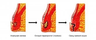
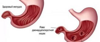
What is a rectal ulcer
An ulcer is an area where there is a defect in the mucous membrane. Its localization can be very diverse, but most often the zone of its formation is the place where excess friction occurs.
Causes of ulcer formation
Rectal ulcers appear for various reasons
There are quite a few reasons that act as a reason for the development of ulcers. Among them are:
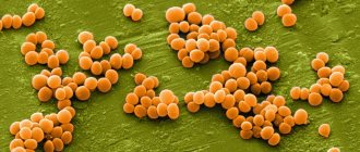
- Infectious nature of ulcerative defect. In this case, it should be noted that the infection can enter the body from the outside through contact with a microorganism that exhibits absolutely pathogenic properties or with opportunistic bacteria. Among the absolute pathogens, exposure to the causative agent of salmonellosis, dysentery and other intestinal disorders is distinguished. As a rule, in this case, inflammation should form, which is accompanied by an imperative urge to defecate, frequent bowel movements and lack of absorption of the liquid part of nutrients. When inflammation of an infectious nature occurs with liquid feces, irritation of the skin occurs, tissue swelling is provoked, which makes the skin even more vulnerable. The inflammatory process is of a non-infectious nature; the development of inflammation is not always caused by the action of bacteria. The cause is inflammatory processes that cause dysfunction of the large intestine and rectum, these include colitis, irritable bowel syndrome, dysbiosis, etc.
- Violation of the integrity of the skin or mucous membrane as a result of traumatic exposure. Mechanical damage is a common cause of ulcer formation. People of any age encounter it. For young people, the appearance of ulcers is most often caused by the use of non-traditional types of sexual contact, the use of foreign objects, etc. For mature and older adults, the cause may be traumatic effects as a result of diagnostic and therapeutic measures. The appearance of the defect is associated with improper use of medical instruments. In addition, as a mechanical damage, there may be an injury associated with a fracture of the pelvic bones or a fall on sharp objects. In rare cases, an ulcer develops due to the mechanical impact of coprolites formed due to poor nutrition or insufficient intake of products that stimulate the ovaries.
- Impairment of adequate tissue circulation. This problem is caused by varicose veins in the hemorrhoidal plexus, which is accompanied by stagnation of venous blood and impaired tissue trophism.
In addition to the main reasons that cause the development of rectal ulcers, there are also provoking factors that increase the risk of developing an ulcer.
These include:
- Lack of physical activity with a predominantly sedentary lifestyle.
- The presence of rectal prolapse, which causes rubbing of the walls.
- Tendency to develop chronic constipation due to an unhealthy lifestyle, including foods containing significant amounts of refined substances in the diet.
- Intestinal dysbiosis, which disrupts the digestion process.
The characteristics of a solitary rectal ulcer are given. The causes, diagnostic methods, differentiation from other diseases, and treatment methods are described. Our own clinical observation is presented.
A single (solitary) rectal ulcer is a disease characterized by the presence of single ulcerative defects of the rectal mucosa, which is clinically manifested by a feeling of incomplete emptying of the rectum during bowel movements and rectal bleeding. The clinical manifestations of solitary rectal ulcer syndrome (SRUC) are varied. Its most common causes are: prolapse of the anterior wall of the rectum (which is why ulcers are more often localized on the anterior wall of the rectum); complete external prolapse of the rectum; the so-called internal prolapse (intussusception) of the overlying mobile parts of the rectum into the lumen of the lower ampullary part - as one of the manifestations of obstructive defecation syndrome. This condition is associated with excessive straining during defecation, in which there is contraction rather than relaxation of the external anal sphincter and puborectal muscles. Changes in the mucous membrane are limited ulcerations caused by trauma to the membrane during muscle contraction and movement of the mucous membrane with its ischemia. Sometimes ulcers are formed due to the fact that to facilitate the act of defecation the patient resorts to a finger aid.
The disease occurs more often in women (in approximately 2/3 of cases). The main symptoms of SSPC are severe defecation disorders in various forms and rectal bleeding with mucus, and symptoms of prolapse are not uncommon.
Sigmoidoscopy allows you to detect one or more ulcers, most often located on the anterior wall at the site of prolapse of the mucous membrane. A biopsy from a rectal ulcer reveals characteristically closing muscle fibers and moderate signs of fibrosis. A specific type of solitary ulcer or several ulcerations with gray-white plaques on the bottom must be differentiated from pseudomembranous colitis. Such ulcers never become malignant.
In the diagnosis of SSPC, along with sigmoidoscopy and colonoscopy, irrigoscopy (including in an upright position of the patient and with straining) or MRI proctography is fundamentally important. These studies are mandatory for obstructive defecation syndrome to identify internal rectal prolapse. This disease is differentiated from ulcerative colitis and Crohn's disease, as well as rectal cancer. Unlike inflammatory diseases, corticosteroids are ineffective for a single rectal ulcer, which can also be a diagnostic criterion.
Treatment of SSPC is based on the effective elimination of constipation (introducing foods rich in dietary fiber into the diet, taking lactulose, prucalopride). Local use of ointments or corticosteroid suppositories is not justified, since rectal ulcers are not a primary inflammatory disease. If the diagnosis is established and the treatment measures are ineffective, it is advisable to refer patients to a consultation with a proctologist. Due to the rarity of this pathology, which is difficult to treat with medication, as well as low awareness and lack of published works in the republic, below is our own clinical observation.
Patient O., born in 1969 She was admitted to the gastroenterology department of the Brest Regional Hospital on 04/01/2011 with complaints of pain on the left flank of the abdomen, in the left iliac region, dense stools periodically mixed with blood and pus once a day after significant straining, and bloating.
From the anamnesis it is known that the patient has been prone to constipation for a long time, which is poorly regulated with a diet enriched in fiber, physical exercise, and only to a small extent responds to laxatives (senna, buckthorn, bisacodyl).
For diagnostic purposes, the patient underwent general blood and urine tests, a biochemical blood test, an immunogram, macroscopic and microscopic examination of stool, FGDS, ultrasound of the abdominal organs and retroperitoneal space, and colonoscopy. Of the pathological abnormalities in the general blood test dated 04/01/2011, slight lymphocytosis was noted - up to 37%. Total colonoscopy from 03/31/2011: in the rectum along the anterior wall 7 cm from the anus and proximally up to 20 cm - 3 flat superficial ulcers up to 1.0-2.0 cm, covered with mucus deposits and a delicate film of fibrin. A rectal biopsy dated April 4, 2012 revealed fibrin, leukocytes, desquamated glandular epithelium, and mucus. A diagnosis of solitary rectal ulcer was made. The patient was treated with intravenous ceftriaxone, mesacol, folic acid, and lactulose. Upon discharge, there is still an admixture of blood and mucus in the stool when straining, and there is no urge to defecate. The patient is recommended to consult a proctologist.
The patient’s next visit to the gastroenterology department of the Brest Regional Hospital occurred in July 2011 after unsuccessful treatment in the proctology department, where a diagnosis of ulcerative colitis was made and conservative treatment was carried out according to the protocols of the Ministry of Health of the Republic of Belarus. The complaints remained the same. General blood test dated July 29, 2011: er. - 4.321012/l, Hb - 118 g/l, l. - 7109/l, c.p. - 0.82, reticuloc. - 9, thrombus. — 185 109/l, p.neit. — 8%, neutral. - 65%, lymph. - 19%, mon. - 8%, ESR - 24 mm/h.
Colonoscopy dated 08/01/2011: the rectum and part of the sigmoid colon up to 70 cm were examined. The shape of the lumen and the relief of the folds are not deformed. The mucous membrane within visibility is pale pink with areas of slight hyperemia and edema. At a distance of about 10 cm from the anus, a superficial ulceration with a thin coating of fibrin up to 3.5-4.0 cm long and about 1.5 cm wide was found on the anterior wall. No rigidity was noted during instrumental palpation. Conclusion: rectal ulcer. The biopsy specimen contains fibrin and leukocytes. She continued taking mesacol and motilac, and added dexamethasone to the treatment, against which she was discharged with improvement - stool without blood and mucus, but the tendency to constipation remained. Long-term use of mesacol, folic acid, lactulose, duspatalin, and sulfasalazine suppositories is recommended. Despite persistent long-term treatment, in March 2013, the patient again sought medical help at the gastroenterology department of the Brest Regional Hospital with complaints of bloating, lack of urge to defecate, stool with severe straining once a day mixed with blood and mucus, and frequent headaches, increased blood pressure due to abdominal discomfort. On March 22, 2013, a colonoscopy was performed, with the help of which, at a distance of 8-12 cm from the anus in the rectum on the anterior wall, a shallow longitudinal tortuous ulcerative defect of the mucous membrane with slightly swollen edges up to 4 cm in length was again detected. Minor deformation and hyperemia of the mucous membrane were noted around the ulcerative defect shells. The biopsy specimen contains fibrin, leukocytes, granulomatous tissue, and a superficial fragment of the colon mucosa.
General blood test dated 03/19/2013: er. - 4.37 1012/l, Hb - 120 g/l, l. - 4.1 10 to 9 degrees / l, ESR - 28 mm/h, thrombus. — 265 109/l, p.neutr. - 3%, s.neutr. - 59%, lymph. - 34%, mon. — 3%. According to the results of the studies carried out - CBC, LBC, HIV ELISA, ELISA for markers of viral hepatitis B and C, ultrasound of the abdominal organs, FGDS - no deviations from the norm were identified. The patient was discharged with recommendations to continue taking suppositories with methyluracil, suppositories with mesalozole rectally, taking resolor (prucalopride) to stimulate propulsive waves in the colon, also due to negative dynamics during endoscopic examination - no scarring and an increase in the size of the ulcerative defect in the rectum - consultation with a proctologist at the Republican Center for Reconstructive Surgical Gastroenterology, Coloproctology and Laser Surgery at the Minsk Regional Clinical Hospital to select further treatment tactics and possible surgical correction. The patient was hospitalized on September 27, 2013 in the proctology department of the Minsk Regional Hospital, where after additional examination (based on the results of irrigoscopy, internal rectal prolapse was detected), a clinical diagnosis was made: obstructive defecation syndrome. Internal rectal prolapse. Solitary ulcer of the rectum. An operation corresponding to the range was performed: rectopexy, sigmoplication and elimination of the deep Douglas pouch, which eliminated constipation and created conditions for healing of the solitary ulcer. Control sigmoidoscopy on January 29, 2014 revealed intact, smooth mucosa with areas of edema and mild hyperemia.
Conclusion: no pathology was identified (examination to the 60 cm mark).
Thus, establishing a diagnosis of a solitary rectal ulcer is based on an assessment of clinical manifestations (long-term constipation, blood and mucus in the stool), as well as endoscopic examination data (sigmoidoscopy, colonoscopy with detection of ulcerative lesions in the distal parts of the colon, lack of effect from anti-inflammatory therapy).
USED LITERATURE 1. McNally P. R. Secrets of gastroenterology. Colon ulcers. - M., 1999. 2. Ivashkin V. T., Rappoport S. I. Handbook of a practical physician on gastroenterology. - M., 1999. 3. Ivashkin V. T. Rational pharmacotherapy of diseases of the digestive system. - M., 2003. 4. Travis S. P. L., Taylor R. X., Misevich J. J. Gastroenterology. Rectal ulcer.— M., 2002. 5. Henry M., Swash M. Coloproctology and the pelvic floor.— M., 1988.— P. 459-460.
Received 03/18/14.
Address for correspondence: Karpitsky Alexander Sergeevich.
Brest regional hospital. 224027, Brest, st. Medical, 7; sl. tel. (8-0162) 42-00-61. Key words:
clinical manifestations, solitary rectal ulcer, endoscopic examination
Author(s):
Karpitskii A. S., Khinich A. P., Dobriyanets A. R.
Medical institution: Brest Regional Hospital
Symptoms
With a rectal ulcer, a person experiences acute or aching pain
The symptoms of ulcers in the rectum are quite diverse, and their nature largely depends on the severity and localization of the process. Among the main manifestations of pathology are:
- Presence of pain syndrome. The pain is sharp or aching in nature. At the initial stages, it occurs periodically and the patient does not attach importance to such changes, but later, in the absence of treatment, it intensifies. The patient’s lifestyle, work patterns, etc. are disrupted. The onset of pain rarely occurs at rest; as a rule, it is associated with gradual filling of the rectal ampulla. More often, pain appears during the act of defecation or after bowel movement. The pain is unbearable, which forces the patient to limit bowel movements and develop fear of the procedure. This is expressed by loss of body weight, lack of appetite, anxiety, etc.
- The appearance of discharge from the anus. At the initial stages of ulcer formation, the nature of the discharge may be layered or with the presence of bloody streaks. In this case, the patient may only notice such a symptom. Subsequently, as the pathology progresses, the symptoms intensify, which is manifested by a large amount of discharge. Their character can be either scarlet, which signals active bleeding, or burgundy or brown with a heterogeneous consistency and the presence of clots. This manifestation is caused by the presence of an old defect, which is characterized by the gradual formation of thrombotic masses. Bleeding tends to increase after defecation, which is usually associated with trauma to the ulcer.
- When an infection occurs, suppuration of the lesion can occur with the development of a threat to life. This condition is accompanied by the release of purulent contents.
- Development of imperative urges to defecate. The patient consults a doctor with complaints of a constant, regular urge to defecate; their manifestation is imperative and is not accompanied by the vision of feces. After straining or the appearance of a small amount of fecal contents, the patient does not feel a feeling of emptying the intestines.
Among the systemic manifestations of rectal ulcers are:
- Development of anemia. Lack of hemoglobin can be caused by several reasons, among them the presence of chronic blood loss due to bleeding ulcers, the initial cause of its formation, which causes disruption of the digestive process.
- Poor general health, development of weakness, malaise, loss of appetite, etc.
- The appearance of skin defects in the anal area, which is caused by constant maceration of the skin, weeping and excessive friction. The addition of a bacterial infection can lead to infection of nearby tissues. This is manifested by intoxication, an increase in body temperature to fever, and the involvement of neighboring sections in the pathological process.
- With a massive inflammatory process, a threat to the patient’s life arises, since the close proximity of blood vessels can spread the infection in a short time.
Nonspecific ulcerative colitis - symptoms and treatment
Tactics depend on the location of the lesion and the severity of the course. In distal forms of the disease, the course is usually mild, so patients can be treated on an outpatient basis. Total or left-sided lesions often have a severe course and therefore require hospitalization and treatment in a hospital setting.
Diet
Diets No. 4, 4B, 4B according to Pevzner are prescribed. The purpose of the diet is to reduce inflammation, fermentation and putrefactive processes in the intestines, and normalize the functions of the intestines and other digestive organs. Dishes are liquid, semi-liquid, pureed, boiled in water or steamed. Very hot and cold dishes are excluded.
Products allowed for ulcerative colitis:
- crackers, soups with a slimy consistency, cooked in low-fat meat, fish broth or vegetable broth, with well-cooked cereals, noodles or noodles, the addition of meatballs is allowed;
- lean meat and fish;
- thoroughly cooked porridge;
- fruit purees;
- fresh grated cottage cheese;
- sugar in small doses;
- jelly and jelly from blueberries, dogwoods, bird cherry, quince, pears;
- strong black tea, maybe with lemon, a decoction of rose hips, black currants, blueberries and bird cherry;
- steam omelette, hard-boiled eggs; butter.
Products prohibited for ulcerative colitis:
- baked goods and confectionery products;
- black bread;
- meat and fish fatty broths;
- sausages, salted and smoked fish, canned food;
- alcohol;
- coffee;
- milk;
- carbonated sweet drinks;
- fresh vegetables and fruits;
- pearl barley and wheat porridge, legumes.
Conservative therapy
Conservative therapy includes:
- aminosalicylates, 5-aminosalicylic acid preparations (5-ASA, mesalazine);
- corticosteroids;
- immunosuppressants;
- biological therapy.
Aminosalicylates are first-line drugs and are used first. To stop an attack of ulcerative colitis, 3-6 weeks of therapy at 1-2 g/kg per day are usually required. After this, anti-relapse treatment is carried out with sulfasalazine (3 g/day) or mesalazine (2 g/day).
For distal lesions (proctitis), preference is given to the forms of the drug in the form of suppositories . For left-sided lesions - drugs in the form of foam for rectal use. In case of total damage, tablet forms of drugs are used.
When treated with these drugs, remission is achieved in 74-81% of cases.
The most pronounced and effective anti-inflammatory agent are glucocorticoids .
The main indications for treatment with glucocorticoids are:
- acute course of severe and moderate severity;
- severe or moderate left-sided and total lesion in the presence of III degree of inflammation according to endoscopic examination;
- ineffectiveness or lack of effectiveness of treatment with aminosalicylates in chronic disease.
In acute severe cases, intravenous administration of glucocorticoids (prednisolone at least 120 mg/day) is prescribed 4-6 times a day. Correction of water and electrolyte balance, transfusion of blood components, and hemosorption are required. A week after intravenous administration, they switch to taking prednisolone orally. During this time, gastroscopy is performed to exclude peptic ulcers of the stomach and duodenum.
For moderate forms, you can limit yourself to oral prednisolone. Typically, prednisolone is prescribed at a dose of 1.5-2 mg/kg body weight per day. The maximum dose is 100 mg (in elderly people 60 mg).
If there are no side effects, then continue taking it for another 10-14 days, then reduce the dose by 10 mg every 10 days. From 30-40 mg they switch to a single dose of prednisolone. From 30 mg, the dose is reduced by 5 mg per week. Typically, the course of hormone therapy lasts from 8 to 12 weeks. In parallel with hormone therapy, aminosalicylates are used until hormones are completely withdrawn.
For proctitis, glucocorticoids are used rectally in microenemas. Usually hydrocortisone is used for this, applied after bowel movements 2 times a day.
Side effects of hormone therapy:
- swelling;
- arterial hypertension;
- osteoporosis;
- various autonomic disorders;
- may contribute to the development of stomach ulcers, and, as a result, gastrointestinal bleeding may occur.
The modern glucocorticoid is budenofalk (containing budesonide). The daily dose is 3 mg budesonide (1 capsule) 4-6 times a day.
When treated with glucocorticoids, hormonal dependence or even hormone resistance may occur, which develops in 20-35% of patients with severe ulcerative colitis. Hormonal dependence is the body’s response to glucocorticoid treatment, in which the inflammatory process resumes after the dosage is reduced.
If there is no effect from hormonal therapy, drugs that have an immunosuppressive effect, as well as biological therapy, are prescribed. The main ones are 6-mercaptopurine and azathioprine - these are first-line drugs. They help reduce the dose of hormones and cancel them in 62-71% of cases. Treatment is usually started along with hormone therapy. The maximum daily dose is 150 mg. A small number of side effects, compared to glucocorticoids, allows the drug to be used for many years.
There is also a similar drug - methotrexate, which is used for intolerance to azathioprine or to accelerate the therapeutic effect. Orally or intramuscularly 30 mg per week. The effect occurs within 2-4 weeks
If there is no effect from azathioprine and 6-mercaptopurine, second-line drugs are used - these are infliximab and cyclosporine A.
If there is no effect from the second injection of infliximab and a 7-day course of cyclosporine A, surgical treatment is indicated.
Anti-relapse treatment
After relief of acute inflammatory processes, anti-relapse therapy with aminosalicylates is prescribed for up to 6 months. If treatment is successful and there are no clinical and endoscopic signs of the disease, therapy is discontinued.
In case of unstable effect from anti-relapse treatment, therapy is extended for an additional 6 months.
If aminosalicylate monotherapy is ineffective, immunosuppressants (azathioprine or 6-mercaptopurine) and hormones are added to treatment.
Patients with ulcerative colitis receiving conservative therapy require constant medical supervision and regular endoscopic examination with biopsy.
Surgery
According to statistics, surgical treatment is indicated for patients in 11-22% of cases. The only method of radical surgical treatment of ulcerative colitis is colproctectomy, that is, removal of the entire colon. The main indications for surgical treatment are:
- lack of effect from conservative treatment;
- development of complications of ulcerative colitis (bleeding, perforation, cancer, toxic dilatation of the large intestine).
Postoperative complications
Occurs in weakened patients against a background of severe disease:
- eventration (intestinal prolapse through a defect in the abdominal wall);
- failure of intestinal stoma sutures;
- serous peritonitis (inflammation of the peritoneum);
- polyserositis;
- abdominal abscesses;
- pneumonia.
Features of treatment depending on the form and course
Left-sided colitis or proctitis of low activity: suppositories and salofalk foam.
Moderate or widespread colitis: salofalk tablets, glucocorticoids.
Severe course: salofalk tablets, glucocorticoids, infusion therapy, iron supplements, transfusion of plasma and red blood cells, colproctectomy (removal of the entire colon with the formation of a permanent ileostomy - an opening in the abdominal cavity to which a colostomy bag is attached).
Fulminant colitis: treatment is the same as for severe colitis.
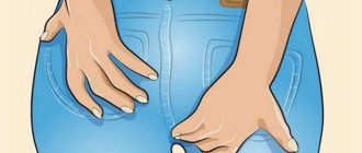
Diagnosis of rectal ulcer
Before diagnosis, the doctor conducts a conversation with the patient, finds out complaints and symptoms
Rectal ulcers require few diagnostic tests to confirm the diagnosis. Among the main methods needed in the study are:
- Conducting an external objective examination. It is important for the doctor not only to find out the main complaints, but also to actually determine their presence. A thorough conversation is carried out with the patient, during which anamnestic data, the conditions under which the ulcer appeared, as well as the presence of possible provoking factors are studied. It is important to know whether there are any intestinal pathologies, malignant processes, etc. in the family.
- After examining the organ, the doctor assesses the size of the formation, its severity, and the involvement of surrounding tissues in the pathological process. The examination includes not only the area of the lesion, but also an assessment of the general condition of the patient, focusing on the examination of the digestive tract, body weight and skin. With severe underweight, the doctor can draw a conclusion about the severity of the process and the presence of anemia.
After an objective examination, additional diagnostic methods begin, which include:
Ulcer treatment
To treat ulcers, antibiotic therapy is often used to kill the causative agent.
Therapy for ulcers that are localized in the rectal area is reduced to affecting the main focus of the pathological reaction. Among the areas of therapy are:
- Drug treatment. Therapy is carried out aimed at excluding an inflammatory reaction, including an infectious one. Antibiotics and antimicrobial agents are used for this. Initially, thorns start with a wide range. In case of a confirmed cause, treatment is started taking into account sensitivity.
- Among the local remedies, suppositories that have a healing effect can be used. They not only form a protective film on the surface of the ulcer, but also improve tissue trophism.
- Venotonic drugs. Their appointment is necessary in case of insufficient functional activity of the vascular wall. With regular use, vascular tone is restored.
- Surgery. This method of therapy is necessary in cases where there is no effect from medications. In addition, operations are indicated when there is a risk of inflammation spreading to adjacent areas. For treatment, sanitation with removal of necrotic tissue or complete excision of the ulcer can be undertaken.
Non-drug treatment
For treatment to be effective, you must adhere to a strict diet.
This direction is different in that it helps, in combination with other methods, to achieve a positive effect from treatment. These methods include:
- Normalization of diet, changes in food quality. Includes a large amount of fiber, vitamins and minerals.
- The consumption of refined products, fried, smoked and salty foods is excluded.
- Taking natural laxatives.
- Increasing physical activity.
- Moderate weight loss, subject to adequate nutrition.