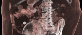
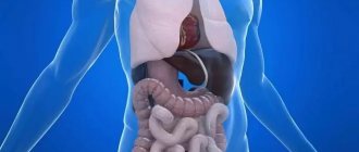
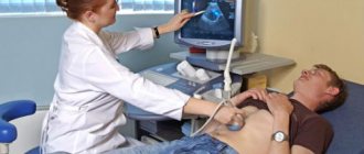
Computed tomography is a non-invasive research method, the essence of which is to obtain detailed images of internal organs using x-rays. CT scan of the intestine allows you to identify certain pathological processes at the earliest stages of development, which are easily treatable. Therefore, this diagnostic procedure is widely used in the field of gastroenterology.
You can get a CT scan of the intestine in Moscow at one of the modern medical centers - the Yusupov Hospital. The clinic is equipped with innovative diagnostic equipment, including a high-class computed tomograph, which makes it possible to detect certain pathologies at the initial stages of development with maximum accuracy.
CT scan of the intestine: the essence of the study
The computed tomography method, used in gastroenterology, consists of combining the use of an X-ray unit and computer processing of the resulting image of the intestine. This technology provides a more complete and clear picture, unlike a conventional x-ray examination.
The effectiveness of intestinal CT allows us to call this procedure a virtual colonoscopy. Such a detailed assessment of the condition of the intestine can only be provided by an invasive method - an endoscopic examination, which is carried out through the rectum. However, most patients perceive colonoscopy negatively, since this procedure is quite painful, its implementation is often accompanied by psychological discomfort, therefore, today, computed tomography of the intestines is considered the most popular diagnostic method.
A computed tomograph is a machine equipped with X-ray tubes and receiving detectors, which are located opposite each other in a ring platform. During the study, the patient is placed in this platform, which resembles a tunnel. During the scanning process, the tomograph platform rotates around the patient's body.
The resulting sections are processed in a computer station, where a detailed image of the intestine is formed.
Types of CT diagnostics
There are 3 main types of CT diagnostics, which differ from each other in technical characteristics:
- Step-by-step CT. The very first type of CT diagnostics. For one revolution of the X-ray tube, one section of the organ occurs. To make the next cut, you need to move the table a certain distance. It takes about 20 minutes. The quality of information is not inferior to spiral computed tomography.
- Spiral CT (SCT). In this tomograph, the X-ray emitter moves in a spiral around its axis with the simultaneous movement of the conveyor table with the patient, which provides information in several planes. Using SCT, it is possible to obtain up to 64 sections of an organ. High resolution makes it possible to see tumors with a diameter of 1 mm. The simultaneous movement of the table and the emitter allows you to reduce the procedure time, but sometimes the moving table causes interference in the images.
- Multislice CT (MSCT). It is a type of spiral CT. The main difference is the high rotation speed of the X-ray emitter and a larger number of special sensors that receive information. Using MSCT, you can obtain up to 320 sections of an organ, which increases the information content and accuracy of the study. It is the shortest study in terms of time, which significantly reduces the radiation load on the body.
Be sure to read:
Intestinal tumors: types, symptoms, treatment methods and prognosis for life
CT scan of the intestine: indications
Most often, computed tomography of the intestine, as well as CT of the rectum, is prescribed to patients at the Yusupov Hospital if the presence of polyps and cancerous formations in this organ is suspected.
Polyps are benign growths that have a high probability of degenerating into a malignant neoplasm. According to experts, early detection and removal of such formations at the initial stages of development prevents malignancy of the pathological process. For this purpose, the use of computed tomography is recommended.
Preventive examination of the intestines should be carried out for both men and women over 50 years of age. A CT scan should ideally be performed every five years, and a colonoscopy at least once every ten years. Persons with a family history of intestinal cancer or chronic polyposis should undergo a CT scan of the intestine after 36-40 years of age and much more often than other categories.
Computed tomography of the intestine is also prescribed for inflammatory processes and bleeding in the organ. CT scan of the intestine provides a quick and painless determination of the source of the problem, assessment of the volume and location of the pathological process.
In addition, intestinal screening is carried out as a control measure during the treatment of diseases in this part of the body, allowing one to evaluate the effectiveness of therapy.
Computed tomography of the intestine: preparation for the study
Before performing a CT scan of the intestine, the patient must cleanse it. In order to ensure better visualization of the gastrointestinal tract, it is necessary to free it from feces and food. No later than three days before the procedure, the patient is recommended to adhere to the following dietary rules:
- exclusion of gas-forming products (whole milk, white cabbage, legumes, apples, raw foods and vegetables in large quantities, sweets);
- avoidance of foods that strengthen the intestines (to prevent constipation);
- avoidance of laxative foods (to avoid bloating).
The day before a CT scan of the intestines, it is necessary to follow a liquid diet: drink large quantities of water, tea, and weak broths. In addition, it is necessary to take cleaning measures, which include the following:
- performing an enema;
- taking carminative drugs and enterosorbents that help get rid of gases in the intestines;
- taking an osmotic laxative;
- the use of radiopaque agents (solutions with iodine and barium), which will allow better differentiation of pathologies with the remains of intestinal contents - prescribed by a doctor before performing a CT scan of the intestine with contrast. The price in Moscow for this type of examination is slightly higher than for a regular CT scan;
- One hour before a CT scan of the intestine, you need to drink an additional 0.5 liters of liquid (purified water).
Computed tomography of the intestine: progress of the procedure
Immediately before the procedure, the patient is asked to remove all things, get rid of jewelry, watches, foreign objects, mobile phone, etc.
Then the subject is placed on the diagnostic table, the operator gives all the necessary instructions: take the required position (on the side, back, stomach). If necessary, to ensure the patient's immobility, which is necessary to obtain high-quality images, the limbs are fixed using special belts.
To perform a CT scan of the intestine, the rectum must be filled with carbon dioxide or air. For this purpose, a specialist inserts a thin tube into the patient’s anus, through which gas is injected using a bulb or pump. This event is necessary in order to maximally straighten the intestinal walls, which normally have bends and folds. Thanks to the “inflation” of the intestine, the view for the radiologist is improved and pathological processes in hidden areas are revealed.
After preparation, the patient is moved into the scanner itself, asked to hold his breath and change his body position for the next sections.
The CT scan takes an average of 15 minutes, after which the specialist removes the gas tube and allows the patient to stand up.
How is the examination carried out?
When heading to the clinic, it is advisable to choose clothes that are spacious enough to allow you to lie still in them for a long time. Chains, hairpins, metal fasteners of clothing and belts can leave shadows in photographs, so it is better to remove jewelry and accessories.
To conduct the examination, the patient is placed on a retractable tomograph table in a supine position. Your hands will need to be placed behind your head. The supine or side lying position can also be used.
CT colonoscopy of the intestine suggests dilation of the intestinal loops with gas. To do this, the patient, who is lying on his side, is inserted into the anus with a special thin tube through which air is supplied into the intestinal lumen using a hand bulb or compressor. The procedure itself is uncomfortable. If the pain during the introduction of gas into the intestine becomes intense, then it is necessary to tell the medical staff about it. To relieve intestinal spasms, the doctor will prescribe an antispasmodic.
An intravenous contrast agent may be administered before the examination begins or after native images are taken. If a bolus injection of the drug is planned, a special catheter is inserted into the patient’s vein, to which a thin tube from the injector is connected.
You will have to lie still for 15-30 minutes, so the patient’s body is fixed using special pillows and belts.
The prepared patient is placed into the machine along with the tomograph table. During the examination, the doctor may ask the patient to hold his breath for a few seconds in order to prevent displacement of organs during breathing and to obtain clearer and more informative images.
When the examination is completed, the doctor checks the quality of the images obtained. If all the images are clear and detailed, then the table along with the patient is pulled out of the tomograph capsule, and the patient is allowed to stand up.
CT scan of the intestines: what does the study show?
Interpretation of images at the Yusupov Hospital is performed by a qualified radiologist. He is engaged in a detailed description of areas that arouse suspicion, deciphering deviations from normal indicators. The results of the analysis are summed up by the attending physician who ordered a computed tomography scan.
A tomogram allows one to distinguish the state of the relief of the intestinal mucosa, the thickness and structure of its walls. In addition, it displays visualization of inflammatory foci, erosive changes, ulcerations, and tumors. CT evidence of a fatty colon mass may be detected.
Computed tomography does not allow detecting the presence of small lesions (less than one millimeter), i.e., identifying signs of pathology at the earliest stages of development. In addition, using CT it is impossible to assess the intestinal mucosa and remove material for subsequent histological examination.
If tumor formations are detected, an additional endoscopic examination, in addition to CT, is immediately prescribed. Colonoscopy provides the opportunity to examine the found tumor and perform a biopsy. Most often, polyps are removed simultaneously with a colonoscopy.
When is an abdominal CT scan indicated?
It is unlikely that I will be able to list absolutely all the cases in which computed tomography of the abdominal cavity turned out to be useful - there are a lot of them. Most often, SCT is prescribed for complex or incomprehensible cases, when the attending physician wants to evaluate in one fell swoop all organs of the obstructive system: liver, spleen, gallbladder, pancreas, adrenal glands, kidneys, upper parts of the ureters, the condition of blood vessels, lymph nodes, the general condition of the intestines, walls the stomach, as well as the spine at this level, the diaphragm, the abdominal wall and other smaller structures. SCT is also used to detect tumors in the abdominal cavity, metastases, stones and many other pathologies.
CT scan, colonoscopy: which is better?
For many patients, CT scanning of the stomach and intestines raises some concerns, the nature of which lies in the use of radio radiation. However, it must be understood that the dose of X-ray radiation is small enough to have a negative effect on the human body. Before prescribing a CT scan, a qualified doctor at the Yusupov Hospital takes into account all identified contraindications and assesses possible risks. To conduct a computed tomography scan, certain indications are required, without which it is not prescribed.
CT colonoscopy loses in many ways. CT has a number of advantages over endoscopic examination, these include:
- non-invasive procedure;
- absence of painful and uncomfortable sensations;
- short duration of the procedure;
- the ability to visualize the intestine as a whole, in contrast to colonoscopy, which allows assessment of only limited areas of the intestinal mucosa;
- visualization of tissues surrounding the intestines, thanks to which diseases of other pelvic organs can be detected;
- the possibility of performing CT in patients with intestinal obstruction, large tumors, severe bleeding, when insertion of the endoscope is difficult;
- easier completion of the study for elderly, debilitated patients;
- independence of the results of computed tomography from the human factor, in contrast to colonoscopy, where an important role belongs to the attentiveness and experience of the doctor.
The disadvantages of the CT method include the impossibility of clarifying the nature of the tumor formation: in order to establish an accurate diagnosis, the patient will still be prescribed a colonoscopy with a biopsy.
An absolute contraindication to computed tomography of the intestine is pregnancy. X-ray radiation can negatively affect the condition of the fetus, causing abnormalities in its development. In addition, bowel CT is not performed on patients under 14 years of age.
Computed tomography of the intestines is also not used for obese patients (if their body weight exceeds 130-160 kg), since they simply do not fit into the tomograph.
In case of acute pain, strong peristalsis, or the active stage of the inflammatory process, a CT scan of the intestine should be performed after the condition improves.
CT: bowel procedure price
The cost of such an examination depends on the following parameters:
- type and class of tomograph;
- number of cuts performed (16-64);
- the need to use a contrast agent and the volume of the administered drug (calculated in accordance with the patient’s body weight).
Computed tomography with contrast in Moscow costs almost twice as much as a regular one, but this study is more informative. CT scan of the small intestine with contrast (price about 10,000 rubles), computed tomography of the rectum (price 9,000 - 10,000 rubles). The cost of such studies without the use of a contrast agent is about 5,000 rubles.
Contraindications to CT diagnostics
| Absolute contraindications | Relative contraindications |
|
|

