Quick transition Treatment of gastritis
Gastritis is a general term that combines several pathological conditions characterized by inflammation and degeneration of the gastric mucosa.
The mucous membrane covers the entire surface of the stomach and plays an important role in digestion. Its glands produce gastric juice, the enzyme pepsin, hydrochloric acid, lipase, hormone-like components, mucus and bicarbonate. These substances are responsible for the breakdown of proteins and fats, protect the body from pathogenic bacteria, and activate metabolic processes.
When inflamed, the mucous membrane produces less acid, enzymes, mucus and other substances that are necessary for the proper functioning of the gastrointestinal tract. There is a risk of developing gastritis.
Gastritis can occur in acute and chronic forms. It is important to see a doctor in time to get a diagnosis and treatment, and to prevent complications.
Forms and complications of gastritis
In the absence of adequate and timely treatment, gastritis can cause complications. These include:
- Stomach ulcer. Peptic ulcers affecting the mucous membrane of the stomach or duodenum. Peptic ulcer disease is provoked by excessive use of painkillers (NSAIDs), and gastritis caused by H. pylori. It is difficult to say where gastritis ends, especially with erosions, and peptic ulcer disease begins. Apparently these are different forms of the same process.
- Atrophy of the gastric mucosa - atrophic gastritis. It occurs due to thinning of the mucous membrane, the formation of fibrosis (microscars) of the membrane during chronic gastritis. With atrophy, the number of active cells in the gastric mucosa that produce enzymes and acid decreases. The absorption of some vitamins is impaired. Atrophic gastritis is a risk factor for oncological transformation and therefore requires special attention.
- Gastric bleeding with erosive gastritis and ulcers. It is characterized by shortness of breath, weakness, dizziness, blood in the vomit and stool, black stools, and pale skin. If these symptoms occur, you should immediately seek medical help.
- Anemia. Most often it occurs due to acute (as described above) or chronic blood loss, for example, with multiple repeated erosions of the stomach. Research suggests that H. pylori-associated gastritis and autoimmune gastritis can affect the body's absorption of iron and vitamin B12 from food, which can also cause anemia.
- Vitamin B12 deficiency and pernicious anemia (pernicious anemia). Autoimmune gastritis does not produce a certain protein that helps absorb vitamin B12, which is necessary for the production of red blood cells and nerve cells. Insufficient absorption of vitamin B12 can lead to the development of pernicious anemia. The same changes occur in the advanced stage of atrophic gastritis of any origin.
- Stomach cancer. Chronic gastritis increases the likelihood of developing benign or malignant neoplasms in the gastric mucosa. For example, gastritis associated with H. pylory increases the risk of adenocarcinoma and lymphoma of the gastric mucosa.
Stomach X-ray or gastroscopy – which is better?
Esophagoduodenoscopy (FGDS) is an endoscopic type of examination, during which a special instrument is inserted through the mouth to examine the condition of the inner wall of the esophagus, stomach, and duodenum.
What is better - X-ray or FGDS? These procedures complement each other and have different indications. Gastroscopy must be done before the x-ray to exclude the presence of perforations in the walls of the organs - otherwise barium will penetrate into the abdominal cavity, which will cause peritonitis.
Gastroscopy is safer, but causes severe discomfort when inserting a probe; not every person manages to swallow it, so many patients are first given antiemetic drugs (Cerucal), and anesthetic sprays are used to treat the larynx. After FGSD, discomfort in the throat may persist for two days.
If during FGSD pathologies of the mucous membrane are identified, then a section of tissue must be taken for histological examination. This diagnostic method is considered the best for identifying cancerous degeneration at an early stage.
Causes and risk factors of gastritis
- Bacterial infection Helicobacter pylori. It is one of the most common types of infections and is transmitted through the fecal-oral route, for example through contaminated food and water. For the development of gastritis, the presence of Helicobacter pylori infection alone is not enough. It is believed that vulnerability to the bacterium is inherited or occurs due to an unhealthy lifestyle (smoking, poor diet), medications.
- Painkillers (non-steroidal anti-inflammatory drugs, NSAIDs). Regular and excessive use of aspirin, ibuprofen or naproxen can cause both acute and chronic gastritis, their toxic effects reduce the production of the main protectors of the gastric mucosa. To distinguish this situation from other types of gastritis, it is called NSAID gastropathy.
- Alcohol. Irritates and gradually destroys the gastric mucosa, exposing it to the aggressive effects of gastric juice. Alcohol most often provokes acute gastritis.
- Age. Older people are at increased risk of developing gastritis because the lining of the stomach thins with age. Older people are also most vulnerable to infections (H. pylori) or autoimmune disorders.
- Stress. Severe stress associated with injuries, burns, severe operations and infections can trigger acute gastritis.
- Exposure to radiation or radiation therapy (due to another medical condition).
- Bile reflux after gastric resection.
- Allergies to foods such as cow's milk and soy (especially in children).
- Autoimmune diseases. As a result of autoimmune processes, the body produces antibodies that attack the cells that form the gastric mucosa. Autoimmune inflammation occurs, and the functions of the protective barrier of the mucous membrane decrease. Gastritis associated with autoimmune disorders is called autoimmune gastritis. It is more common in people with other autoimmune disorders, including Hashimoto's disease and type 1 diabetes. Autoimmune gastritis may also be associated with vitamin B12 deficiency.
- Other diseases. The risk of gastritis may be increased by other medical conditions, including Crohn's disease, sarcoidosis, parasitic infections, and HIV/AIDS.
How to prepare for the examination
X-rays do not require special preliminary preparation, provided that the organs of the digestive system are functioning normally. The main requirement is that you need to eat 7–9 hours before the procedure; you can drink some water before going to bed; in the morning you should not drink or eat.
If constipation occurs, the patient should be given 2 cleansing enemas or gastric lavage.
If gastrointestinal dysfunction is observed, the examination must be carried out on an elderly person, then preliminary preparation will take several days.
Diet before contrast X-ray of the stomach – duration 3–5 days:
- exclude foods that provoke increased gas formation - legumes, cabbage, carbonated drinks;
- you cannot eat yeast baked goods, sweets, fatty foods, dairy products;
- The basis of the diet should be porridge with water, meat and low-fat fish, boiled or steamed.
Normal X-ray characteristics - the stomach has a hook-shaped shape, the contrast agent is distributed evenly, there are no kinks, defects or disruption of the integrity of the mucous membrane.
Stages of gastritis
- Hyperemia. At the first stage of gastritis development, hyperemia (redness) of the gastric mucosa is observed. This is a protective vegetative-vascular reaction - dilation of blood vessels and increased blood flow in response to a negative effect on the mucous membrane. Hyperemia is accompanied by edema, this is a sign of the development of inflammation.
- Chronic inflammation, metaplasia, dysplasia. The production of hydrochloric acid decreases, the mucous membrane thickens. Hypertrophy is typical for people who abuse alcohol. Inflammation is characterized by the accumulation of leukocytes in the stomach wall; prolonged inflammation can change the structure of the gastric epithelium, it can become similar to intestinal epithelium, this phenomenon is called metaplasia and may be associated with an increased risk of cancer. But the risk is especially high if a biopsy reveals a violation of the structure of the tissue and cells of the stomach - dysplasia.
- Atrophy. Prolonged inflammation causes thinning of the gastric mucosa, recovery processes slow down, atrophic changes in the mucosa are observed - epithelial cells die and are replaced by scar tissue.
- Erosion and ulcers are a frequent companion to gastritis. Focal and profound changes develop due to a decrease in the performance of the mucous glands, thinning of the protective layer, in most cases this is a consequence of exposure to H. pylori.
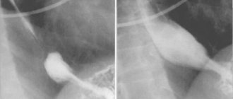
What does a fluoroscopy of the stomach show?
X-ray with contrast allows not only to detect the presence of pathological changes in organs in the digestive system, but also to track the dynamics of the development of the disease and the effectiveness of therapy.
What pathologists does an x-ray of the stomach reveal:
- ulcers - contrast is distributed unevenly, fills niches;
- reduction or increase in the volume of the stomach cavity;
- tumors, polyps, or a predisposition to their occurrence;
- deformation or displacement of the walls of organs or the stomach itself relative to other organs - pathology develops with a hernia of the diaphragm or abdominal wall;
- violation of the lumen of the stomach, duodenum - this pathology occurs when a malignant tumor develops inside or outside the organ;
- foci of inflammation.
Fluoroscopy in the Trendelenburg position is performed if a diaphragmatic hernia is suspected.
Treatment of gastritis
Treatment for gastritis depends on the cause. Acute gastritis caused by taking NSAIDs or alcohol abuse does not require drug therapy; it is enough to exclude these triggers.
In other cases, your doctor may recommend:
- Antibiotic therapy against H. pylori.
- Drugs that block the production of hydrochloric acid (a component of gastric juice) and promote healing of the mucous membrane (proton pump inhibitors* - omeprazole, lansoprazole, rabeprazole, esomeprazole, dexlanzoprazole, pantoprazole.
- Antacids** (neutralize stomach acid, relieve pain).
* - Long-term use of proton pump inhibitors, especially at high doses, may increase the risk of hip, wrist and spine fractures, and an osteoporosis prevention program may be required. ** - Side effects - constipation, diarrhea.
Features and advantages of the gastritis treatment method at the Rassvet clinic
Diagnosis and treatment of gastritis at the Rassvet clinic is carried out in the gastroenterology department. We use evidence-based methods based on international clinical guidelines. Your primary treatment will begin only after a physical examination and all necessary tests and diagnostic tests have been completed.
Important. The influence of certain foods or dietary systems on the risk of gastritis has not been proven by research.
At the Rassvet Clinic, we first of all distinguish gastritis from functional dyspepsia. Gastritis is often asymptomatic, but it is necessary to treat it, since it is a slow but sure road to stomach cancer. Functional dyspepsia, on the contrary, is accompanied by many complaints, but endoscopic examination and biopsy do not reveal pathology.
How gastritis is treated at the Rassvet clinic
To clarify the diagnosis, we use the most modern and accurate equipment and logistics methods. For example, we have built a system for diagnosing gastritis and determining cancer risk according to the OLGA classification. Our endoscopes allow you to perform gastroscopy with multiple magnification and examine the mucous membrane through light filters, taking a biopsy from the most suspicious areas. The biopsy samples themselves are also assessed by the histologist using the OLGA scale, and as a result we obtain a figure that reflects the risk of oncological transformation in the coming years. Further treatment tactics depend on the value of this figure.
We value the comfort of patients, so in Rassvet you can undergo an examination under anesthesia in the shortest possible time.
Recommendations from a gastroenterologist at the Rassvet clinic for patients with gastritis
Timely consultation with a doctor and proper treatment will help keep the disease under control. A strict diet is not needed. However, the rules of a healthy diet should be followed - do not overeat, avoid foods that irritate the mucous membranes (smoked, fried, fatty), and give up alcohol. If you are forced to take painkillers that increase the risk of gastritis, check whether they can be replaced with a drug that is less aggressive on the gastric mucosa (acetaminophen, paracetamol).
Author:
Amelicheva Alena Aleksandrovna medical editor
How is fluoroscopy of the stomach performed?
At the initial stage, a survey X-ray is taken, which allows the doctor to identify the presence of pathologies and inflammatory processes in the pharynx, parts of the esophagus, and other organs. If no abnormalities are found, the stomach is examined with contrast.
How to do an x-ray:
- The patient is given a contrast agent to drink, which allows the condition of the internal organs to be seen more clearly. To do this, they use a special barium for X-rays - a solution that tastes like chalk, the person takes it after the procedure begins.
- Barium helps to delay X-rays, and a minimum of 250 ml of solution is required for the procedure, depending on what needs to be examined.
- If it is necessary to study the condition of the large intestine, the solution is administered by enema.
- The study is carried out using a trochoscope or using a special x-ray stand. The patient needs to change position several times so that the doctor can see the condition of the stomach in real time and record the results on an image or information medium.
- The duration of the procedure is 20–45 minutes.
Important! Is barium harmful to the body? The substance itself is harmless to humans; the rays themselves pose a danger, so if there is an alternative, most doctors choose safer types of diagnostics. Constipation often occurs after an x-ray.
X-ray according to Trendelenburg
To conduct the study, the patient should take a special position - the pelvis should be elevated and at an angle of 30-35 degrees relative to the head.
In this position, the intestine descends into the chest cavity, barium penetrates into the intestine, which allows you to clearly see the contours of the organ. The procedure opens access to the pelvis; with its help, you can not only identify a hernia, but also remove it surgically.