The article was prepared by a specialist for informational purposes only. We urge you not to self-medicate. When the first symptoms appear, consult a doctor.
Standard laboratory diagnostics do not detect many intestinal diseases. Some serious pathological processes occurring in the organ require colonoscopy. In the article we will analyze why this procedure is needed and whether there is any alternative to it.
What it is?
Colonoscopy is an endoscopic examination of the large intestine. It allows you to detect pathology throughout the entire large intestine from the rectum to the cecum, as well as examine the terminal part of the small intestine (ileum).
Colonoscopy is the leading method for early diagnosis of colon cancer.
The method allows not only to conduct an examination, but also to remove precancerous formations (polyps) during the procedure. The examination is performed using a colonoscope - a flexible tube with an optical system and a manipulator. The colonoscope is about 160 cm long and about the thickness of your index finger. The device is inserted into the anus and moves along the intestines, transmitting an enlarged image of the intestinal mucosa to the monitor screen.
What can a colonoscopy detect?
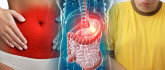
The doctor recommends undergoing an examination if it is necessary to clarify the cause of some symptoms. For example, when blood is detected in the stool. This can be either visible traces of blood (black stool) or a positive stool test for occult blood.
Colonoscopy is indicated for frequent bowel problems (constipation, diarrhea), abdominal pain, increased flatulence (bloating), and sudden weight loss.
The study is necessary when diagnosing iron deficiency and early forms of intestinal cancer. Preventive colonoscopy is recommended once every 10 years for all people over 45 years of age.
Is it painful to have a colonoscopy?
Having a colonoscopy without anesthesia may cause discomfort or pain. This depends on the patient’s individual pain sensitivity threshold.
For some patients, the procedure may cause mild discomfort, while for others it may cause intense pain.
Before insertion into the anus, the tip of the colonoscope is treated with a special gel to facilitate its insertion. Discomfort or pain during a colonoscopy usually occurs due to the distension of the intestine when air is supplied, which helps to expand and better view the mucous membrane.
An alternative is to perform a colonoscopy while you sleep. Drug sedation ensures an absolutely painless and comfortable procedure.
What are the indications for colonoscopy?
Colonoscopy can be performed either routinely or urgently.
Why is it better to do the procedure at the Central Clinical Hospital of the Russian Academy of Sciences?
When choosing a clinic for an informative, but not very pleasant (without anesthesia) examination, you should first of all focus not on the cost of the procedure, but on factors such as the experience and qualifications of doctors, hospital equipment, and patient reviews. The Central Clinical Hospital of the Russian Academy of Sciences offers many advantages:
- painlessness of the procedure;
- high-precision modern equipment;
- high level of qualification of specialists;
- professionalism of experienced doctors in making a diagnosis and prescribing effective treatment;
- affordable price of the procedure.
The clinic staff considers tactfulness, attention, willingness and ability to help to be a priority in relations with patients. Already at the first meeting, during the consultation, the doctor simply and clearly conveys important information about colonoscopy - how to prepare for it, how long the examination will last, what can and cannot be done the day before and on the day of the procedure. This creates a certain mood and inspires trust. This means that the result of a well-thought-out treatment method after an informative, most gentle diagnosis will be wonderful.
Routine colonoscopy is performed when:
· bloody discharge from the rectum;
· periodic pain in the lower abdomen that bothers the patient for a long time;
· regular loose stools;
· polyps or suspected neoplasm in the large intestine;
· nonspecific ulcerative colitis and Crohn's disease;
iron deficiency anemia;
· presence of cases of colon cancer in the family;
· for taking tissue samples for histological examination (biopsy).
Rehabilitation
After a colonoscopy, a hospital stay is not expected; the patient leaves the hospital. Drugs administered for anesthesia are eliminated from the body within several hours. For the first half hour or hour, sudden movements should be avoided, and the feeling of fullness in the intestines is a normal possible reaction, it passes quickly.
How to prepare for a colonoscopy?
You can prepare for the test at home. A few days before the study, the patient is prescribed a slag-free diet and bowel cleansing using special medications or enemas.
Important:
1. The patient must tell the doctor in advance about all concomitant diseases and medications taken, especially blood thinners.
2. 3-4 days before the study, you need to stop taking iron supplements.
3. If the patient is prone to constipation, it is necessary to take laxatives daily for a week before the colonoscopy.
4. If the patient regularly took a laxative before the procedure, it is necessary to increase the dose of the drug by 1.5 times.
5. It is necessary to purchase special intestinal cleansing products from the pharmacy.
If these requirements are met, the colonoscopy will be successful. Insufficient bowel preparation may make the diagnosis inconclusive and the study will have to be repeated.
Contraindications
Colonoscopy is a low-traumatic diagnostic method, but it has a number of contraindications in which the procedure cannot be performed.
These include:
- State of shock with a drop in blood pressure below 70 mm. rt. Art.
- Acute respiratory infections, inflammation of the respiratory system, accompanied by an increase in body temperature.
- Intestinal infections in the acute phase.
- Peritonitis.
- Heart failure.
- Blood clotting disorder.
- Crohn's disease, acute stage of colitis with massive intestinal damage.
- Diverticulitis in the acute phase.
- Severe disturbances in a person’s well-being.
In addition, there are relative contraindications to the procedure:
- Massive anal bleeding.
- Anal fissures.
- Acute stage of hemorrhoids.
- Paraproctitis.
- The period of bearing a child.
- Presence of a large hernia.
- Early recovery period after abdominal surgery.
- Diverticulitis.
- Poorly conducted preparation for cleansing the intestines, etc.
Doctors should seriously evaluate the possible risks of performing a colonoscopy if the patient has the following diseases and conditions:
- Heart diseases.
- Allergy to medications.
- Lung diseases.
- Diabetes.
- Treatment with drugs that affect blood clotting processes.
The doctor must be aware of the medications the patient is receiving. You may have to stop taking them and resume taking them after the procedure.
Video: Live healthy! “Colonoscopy - what is this procedure and who should undergo it?”:
Diet on the eve of the examination
The day before your colonoscopy, you should only eat easily digestible foods, such as clear vegetable broth.
Eating after 12.00 is prohibited. Only liquid intake (weak tea or still water) is allowed and recommended.
On the day before the study, eating is prohibited - you cannot have breakfast!
Colon cleansing with special preparations
It is very important to properly prepare your bowels for a colonoscopy. For this purpose, special laxatives have been developed that help to thoroughly cleanse the intestines without resorting to enemas or outside help.
What to do next
Colonoscopy is an informative diagnostic method
Upon completion of the manipulations, the patient receives a protocol with the results of the study, as well as recommendations for subsequent instructions. When local anesthesia is applied, the person is able to move independently and can therefore be free immediately.
After general anesthesia, it is necessary to spend an additional amount of time in a medical institution to gradually recover from the state of sleep under the supervision of specialists. After the procedure, eating and drinking are allowed.
Preparing for a colonoscopy in patients with constipation
· 7 days before the colonoscopy, you need to change your diet.
· If you use laxatives, you should continue to use them throughout the entire period of preparation for a colonoscopy.
· If the patient suffers from prolonged constipation (stool once every 6–10 days), it is necessary to double the dose of the usual laxative. On the recommendation of a gastroenterologist, a combination of stimulating and osmotic drugs can be used.
· 2-3 days before the start of preparation for the procedure, you need to stop taking iron supplements, because they contribute to constipation and impair the visualization of the mucous membrane.
How is the research going?
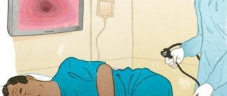
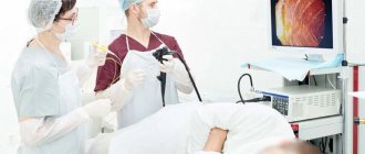
Colonoscopy is performed by a certified endoscopist on an outpatient basis. The patient takes off his clothes below the waist and puts on special disposable underpants, then he lies down on the couch on his left side and presses his legs to his stomach.
The endoscopist gradually inserts the colonoscope into the anus and advances it into the colon. To better visualize the mucous membrane and advance the device, the doctor expands the intestines, supplying air. Next, he advances the colonoscope to the border of the large and small intestine and examines the terminal part of the small intestine (ileum). The doctor carefully assesses the condition of the intestinal mucosa by slowly pulling the colonoscope in the opposite direction.
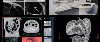
A colonoscope allows you to take a series of images of the intestinal mucosa, which the doctor carefully examines on a monitor. If suspicious areas are detected, the endoscopist takes tissue samples (biopsy), which are then submitted for histological examination. If the doctor needs to more carefully examine minor changes in the mucous membrane, then he changes the composition of the light from the colonoscope lamp.
Colonoscopy, CS, hose, intestine and other names have this study among patients. The research method allows you to examine the lower intestines. The endoscope is passed through the anus, examining from the rectum to the ileocecal angle (the transition of the small intestine to the large intestine) with examination of the final section of the ileum (small) intestine.
CS (Colonoscopy) is a method of examining the rectum, large intestine (from the sigmoid to the dome of the cecum) and the distal jejunum (the final section of the small intestine) using an endoscope (a medical device that allows you to look inside the lower parts of the gastrointestinal tract through the anus ). During CS, you can examine: the rectum, sigmoid colon, descending colon, transverse colon, ascending colon, cecum, ileocecal junction (the natural valve between the small and large intestines - the Bauhinian valve) and the distal small intestine throughout 10-15 cm. As with gastroscopy, the condition of the mucous membrane covering these hollow organs is assessed.
Colonoscopy is used to diagnose the following diseases
- Inflammatory diseases of the large intestine: Crohn's disease;
- Nonspecific ulcerative colitis;
- Other infectious and non-infectious colitis;
Colonoscopy can detect
- Inflammation;
- Atrophy;
- Polyps;
- Neoplasms (tumors, cancer);
- Erosion;
- Ulcers;
- Foreign bodies;
- Diverticula;
- Vascular tumors - hemangiomas;
- And other rarer finds.
Indications for performing a Colonoscopy are:
- Determination of the nature of the pathology revealed by other examination methods (X-ray, Irigoscopy, MRI, MSCT);
- Confirmation and/or clarification of the diagnosis;
- Clarification of the localization of the pathological process;
- Clarification of the prevalence of the pathological process;
- Biopsy;
- Evaluation of treatment effect;
- Therapeutic manipulations;
- Determining the source of bleeding;
- Stop bleeding;
- Foreign body removal;
- Diagnosis and treatment of anastomositis in the early postoperative period;
- Intraoperative assistance.
- Screening for colorectal cancer.
There are the following contraindications for CS. As they say: “Consult a specialist!”
Contraindications for Colonoscopy
1) Absolute contraindications (the risks of performing a CS exceed the diagnostic value of the study):
- Myocardial infarction in the acute stage (2 weeks after the heart attack);
- Stroke in the acute period;
- Cardiovascular and/or respiratory failure of the 3rd degree;
- Uncontrollable attack of bronchial asthma;
- Severe and extremely severe somatic condition;
- Hemorrhagic shock and other types of shock;
- Peritonitis;
- Intestinal perforation;
- Severe form of developing colitis with toxic magacolon.
In all these cases, CS is possible only for health reasons. When the results of the study exceed the risks from possible complications. The decision to perform a CS is made by a council of doctors together with an endoscopist, weighing all the pros and cons.
2) Relative contraindications:
- Arrhythmias (paroxysmal);
- Angina attack;
- Crohn's disease and UC in severe cases;
- Abdominal abscesses and infiltrates with abscess formation;
- Abdominal aortic aneurysm;
- Hydrothorax;
- Tense ascites;
- Paraproctitis;
- Hemorrhoids and anal fissures;
- Diverticulitis is inflammation of the diverticula of the large intestine;
- Inguinal and umbilical hernias with the possibility of loops of the large and/or small intestine coming out and their subsequent strangulation;
- Within 14 days after abdominal surgery on the abdominal organs.
Relative contraindications increase the risks during CS. But at the same time, the diagnostic value of the study is higher than these risks. The issue of risk assessment and CS is decided by the endoscopist.
3) Diseases that significantly limit Colonoscopy, impair tolerability and reduce the scope of the study:
- Adhesive disease;
- Severe diverticulosis;
- Hirschsprung's disease;
- Umbilical and inguinal hernias.
Risks arising during colonoscopy
- Related to preparing for the study:
- Violation of water and electrolyte balance;
- Individual intolerance to the drug;
- Weakening the effect of oral medications.
- Related to premedication and local anesthesia:
- Allergic reactions to lubricating gel or anesthetic gel (Lidocaine). Angioedema-type reaction and/or other allergic reactions accompanied by swelling of the mucous membrane of the respiratory tract with asphyxia and respiratory failure;
- Cardiac arrest or arrhythmia (Lidocaine).
- Related to the administration of anesthesia:
- Breathing disorders;
- Cardiac dysfunction;
- Exacerbation of chronic diseases;
- And others (better ask your anesthesiologist).
- Related to the endoscope:
- Perforation of the colon. More often, ruptures occur in places of pathological narrowing and in the region of the splenic angle and rectosigmoid.
- Bleeding both into the lumen of the colon and into the abdominal cavity. Into the intestinal lumen, more often when the mucosa is changed as a result of the presence of ulcers or tumors. Into the abdominal cavity in case of rupture of adhesions or injury by an endoscope to the liver or, more often, the spleen;
- Pneumatosis intestinalis, due to excessive inflation with air.
- Related to manipulations during the study:
- Bleeding during biopsy, usually in the area of the bottom of ulcers and fissures;
- Perforations.
- Related to the quality of processing of endoscopic equipment:
- Infection;
- Allergic reactions to detergent residues.
Pros and cons of Colonoscopy as a diagnostic method
«+»
- “Gold standard” for diagnosing many diseases of the large intestine and rectum;
- Possibility of collecting material for morphology (biopsy);
- Possibility of carrying out therapeutic manipulations;
- High information content and accuracy;
- Screening for early cancer and precancerous conditions.
«-»
- Although small, the invasiveness of the technique (compensated by the high quality of modern endoscopic technology and staff skills);
- Unpleasant sensations (compensated by conducting research under anesthesia);
- There are risks (compensated by compliance with standards and protocols for conducting research and processing equipment).
A few words about “unpleasant sensations”
When performing a CS, the patient experiences the following “indescribable” sensations:
- Pulling sensations in the abdomen, discomfort from the sensation of “stretching the intestines” can sometimes reach the pain threshold (associated with the passage of an endoscope and the tension of the intestines when straightening and/or collecting loops). Pain usually occurs when the loops are excessively stretched by the endoscope or when the intestinal loops are fixed by adhesions to each other;
- The feeling of intestinal distension, “swelling” is the same as with flatulence (gas) in the intestines (associated with the use of inflation with air to straighten the intestinal lumen and folds). Pain can occur when the intestines become overly inflated with air due to overstretching of the intestinal wall;
- There may be sensations of movement and/or movement inside the abdomen (occur when advancing and manipulating the endoscope);
- Sometimes there is a feeling of urge to defecate (associated with residual contents in the intestine after preparation and/or distention with air).
There are no other sensations during a CS. All sensations are strictly individual and depend on many factors. Painful sensations usually occur due to: poor preparation, the presence of diseases limiting the performance of colonoscopy, or violation of the technique of performing the examination itself. But if you listen to the recommendations of medical personnel and prepare well for the study, then the procedure takes place with the minimum required time and a minimum of discomfort.
How should you behave during a Colonoscopy?
- Prepare well for your research. The procedure is carried out after cleansing the intestines. For cleaning, special “lavash” solutions (saline or PEG-based polyethylene glycol), laxatives or cleansing enemas (more often used in a hospital) are used.
- Try to breathe deeply and evenly, relax your stomach, and follow the commands of the medical staff. During the examination, you may need to change your body position on the couch (on your left side, back, right side).
- If you experience pain and/or severe discomfort, notify the medical staff. Do not tolerate it under any circumstances!
- To facilitate the passage of the endoscope and reduce discomfort/pain, manual assistance from a nurse may be required; follow her instructions.
Only point 1 concerns everyone who performs a Colonoscopy under anesthesia. The rest of the sensations and interactions with the medical staff will go unnoticed by you.
Finally, a few words about the quality of Colonoscopy
- Quality bowel preparation!
- Follow a diet free of ballast substances 3 days before the start of preparation for the CS.
- Strict adherence to the regimen of taking bowel cleansing medications recommended by the doctor for preparation.
- Use of defoamers (simethicone) when taking the last portion of lavage solution.
- All this improves the quality of preparation and simplifies the examination of the mucous membrane of the large intestine.
- “Fast” does not equal “Quality”. A complete examination of the large intestine is performed upon reaching the dome of the cecum (ideally, the colonoscope is inserted into the ileum through the bauhinium valve). The examination is carried out both when the endoscope is inserted and when it is removed. A qualitative indicator is a detailed examination of the large intestine while withdrawing the endoscope for at least 6 minutes. Only during this time is it possible to examine and find the greatest number of changes in the mucous membrane of the large intestine.
- Qualitative indicators are the achievement of the level of the dome of the cecum during examination and photographic recording of the achievement of the dome and altered areas of the mucosa.
- The use of auxiliary techniques: chromoendoscopy, zoom endoscopy, narrow-spectrum examination (NBI, i-scan, etc.) and other techniques. These techniques increase the detail and contrast of the endoscopic image.
- Use of endoscopes with high image resolution, HD and higher. Availability of photo and video recording of the resulting images of the mucous membrane.
- Carrying out a biopsy. The final endoscopic conclusion is a conclusion confirmed morphologically based on the results of the pathologist’s assessment of biopsy specimens.
- Application of modern endoscopic classifications when interpreting results and writing a study protocol.
The author of the article will undergo a screening CS at the age of 45 years (recommendations of the Russian Endoscopic Society and the American Society of Gastroenterology).
Side effects of colonoscopy.
Colonoscopy is one of the safest endoscopic methods.
Perforation (perforation), which may occur as a result of examination or removal of polyps, or the development of bleeding are extremely rare events.
Sometimes it is possible to develop individual intolerance to the administration of medications during the study. Therefore, the patient is under the supervision of a doctor and his assistants throughout the procedure, who monitor vital signs (pulse, blood pressure, oxygen saturation).
Immediately after the procedure, the patient may experience discomfort and bloating in the abdomen due to the air that was introduced during the examination.
If after the manipulation the patient is bothered by tolerable spasmodic pain due to the passage of gases and residual water, there is no need to worry. These phenomena go away on their own.
If after a colonoscopy you experience bleeding from the rectum, severe pain in the lower abdomen, vomiting, or fever, you should immediately consult a doctor.
If the colonoscopy was performed “in a dream,” then the patient cannot drive a vehicle for 24 days after the examination, and he will need accompanying persons to get home.
At the Ilyinskaya Hospital, endoscopic removal of polyps is possible directly during a diagnostic colonoscopy - this allows you to avoid repeated preparation for the study, repeated anesthesia (drug sleep) and repeated performance of the study itself. If, when small polyps are detected (up to 5 mm), you prefer to remove them during a diagnostic colonoscopy, without breaking this procedure into two stages, then you need to inform your doctor about this in advance!
Plan of tests and examinations during pregnancy by week
Pregnancy check-ups per week is a schedule developed by doctors for conducting tests and visiting close specialists. It is based on the recommendations of the Ministry of Health and Social Development of the Russian Federation and is aimed at maintaining health and well-being during pregnancy.
It is important that the expectant mother visits a doctor, follows the doctor's recommendations and undergoes routine examinations. A gynecologist monitors the progress of pregnancy, the risk of possible complications, the development of the fetus and the condition of the pregnant woman herself.
studies and a series of tests help to identify possible problems in a timely manner and provide the necessary assistance.
In a normal pregnancy, examination of pregnant women in the first trimester includes visiting a gynecologist once a month in the first trimester, in the second trimester - from 36 weeks onwards once every 2-3 weeks, and before giving birth, the gynecologist examines the pregnant woman every week.
Examination plan for a pregnant woman in the antenatal clinic
To receive legal assistance in overcoming pregnancy, you must register with the Prenatal Clinic. It is better to do this at 6-8 weeks of pregnancy (be sure to take your passport and insurance policy with you).
Examinations in the first trimester (from 1 to 13 weeks)
In the first trimester, the gynecologist will refer the pregnant woman to a highly specialized doctor for a mandatory examination:
- ENT - examines for chronic or infectious diseases of the respiratory and hearing organs, smears of the mucous membrane;
- Dentist - examines the oral cavity, teeth;
- Ophthalmologist - gives recommendations on the method of delivery (in the case of high intraocular pressure, a cesarean section may be recommended);
- Cardiologist - examines the heart;
- radiologist - all family members with whom the pregnant woman lives must do fluorography and transfer this data to the therapist; The pregnant woman herself is prohibited from having x-rays or fluorography;
- therapist - gives an opinion on the results of the examination.
The gynecologist measures blood pressure, height, weight and pelvic size and enters this data into the pregnant woman’s medical record. Gives recommendations on taking vitamins, proper nutrition, daily routine and prescribes directions for testing:
- general urine analysis. The analysis allows you to quickly assess the general health of a pregnant woman and how her kidneys are functioning. For analysis, morning urine is collected (at night the kidneys work more actively, the urine becomes more concentrated, so this diagnosis is more accurate);
- smear through the flora. A smear on the flora reveals possible infections and inflammations. In case of anomalies, the gynecologist prescribes additional examinations. A test for sexually transmitted diseases (STDs) is often ordered if the smear test is poor. Some infections are dangerous for the normal development of the fetus. In this case, they are treated taking into account the duration of pregnancy and the quality of the drug (the choice of drug is not general, but local);
- general blood analysis. A blood test during pregnancy is carried out at registration and repeated in each trimester. Thus, the doctor has the opportunity to monitor the patient’s condition. Based on the results of a clinical blood test during pregnancy, determine the number of leukocytes, platelets, hemoglobin, assessed by SOE and other indicators; blood chemistry. The analysis helps to identify failure of important organs: the liver and pancreas. High concentrations of creatinine and urea are found in renal failure. High bilirubin levels associated with liver problems. The functioning of the pancreas is monitored by glucose levels so as not to miss the possible development of gestational diabetes;
- blood clotting test. If there are no deviations, the test is carried out once per trimester;
- blood type and Rh factor. Even if you know your blood type, you should still do this test. In the event of possible major blood loss or unplanned surgery, this information can save the life of mother and child. If a woman has a negative Rh factor, and the father of the child is positive, then a Rh conflict may occur. This means that the mother's body perceives the baby as a foreign body and produces antibodies to eliminate it. This reaction can lead to anemia, miscarriage, or death of the fetus in the womb. If the mother's Rh factor is negative, then the blood is donated by the future father. If the Rh factor is positive, a pregnant woman undergoes an antibody test once a month until the 32nd week of pregnancy. After the 32nd week of pregnancy and twice a month until the end of pregnancy. If this is your first pregnancy and there are no antibodies before the 28th week of pregnancy, doctors suggest administering a serum that subsequently blocks the production of antibodies;
- tests for HIV, syphilis, hepatitis. These tests are carried out at the beginning of pregnancy and at 30-35 weeks (due to the long incubation period of diseases);
- TORCH infection. The TORCH test shows the expectant mother’s immunity to toxoplasma, rubella, cytomegalovirus, herpes and some other infections. They are very dangerous for the intrauterine development of a child. If antibodies are not detected, the pregnant woman should take all preventive measures to avoid getting sick while carrying a child;
- double test. Includes ultrasound and blood tests for levels of human chorionic gonadotropin (hCG) and plasma produced protein (PARP-A). These tests show whether a child is at risk of developing a chromosomal abnormality (Down syndrome).
Examinations in the second trimester (from 14 to 27 weeks)
In the second trimester, the gynecologist begins to measure and record the height of the uterine fundus and the volume of the abdominal cavity. A pregnant woman visits a gynecologist more often: once every two to three weeks. If the condition of the expectant mother is normal, the doctor prescribes what examinations the pregnant woman should undergo:
- at 18-21 weeks. At 18-21 weeks, she undergoes a second screening, the “triple test”, with the help of which doctors more accurately determine the risk of possible pathology of the child;
- at 18-21 weeks they do a second routine ultrasound, assess the condition of the placenta and amniotic fluid, observe how the baby develops, by this time you can already know the gender;
- do a general urine test;
- They do a second examination for syphilis.
The doctor may order additional repeat blood, urine, and other tests if necessary to What tests are given to a pregnant woman in the third trimester?
- In the period from 30 to 34 weeks, do a third ultrasound examination, determine the size and approximate weight of the baby, observe its position in the uterus, examine the condition of the placenta, whether there is an umbilical cord, and monitor the quantity and quality of amniotic fluid.
- Within 32-35 weeks, do a CTG (cardiotocography), study the functioning of the embryo’s cardiovascular system, and observe its motor activity. This test helps determine how the child is feeling and whether he or she has developmental problems.
- From 36 weeks until delivery, a gynecologist examines the pregnant woman every week.
- At week 30, the doctor gives a referral for a general urine test, a general blood test, a biochemical blood test, an HIV and hepatitis test, a third test for syphilis and a smear for cytological examination.
What is an exchange card
At the end of the second trimester, between the 22nd and 23rd weeks of pregnancy, the gynecologist gives the expectant mother a smear.
You might be interested in: Conjunctivitis. allergic, bacterial and viral conjunctivitis. causes, symptoms, diagnosis and treatment of the disease.
This is a medical document in which the doctor records the course of pregnancy and the woman’s health status.
A pregnant woman should carry it with her passport and OMS guidelines if she is in urgent need of medical attention. This becomes especially important in the last trimester.
The exchange card must consist of three mandatory parts:
- The first part contains information entered by the gynecologist at the prenatal clinic. Here you can find out the personal data of a pregnant woman, information about chronic and past diseases, previous pregnancies and births, all measurement data during a medical examination, copies of ultrasound, CTG, examination results, recommendations after examination by other specialists.
- The second part contains information entered by the maternity hospital doctor. Before discharge, it is written down how the birth went, the prenatal period, if there were complications, and if necessary, recommendations for treatment are written down. This information is necessary for the gynecologist at the maternity hospital when a woman comes for her first examination after childbirth.
- The third part contains information about the born child. Enter height, weight, degrees on the Apgar scale. This information is important for the pediatrician, who examines the baby and copies all the data about the child, starting with the exchange card and ending with the medical record of the little patient.
Brief table of examinations during pregnancy by timing:
Duration of pregnancy (weeks) Type of examination and tests15
| 5-7 | First visit to the gynecologist, confirmation of pregnancy. |
| 7-11 | Visit to the gynecologist, registration at the prenatal clinic. Measuring pressure, pulse, temperature, pelvic size, weight and height of a pregnant woman. What tests need to be done: - Coagulogram; - general urine analysis; - general blood analysis; — chemical blood test; — blood test for TORCH infection; — blood group analysis and Rh factor analysis; - blood test for HIV, hepatitis B and C, syphilis; - cytological smear. |
| 2 weeks after registration | Visiting a doctor: - dentist; - ophthalmologist; - ENT specialist; - cardiologist; - therapist. |
| 11-14 | First scheduled ultrasound, First examination (“double test”) |
| 16 | Examination by a gynecologist |
| 16-18 (allowed up to 20 weeks) | Second routine Ultrasound examination and second screening (“triple test”) |
| 20 | — gynecological examination; - general urine analysis; - second study of syphilis. |
| 22 | Gynecologist |
| 24 | — Gynecological examination; — Examination of the legs for varicose veins; - Analysis of urine. |
| 26 | Gynecologist |
| 28 | - Gynecologist; - Examination of legs for varicose veins; - Urine test. |
| 30 | - gynecologist; - Analysis of urine; - blood analysis; - blood analysis; - third test for syphilis; - HIV test; - flower tampon. Third planned ultrasound |
| 33-34 | Fetal cardiography (FCC) |
| 35 | — Gynecologist — examination for varicose veins — urine test — fetal CTG. |
| 37 (once a week until delivery) | Gynecologist |
- 22330