RADIATION ANATOMY OF THE ESOPHAGUS
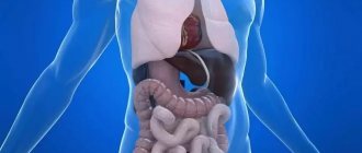
Radiation anatomy of the abdominal organs
NORMAL ANATOMY OF THE ESOPHAGUS
The esophagus is a muscular organ that connects the pharynx to the stomach. The shape of the esophagus can be compared to a tube, flattened in the anteroposterior direction.
The esophagus is located almost vertically anterior to the spinal column. The length of the esophagus in women is 230-250 mm, in men - 250-300 mm, in newborns - 110 mm, by one year it reaches 155-180 mm, by 3 years - 175-210 mm.
The beginning of the esophagus in a newborn is at level CIV, by 12 years - Cv, in an adult - CV1, and in older people - CVII. Its lower limit in adults reaches the level of Thx_x1, in newborns - ThK_x. The width of the lumen of the esophagus at the level of the upper border in adults is 19 mm, at the level of the lower border - 22 mm, and at the level of the thoracic region - 21-30 mm. In newborns, the width of the esophagus is 8-10 mm.
The esophagus deviates noticeably from a straight line. On the neck, located behind the trachea, it passes slightly to the left of the midline of the body. Behind the esophagus is the vertebral column, from which the esophagus is pushed anteriorly by the aorta at the level of ThIx. Having entered the chest cavity through its upper opening along with the trachea, the esophagus intersects with the left main bronchus at the border of ThIV_v, passing behind it. Then it deviates slightly to the right and only in front of the esophageal opening of the diaphragm is again located to the left of the median sagittal plane, while the descending aorta passes significantly to the right and more dorsally. Thus, the aorta and esophagus bend around each other in the form of a very gentle spiral. Through the esophageal opening of the diaphragm, located slightly to the left of the median plane, the esophagus, together with the vagus nerves, enters the abdominal cavity.
The esophagus is connected to the serous membranes: at the top it is covered by the left mediastinal pleura, below (under the root of the lung) - by the right mediastinal pleura; The pericardium is adjacent to the lower part of the thoracic esophagus in front.
The esophagus has three sections: cervical, thoracic and abdominal. The cervical region begins at the level of the pharynx and ends at the upper opening of the chest, where the thoracic region, located in the posterior mediastinum, originates. At the level of the esophageal opening of the diaphragm, the latter passes into the abdominal section, which lies in the abdominal cavity and ends at the cardial section of the stomach.
Most often, 4 physiological narrowings are detected in the esophagus (some of them appear by 2-3 years of age), the severity of which is quite individual:
1st - at the junction of the pharynx and esophagus;
2nd - at the aortic arch (at the level of Th]n);
3rd - at the left main bronchus (at the level of Thv);
4th - in the area of the esophageal opening of the diaphragm (at the level of Thx).
The division of the esophagus into segments according to Brombar is widely accepted. In adults, 9 segments are distinguished: supraortic, aortic, interaortobronchial, bronchial, subbronchial, retrocardial, supradiaphragmatic, diaphragmatic and abdominal.
The supraortic (tracheal) segment begins with the entrance to the esophagus at the lower edge of the cricoid cartilage and ends at the upper edge of the aortic arch. This segment of the esophagus is mobile. When the head is tilted forward, the level of the entrance to the esophagus drops to CVII, and with a significant tilting of the head, the cricoid cartilage, together with the entrance to the esophagus, rises to the level of Cv_VI.
The aortic segment is located at the ThIV level and its length corresponds to the diameter of the aortic arch. The second physiological narrowing of the esophagus, caused by the pressure of the aortic arch, is localized in this segment; it has the appearance of an arcuate depression located on the anterior wall. With an abnormally located right-lying aorta, this depression is determined by the right and posterior contours of the esophagus.
The interortobronchial segment extends from the lower surface of the aortic arch to the upper outer wall of the left main bronchus and projects onto the upper part of Thv.
The bronchial segment is located corresponding to the bifurcation of the trachea and the lower part of the body of Thv. Here is the third physiological narrowing of the esophagus, caused by pressure from the left main bronchus. The more vertically the left bronchus is located, the wider this depression is.
The subbronchial segment starts from the level of the lower wall of the left main bronchus and ends at the upper contour of the left atrium, projecting onto Thv|. It is located behind the left atrium and anterior to the descending aorta. An enlarged left atrium or tracheobronchial lymph nodes displace this segment of the esophagus to the right and posteriorly, and an aortic aneurysm - to the right and anteriorly.
The retrocardial segment of the esophagus begins at the level of the upper contour of the left atrium and ends according to its lower contour, projecting at the level of Thvl]VIII. It is directed from front to back, its front surface is adjacent to the left atrium, and its back surface is in contact with the anterior left surface of the aorta. An enlarged left atrium deviates this segment of the esophagus to the right and posteriorly, and an enlarged descending aorta deviates to the right and anteriorly.
The supradiaphragmatic segment of the esophagus extends from the inferior contour of the left atrium to the diaphragm, which corresponds to the level of ThIx. This segment is the widest part of the esophagus and can acquire a spindle-shaped shape, forming the ampulla of the esophagus; surrounded by loose fiber, making it mobile. On the left it comes into contact with the mediastinal pleura, with inflammatory changes in which deformations and displacements of this segment to the left are observed.
The diaphragmatic segment is located in the esophageal opening of the diaphragm at the level of Thx. It corresponds to the fourth physiological constriction, which plays the role of a functional compressor.
The abdominal segment is located from the diaphragm to the cardial opening of the stomach and is projected at the level of Thx_XI. It is directed from back to front and from right to left, adjacent to the back of the left leg of the diaphragm, to the right and front to the liver, to the left to the cardiac part of the stomach, forming the angle of His with the vault of the stomach.
In children, there are 6 such segments, since the aortic, interaortobronchial, bronchial and subbronchial segments are combined into one - aortobronchial.
The esophagus has 4 main walls: anterior, posterior, right, left. There are also intermediate walls (antero-right, posterior-left, etc.). The thickness of the esophageal wall in adults is on average 3-4 mm, in children under 1 year of age - up to 1 mm.
The wall of the esophagus consists of four layers: mucosa, submucosa, muscularis and adventitia.
The mucous membrane of the esophagus forms longitudinally arranged folds, so the shape of its lumen in a cross section resembles an asterisk. The submucosa is well expressed, ensures the mobility of the mucous membrane and promotes the formation of folds.
The muscular lining of the esophagus consists of an inner circular and outer longitudinal layer. The inner circular layer at the border with the pharynx thickens and forms a sphincter - a constrictor of the esophagus. Longitudinal muscle fibers in the cervical part of the esophagus form three bundles, of which the more powerful one occupies the anterior surface. The muscle layer of the posterior wall of the esophagus at this level is represented only by circular fibers, which contributes to the formation of border diverticula. Downwards, the outer muscular layer passes into the outer muscular layer of the stomach.
The adventitia forms the framework of the esophagus. Its surface layers gradually pass into the peri-esophageal tissue and connective tissue of neighboring organs and fix the esophagus to the spine.
The esophagus is supplied with blood from several sources, while the arteries feeding it form abundant anastomoses among themselves. The cervical part of the esophagus is supplied with blood by vessels arising from the inferior thyroid artery, the thoracic part - by the esophageal and bronchial branches of the thoracic aorta. The abdominal part of the esophagus is fed by vessels arising from the left gastric and inferior phrenic arteries.
The veins of the esophagus form the superior and inferior plexuses. The superior plexus occupies the upper third of the organ, and the inferior plexus occupies the middle and lower thirds. The veins of the superior plexus belong to the superior vena cava system. The veins of the lower plexus anastomose with the splenic and gastric veins, which carry blood to the portal vein. Thus, so-called portocaval anastomoses are located in the wall of the esophagus. That is why varicose veins can be observed in the esophagus and in the upper parts of the stomach with portal hypertension.
The lymphatic vessels of the cervical part of the esophagus flow into the subclavian trunk and anastomose with the lymphatic vessels of the pharynx, the vessels of the thoracic part flow directly into the thoracic duct. The lymphatic plexuses of the abdominal esophagus anastomose with the lymphatic plexuses of the upper stomach.
The esophagus is innervated by the branches of the right and left vagus nerves, which form the esophageal plexus, which anastomoses with the fibers of the sympathetic nervous system.
X-RAY ANATOMY OF THE ESOPHAGUS
To study the esophagus, it is necessary to use contrast media. Such media are air (or other gases), a suspension of barium sulfate or water-soluble contrast agents (urografin, verografin, omnipaque, etc.). The contrast agent, its quantity and concentration are selected depending on the task facing the researcher.
When contrasted, the esophagus appears as a longitudinally located ribbon-like shadow of uneven width, located in the neck, in the chest and partially in the abdominal cavity (Fig. 11.25-11.27).
The contours of the esophagus are always smooth and clear with the presence of depressions corresponding to physiological narrowings. Depending on the degree of filling, the contours of the esophagus change: when filled tightly, they are slightly convex, and as they empty, they flatten. Unstable wave-like or fine-toothed contours of the esophagus are observed with segmental contractions of muscle groups and limited relaxation of the walls. The elasticity of the walls of the esophagus is determined by the state of the contours, lumen and wall thickness at various degrees of filling and peristaltic movements, which are detected by fluoroscopy. Violation of the elasticity of the walls of the esophagus is accompanied by straightening of the contours, lack of variability of the lumen and contours (rigidity).
The relief of the mucous membrane of the esophagus is represented by continuous, longitudinal, elastic folds running parallel to each other. On each of the four walls there are two folds of the mucous membrane, however, as a rule, 3-4 folds can be identified (see Fig. 11.26).
In young children, folds are detected only in the abdominal region. The thickness of the folds of the esophageal mucosa in adults ranges from 1 to 3 mm, in children under 1 year of age - up to 1 mm. The thinnest folds are located in areas of physiological narrowing; the greatest thickness of the folds is noted in the supradiaphragmatic segment. With increased tone of the esophagus, the folds of the mucous membrane are high, thin, and tortuous, and with decreased tone, they are flattened. At the level of ThVI|V||| due to rotation of the esophagus, a crossover of the folds of the mucous membranes of the opposite walls occurs as a result of their projection layering, which is most clearly defined in the right oblique projection. As a result of the mobility of the mucous membrane, due to the severity of the submucosa, its folds change thickness during x-ray examination. The air swallowed during contrasting of the esophagus in the form of bubbles can give round or oval clearly defined enlightenments, simulating pathological formations (tumor, varicose nodes). Unlike pathological formations, air bubbles disappear during the examination.
The motor-evacuation function of the esophagus is the main one. In the esophagus, active and passive movements are distinguished. Active include peristaltic movements of the esophagus, which depend on the tone of the walls and the state of the nervous system, while passive include transmission pulsatory, phonatory, and respiratory.
Rice. 11.25. X-ray of the esophagus in a direct projection.
1 — aortic arch; 2 — displacement of the esophagus by the aortic arch.
Rice. 11.26. X-ray of the esophagus in lateral projection.
Rice. 11.27. Radiographs of the esophagus at the level of the diaphragmatic opening.
a — the picture was taken while inhaling; b — the picture was taken while exhaling.
The tone of the esophagus is assessed by the width of its lumen, the speed of passage of barium suspension and the nature of the folds of the mucous membrane. The tone and elasticity of the walls ensure the expansion of the esophagus during the passage of a bolus and its contraction after emptying (see Fig. 11.27). With normal esophageal tone, a liquid barium suspension passes through the esophagus in a vertical position of the patient in 3-5 seconds, in a horizontal position - in 8-10 seconds. An increase in the tone of the esophagus is accompanied by its shortening, narrowing of the lumen, and a decrease in the time of passage of the barium suspension, but with a sharp increase in tone and the occurrence of a spasm, this time increases. With decreased tone, the esophagus lengthens, its lumen expands, and the transit time of the barium suspension increases.
Active movements of the esophagus are also caused by contractions of the pharyngoesophageal and esophageal-gastric junctions, which act as true compressors. Their function is regulated reflexively. Normally, the barium suspension in the pharyngoesophageal and esophageal-gastric junctions passes in a narrow stream without stopping.
Peristalsis occurs when food enters the esophagus from the pharynx and follows the act of swallowing. There are primary and secondary peristaltic waves. Primary waves are associated with the swallowing reflex: they arise during the act of swallowing, are regulated by the central nervous system and do not depend on local mechanisms. They are directed from the pharynx to the stomach and have the correct rhythm. Secondary peristaltic waves occur outside the act of swallowing and are caused by irritation of intraesophageal receptors. The amplitude of secondary waves is weaker than the primary ones. During X-ray examination in a vertical position, due to the rapid movement of liquid barium suspension, peristaltic waves are rarely detected normally. In a horizontal position of the patient, especially in a position with an elevated pelvis, peristaltic activity intensifies, the waves are frequent and deep. Tight filling of the esophagus with a thick barium suspension also causes increased peristalsis. The occurrence of both primary and secondary peristaltic waves is accompanied by a decrease in tone in the area of the esophageal-gastric junction and promotes the flow of contrast mass into the stomach. Unlike peristaltic waves, during segmental contractions the tone in the area of the esophagogastric junction does not decrease.
X-ray of the digestive system (esophagus, stomach, colon)
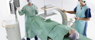
X-ray examination of the digestive system.
This group of studies includes:
- fluoroscopy and radiography of the abdominal cavity,
Indications for x-ray examination of the digestive system:
- Preparing for planned surgery
- Detection of organic diseases (tumors) of the esophagus, stomach, 12 pcs, colon, assessment of the volume of the lesion.
- Detection of foreign bodies in the esophagus, stomach, small and large intestine.
- Obstruction of the small and large intestines.
- Dynamic monitoring of the state of the digestive canal during treatment and (if necessary) at different times after completion of treatment.
- Assessment of parts of the colon after surgery and stoma removal (before surgery to reconstruct the colon).
Research methods for the digestive system:
Fluoroscopy and radiography of the abdominal organs are performed in 2 projections - frontal and lateral, in a vertical and/or horizontal position. No special preparation is required.
For foreign bodies of the esophagus, stomach - to identify radiopaque (visible in an x-ray image) foreign bodies (dentures, metal objects, compact large meat bones, etc.), a survey fluoroscopy is performed followed by radiography. A complete radiological characterization of a foreign body of the esophagus can only be obtained when studying data from two radiographs: in the lateral and direct projections of the neck or chest (depending on the location of the foreign body).
Preparation and time of the study:
- Fluoroscopy and radiography of the abdominal organs do not require special preparation; the examination time is 10-15 minutes.
- Examinations of the esophagus and stomach to identify foreign bodies are carried out without special preparation.
Study time: 30-60 min.
Advantages of X-ray examination:
— non-invasive diagnostic technique
— the ability to detect in real time (with fluoroscopy) the presence of pathological changes, foreign bodies of the esophagus, stomach, small and large intestine, their localization and prevalence.
- with radiography - less radiation exposure due to the short duration of the exposure (image).
Disadvantages of X-ray examination:
— With fluoroscopy, there is no objective documentation of the detected changes, except for the doctor’s direct recording of what he saw on the screen, and the dose load on the patient also increases (but with a digital X-ray machine, the difference is almost minimal compared to radiography).
— Determination of the thickness of the stomach wall can be done using ultrasound and CT.
- when identifying intra-abdominal tumors and their connection (germination) into the walls of the gastrointestinal tract, ultrasound of the abdominal cavity can determine thickening of the wall of the stomach or intestine, and identify metastases in the lymph nodes or liver.
— if there are signs of free gas in the abdominal cavity (for example, with abdominal trauma), an expanded diagnostic study (MSCT of the chest and abdominal cavity) is advisable.
