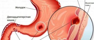
Perforation, or perforation, of stomach and duodenal ulcers is a breakthrough of the ulcer into the free abdominal cavity with the entry of gastroduodenal contents into it. In 75% of cases, a perforated ulcer is located in the duodenum, most often observed in men aged 20–40 years with a short history of ulcers (up to 3 years). Sometimes perforation of an ulcer can occur in people who have never previously complained of epigastric pain and were not aware of the presence of an ulcer. In young people, perforation of duodenal ulcers predominates, and in middle-aged and elderly people, perforation of gastric ulcers predominates. Perforation of ulcers is more often observed in autumn and spring.
Sign up for a consultation First consultation is free!
Symptoms of perforated ulcer
The main complaint when an ulcer perforates is pain in the upper abdomen. In most patients, perforation of stomach and duodenal ulcers begins suddenly, accompanied by sharp pain in the abdomen. The pain can be so severe that patients compare it to a “dagger blow.” They are permanent in nature, localized first in the epigastric region or in the right hypochondrium, and then spread relatively quickly throughout the abdomen, often along the right lateral canal. In 30-40% of patients, pain radiates to the shoulder, scapula or supraclavicular region: on the right - with perforation of pyloroduodenal ulcers, on the left - with gastric ulcers. When an ulcer perforates, general symptoms are also observed: dry mouth, thirst, nausea. In 30-40% of patients, reflex vomiting occurs, which becomes more frequent as peritonitis progresses.
Perforation of a gastric or duodenal ulcer
Perforation (perforation) of a gastric or duodenal ulcer (DUU) is a through destruction of the wall of these organs by a destructive process with the release of contents into the free abdominal cavity. PJCD is observed in 1.5-2 people per 10 thousand population. In one of our series of observations, its share in the structure of somatic diseases was 0.1%, among surgical diseases - 0.8%, and among acute surgical diseases of the abdominal organs - 2.6%
Perforation of gastroduodenal ulcers is the second life-threatening complication of peptic ulcer disease after gastrointestinal bleeding. The frequency of perforation of gastroduodenal ulcers, as well as the incidence of peptic ulcer disease, is increasing in Russia. If earlier for 79 years (1873-1952) reports were published in the domestic literature on the treatment of 20,688 patients with perforated ulcers (I. Naimark, 1958), then since the 90s of the last century more than 33 thousand are operated on annually in Russia alone patients with a similar complication. Most often, perforation of an ulcer occurs at the age of 20-40 years. Among men, perforation is observed 7-8 times more often than in women, but with age this difference decreases.
There is perforation of ulcers into the free abdominal cavity (87%) and covered (9%). In the first variant, perforation with the rapid development of diffuse peritonitis occurs on the anterior wall of the duodenum or prepyloric stomach (up to 90%), rarely on the body of the stomach. Perforation of an ulcer of the posterior wall of the duodenum or stomach is much less common (up to 5-10% of cases), while perforation, which is called atypical, (up to 4%) can occur in the tissue of the retroperitoneal space, in the lesser omentum. With a covered perforation, the hole itself in the wall of the intestine or stomach is covered by the liver, omentum, mesentery of the colon, or a blocked piece of food. In such rare cases, it is possible to cure a perforated ulcer without surgery due to the delimitation of the inflammatory process from the free abdominal cavity.
Perforation can occur in cases of both acute and chronic ulcers, one or several at the same time. The etiology and pathogenesis of perforated gastric or duodenal ulcers, as the cause of this disease, are still far from being fully disclosed, despite undoubted successes in this regard over the past decades. It must be assumed that in most cases, perforation is preceded by an exacerbation of peptic ulcer disease, manifested by a deepening of the inflammatory process and disruption of microcirculation with the development of local tissue necrosis with progressive impairment of local and humoral immunity. Similar processes, but apparently more pronounced, occur during perforation of “silent” or acute gastroduodenal ulcers.
The factors that provoke perforation of an ulcer are alcohol intake, trauma (cases of perforation during probing and X-ray examination of the stomach have been described), physical activity, psycho-emotional stress, overfilling the stomach with food, and taking non-steroidal anti-inflammatory drugs. It has been noted that perforation often develops during the daytime. The peak of perforation of the ulcer and duodenum was observed in the afternoon, and of the stomach - at noon or midnight.
Clinic and diagnostics
When an ulcer perforates into the free abdominal cavity, the contents of the stomach or duodenum pour into it, acting primarily on the peritoneum as a chemical irritant. Over time (after 12 hours or more), bacterial flora begins to multiply abundantly in the peritoneal exudate with the development of purulent peritonitis. With covered perforation of the ulcer, the contents that initially leak in small quantities are localized mainly near the perforation, causing a “chemical burn” of the peritoneum and a local initially aseptic inflammatory process. If the course is favorable, recovery without surgery is possible. When an infection attaches (in the case of a large amount of primary contents from the stomach or intestines or repeated perforation) and the inflammatory process remains isolated from the free abdominal cavity, a subhepatic abscess develops. A variant of the course of covered perforation of an ulcer is also possible according to the type of development of diffuse peritonitis, when there is no sufficient delimitation of the inflammatory process from the free abdominal cavity or it is temporary. With atypical perforation of an ulcer, the course of the process is usually similar to that with covered perforation.
In the clinical picture of a perforated ulcer, three periods (stages) are conventionally distinguished: shock, imaginary well-being and general peritonitis. Usually, a few days before perforation, patients note an exacerbation of the disease with corresponding symptoms: the appearance or intensification of pain in the epigastric region, becoming cramping in nature, nausea, vomiting, low-grade fever, etc. Authors of past years called these signs an alarming signal, a pre-perforation state (Kraus , 1865; G. Petrashevskaya, 1912; etc.).
Actually, perforation of the ulcer is manifested by sudden severe pain in the epigastric region, the severity of which is compared to a dagger blow to the stomach. The pain, as a rule, is so severe that the patient sometimes falls, turns pale, and breaks out in a cold sweat. In most cases, this symptom begins the first period - the shock stage, lasting up to 6 hours. The pain radiates to the right shoulder and right shoulder blade. As the contents of the stomach or intestines spread, usually along the right lateral canal of the abdominal cavity, pain from the epigastric region moves along the right flank of the abdomen to the iliac region, subsequently affecting the entire abdomen. The pain continuously and persistently increases, forcing the patient to take a forced position - lying on his side or back with his legs pulled up to his stomach. The patient's face is haggard, the expression is pained, frightened. Bradycardia is characteristic, breathing is shallow and rapid.
The stomach does not participate in breathing. Even with the most careful and superficial palpation, the second cardinal symptom of perforation is determined - a “board-like” muscle tension of the entire anterior abdominal wall, more pronounced in the area of the perforated ulcer itself. Symptoms of peritoneal irritation are sharply positive in all parts of the abdomen. In most cases, with percussion of the abdomen, “liver dullness disappears”; intestinal motility during auscultation is often weakened or not determined. During digital rectal and vaginal examination, severe pain in the pelvic peritoneum is noted.
The second period - the period of imaginary improvement - lasts approximately 6 hours and is characterized by a decrease in pain, improved well-being, and a decrease in muscle tension in the anterior abdominal wall. The patient often becomes euphoric, moves actively, considers his condition to be much better, and refuses examination, much less surgery. But he begins to show signs of intoxication: bradycardia is replaced by moderate and increasing tachycardia, body temperature rises, intestinal paresis and leukocytosis develop. Palpation of the abdomen remains painful, the Shchetkin-Blumberg sign is positive.
The third period - the period of diffuse peritonitis - occurs most often 12-18 hours after the perforation of the ulcer. The clinical picture quickly changes for the worse. Abdominal bloating and intestinal paresis increase. Repeated vomiting occurs, causing dehydration and electrolyte shifts. Body temperature rises to 38 C__, tachycardia increases to 100-120 beats/min, hypotension is noted. Shortness of breath increases.
On examination, a dry tongue and swollen abdomen are noteworthy. On palpation, the latter is tense and sharply painful in all parts. The Shchetkin-Blumberg symptom is positive. Intestinal peristalsis cannot be heard. Percussion reveals fluid in the abdomen. Oliguria increases, up to anuria. Leukocytosis is detected in the blood with a shift in the leukocyte formula to the left. Due to dehydration, hematocrit and hemoglobin, urea and creatinine increase, hyperkalemia and metabolic acidosis develop. Signs of systemic inflammatory response syndrome begin to dominate, with a likely outcome of sepsis, septic shock and multiple organ failure. In such cases, the prognosis is always serious even with the most vigorous and modern treatment methods.
It is believed that the diagnosis of a perforated ulcer is not difficult. When there is a classic triad of signs - a sudden onset with “dagger” pain in the abdomen (95%), “board-like” tension in the muscles of the anterior abdominal wall (92%) and a history of ulcers (80%), indeed, we can confidently speak about perforation of the ulcer. But prehospital and hospital errors, sometimes reaching 40% and 10%, respectively, indicate the difficulties of diagnosis. More often this occurs in patients without a previous ulcer history, with perforation of “silent” or acute ulcers, covered or atypical perforations.
Of the instrumental examination methods, the first priority is a survey radiography of the abdominal cavity, which allows one to identify free gas under the dome of the diaphragm. It is important that the study is performed in an upright position of the patient or, if his condition is severe, in a later position on the left side. Clear evidence of the presence of pneumoperitoneum can be obtained in 75% of patients. In doubtful cases, pneumogastrography is performed (500-600 ml of air is pumped into the stomach through a probe) or gastroscopy followed by X-ray control. The actual X-ray contrast examination of the stomach and duodenum with water-soluble contrast agents is rarely used, although in typical cases it confirms organ perforation due to contrast leakage beyond the contour of the organs being examined. Some foreign surgeons are guided by the results of such a study when determining indications for conservative treatment of perforated ulcers.
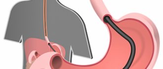
Gastroscopy allows you to detect direct and indirect signs of ulcer perforation. In particular, the endoscopist can, through the perforation hole, observe the organs of the abdominal cavity adjacent to the ulcer (omentum, intestine, liver), see that the perforation hole is covered with a piece of food from the inside, and omental tissue from the outside. Indirect signs of perforation are pronounced inflammatory changes around the ulcer itself, the inability to sufficiently inflate the stomach with air (it exits into the abdominal cavity through the perforation hole), as well as pronounced, difficult to overcome pylorospasm with a perforated duodenal ulcer.
If there are remaining doubts about the exact diagnosis, in recent years they have increasingly begun to resort to laparoscopy, which allows one to quickly make a differential diagnosis with other acute surgical diseases of the abdominal organs and confirm or refute the diagnosis of a perforated ulcer. It is appropriate to note that today differential diagnosis of these diseases is not carried out without preliminary ultrasound of the abdominal organs and retroperitoneal space. The importance of laparoscopy is increasingly increasing due to its use for the treatment of perforated ulcers.
Treatment
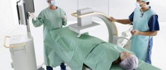
The diagnosis of a perforated gastric or duodenal ulcer is an absolute indication for emergency surgery. If the patient categorically refuses surgery or there are no technical conditions for its implementation, one should not forget about conservative treatment and use the Taylor method. The essence of the latter is as follows: Fowler's position of the patient, constant aspiration of stomach contents through a nasogastric tube, detoxification, antibacterial and antiulcer therapy (including anti-Helicobacter drugs), hunger, cold on the stomach.
In the foreign literature, the indications and contraindications for this method of treatment, primarily for perforated duodenal ulcers, have been studied and substantiated in sufficient detail. In particular, the indications include cases of ulcers that are not determined by X-ray contrast studies (there is no leakage of gastrografin beyond the duodenum); cases with no peritonitis; hospitalization period is more than 24 hours from the moment of perforation; severe concomitant diseases. Contraindications to conservative treatment include a long history of ulcers, perforation of an ulcer while taking steroid drugs, perforation of a gastric ulcer, the presence of signs of peritonitis, uncertainty of the final diagnosis, perforation of a duodenal ulcer in a hospital setting while undergoing adequate drug therapy. Such conservative treatment is effective in 81% of patients, the mortality rate is 2-5%. It has been noted that patients over 70 years of age tolerate conservative treatment worse (Crofts et all, 1989; Rigg et all, 1990; Jamieson, 2000).
The type and extent of surgical intervention for a perforated gastric or duodenal ulcer depend on the duration of hospitalization from the moment of perforation, the severity of peritonitis, the localization of the ulcer, the local condition of the tissues in the area of the perforation, the age of the patient, the presence and severity of concomitant diseases, the experience and technical equipment of the surgeon.
Simple suturing of a perforated ulcer has been and remains the dominant type of intervention in emergency surgery. At the same time, we should not forget that the main task is to save the patient’s life, and not to obtain good long-term results. In other words, without the slightest increase in risk to the patient’s life, it is necessary to choose from three alternative methods the optimal surgical option for a particular patient.
Suturing of a perforated ulcer of the stomach and duodenum is indicated:
- in cases where there are no conditions for performing a radical operation: the presence of diffuse fibrinous-purulent peritonitis, insufficient qualifications of the surgeon, lack of technical capabilities to perform a more complex operation;
- with perforation of acute ulcers in young people without a history of ulcers or with a short period of time;
- for perforation of ulcers in elderly and senile patients with severe concomitant diseases;
- if there are technical difficulties for radical surgery for perforation of high (cardiac) or postbulbar ulcers.
As for the suturing technique itself, double-row (through all layers and seromuscular) and single-row (seromuscular) sutures applied in a direction transverse to the axis of the organ are acceptable. To apply the inner row of sutures, either chrome-plated catgut or monofilament absorbable sutures are used. To apply the outer row, silk, nylon or modern synthetic monofilament threads are used. In the vast majority of cases, suturing is supplemented with omentoplasty.
In rare cases where it is impossible to bring the callous edges of a perforated ulcer together, they resort to tamponade of the perforation hole with an omentum according to P. Polikarpov. The operation is completed with thorough sanitation of the abdominal cavity, its drainage and nasointestinal intubation.
Primary gastrectomy is performed:
- with perforation of callous ulcers of both the stomach and duodenum in persons with a long history of ulcers and frequent relapses;
- when ulcer perforation is combined with bleeding, stenosis or penetration;
- with repeated perforation of the ulcer.
Of the gastric resection methods, preference should be given to the Billroth 1 method.
Vagotomy with pyloroplasty is used for perforation of only a duodenal ulcer. In this case, selective proximal vagotomy is combined with simple suturing of the ulcer. In widespread clinical practice, preference is given to truncal vagotomy, ulcer excision and Finney pyloroplasty. Different researchers interpret the indications for this method very broadly: from refusing other operations in all cases of perforation, including the early stages of peritonitis, to using it only for chronic perforated ulcers without pronounced anatomical and morphological changes in the area of perforation.
Vagotomy with anthrumectomy is used for perforation of an ulcer of the duodenum only with large inflammatory changes in the area of perforation, phenomena of pyloric stenosis or ulcer penetration.
In the last decade, reports of laparoscopic suturing of perforated gastric or duodenal ulcers have increasingly begun to appear. The collective experience of domestic authors who presented reports at two congresses in 2003 amounts to 610 laparoscopic suturing of perforated gastroduodenal ulcers. A return to the old method of treatment with today's technical equipment should be considered progress in emergency surgery, taking into account the available possibilities for drug treatment of peptic ulcer disease.
After performing palliative operations, modern antiulcer and anti-Helicobacter treatment is mandatory from the first day. The volume and composition of infusion-transfusion therapy is determined by the prevalence and nature of peritonitis, that is, the time period from the onset of the disease to surgery. With a timely operation, postoperative mortality is 3-5%; with late hospitalization (more than 24 hours), postoperative mortality reaches 20-30%.
A feature of surgery for perforated gastroduodenal ulcers in children is the absolute need to perform a minimal operation; gastric resection should not be used in them.
Igor BELOV,
Deputy Chief Physician for
surgical care clinical
Hospital of the Central Medical Unit No. 119,
Professor.
Diagnostics
In the general blood test, leukocytosis is observed, a neutrophilic shift of the leukocyte formula to the left.
Plain x-ray of the abdomen reveals free gas in the abdominal cavity (pneumoperitoneum). In the absence of pneumoperitoneum on a plain radiograph of the abdomen, after it has been emptied, 500–700 ml of air is injected into the stomach using a probe, which partially passes through the perforation into the free abdominal cavity and is found under the diaphragm.
Also, a perforated ulcer is detected using gastroscopy.
Our leading specialists
All specialists
Evseev Maxim Alexandrovich
- Deputy Chief Physician for Surgery
- Surgeon
- Bariatric surgeon
- Oncologist
- Doctor of Medical Sciences
- Professor
- Doctor of the highest category
Mirgatia Irakliy Olegovich
- Head of the surgical department
- Surgeon
- Oncologist
Vladykin Alexey Leonidovich
- Surgeon
- Bariatric surgeon
- Oncologist
- Candidate of Medical Sciences
- Doctor of the highest category
Alekseev Mikhail Sergeevich
- Surgeon
- Leading doctor price list
- Doctor of Medical Sciences
Fedotov Stanislav Viktorovich
- Head of the operating unit
- Surgeon
- Bariatric surgeon
- Leading doctor price list
- Doctor of the highest category
Balarev Anton Sergeevich
- Surgeon
- Oncologist
- Candidate of Medical Sciences
- Doctor of the highest category
Efimkina Jennet Orazmammetovna
- Surgeon
Rozumny Ilya Arkadievich
- Surgeon
Kharitonov Andrey Ivanovich
- Surgeon
- Doctor of the highest category
Titkov Boris Evgenievich
- Chief Physician of the Hospital Center
- Surgeon
- Oncologist
- Doctor of Medical Sciences
- Professor
- Academician AMTN
- Surgeon of the highest qualification category
Advantages of the Hospital Center
Individual treatment regimen for each patient
For each patient, it is mandatory, even at the pre-hospital stage, that an individual treatment regimen is developed, taking into account all the characteristics of the body: age, health status, medical history, etc. – this approach allows you to minimize risks both during surgery and in the postoperative period, and as a result, ensure the fastest possible rehabilitation with a minimum period of hospital stay.
Multidisciplinary approach
The medical staff of the Hospital Center is a single team made up of expert doctors of different specializations, which allows for a multidisciplinary approach. We treat the patient, seeing in front of us not a list of diseases he has, but a person whose problems are interconnected and interdependent. The therapeutic measures taken are always aimed at overall improvement of the patient’s health, well-being and quality of life, and are not limited to eliminating the symptoms of a specific disease.
Surgical treatment of any level of complexity
The operating doctors of the Hospital Center are proficient in advanced and high-tech methods of performing operations. The combination of highly qualified doctors and innovative equipment allows for surgical treatment of the highest level of complexity.
High-tech, minimally invasive treatment methods
The basis of the treatment methodology carried out at the Hospital Center is the principles of minimizing risks for the patient and the fastest possible rehabilitation.
The implementation of such an approach is possible only by using the most high-tech techniques, modern equipment and the application of the latest achievements of medical science.
The qualifications of doctors combined with modern equipment allow us to successfully implement this approach to treatment.
Fast-track surgery
Fast-track is a comprehensive technique that allows you to minimize the length of a patient’s stay in the hospital without compromising the quality of treatment.
The approach is based on minimizing surgical trauma, reducing the risk of postoperative complications and accelerating recovery after surgery, which provides our patients with a minimum hospital stay.
Thanks to this approach, even complex operations such as cholecystectomy require a hospital stay of no more than 3 days.
Personal medical supervision in the postoperative period
To completely exclude the development of possible complications, the early postoperative period is spent in all patients, regardless of the complexity of the operation, in the intensive care unit under the individual supervision of a resuscitator.
The transfer of the patient to the ward is carried out only in the complete absence of even the minimal possible risks.
Informing relatives 24/7
We are as open as possible and show care not only for the patient, but also for his loved ones. Information about patients' health status is provided to relatives seven days a week, 24 hours a day.
Visiting patients is also possible at any convenient time.
Highly comfortable single and double rooms
Patients are offered spacious, comfortable rooms for single and double occupancy, equipped with everything necessary for rest and recovery.
In the children's department, our young patients are accommodated together with their parents.
Tax deduction
According to the tax legislation of the Russian Federation, each patient has the right to compensation of up to 13% of the amount spent on his treatment, as well as the treatment of close relatives.
Our specialists will prepare for you a package of documents for the tax office for a refund of 13% of the amount of treatment expenses, and will also give recommendations on various ways to interact with the tax office.
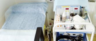
X-ray of the stomach with barium
Duration of the study
: 15 minutes.
Preparing for the study
: There is
Contraindications
: There is
Preparation of the conclusion
: 30-60 min.
Restrictions
: There is
What is a barium x-ray?
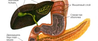
X-ray with barium sulfate is aimed at studying the condition of the gastrointestinal tract. The contrast agent makes it easier to examine the internal organs, their relief, contours, shape, and size.
The essence of the method
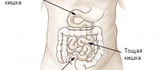
Hollow organs are difficult to fully visualize, so a barium mixture is injected before scanning. It colors tissues that are shaded in X-ray images. Thus, the examination picture clearly examines the walls of the large intestine, parts of the small intestine, stomach, and esophagus, and foci of pathologies are identified.
Indications
The doctor issues a referral for diagnostics for the following complaints:
- Abdominal pain.
- Unstable bowel function.
- Changes in the color and structure of stool.
- Blood in the stool.
- A sharp decrease in body weight.
- Intestinal gases.
Preparation
If barium sulfate is planned to be administered before an x-ray, the patient should prepare for the procedure in advance:
- Follow the diet for 2-3 days. You need to stop taking flour, dairy, spicy, smoked foods, legumes, black bread, and carbonated drinks. Food should be easily digestible.
- Cleanse your intestines with laxatives the day before. The dosage and number of doses is calculated according to the instructions in accordance with the weight. With proper cleansing, an enema is not necessary.
Barium X-rays are performed on an empty stomach in the morning, so you should not eat breakfast before the scan. You should leave your jewelry at home and wear loose-fitting clothes without metal inserts.
How is barium x-ray performed?
First, the doctor injects a contrast agent. Typically 400-600 ml of barium sulfate solution is given in liquid form. The patient drinks it and waits 10-15 minutes. Next, the subject takes the desired position, scanning begins, which lasts 50-60 minutes. During this time, the patient lies motionless, and the device takes from 6 to 10 pictures to assess the state of the gastrointestinal tract in dynamics. If necessary, the doctor asks you to turn over. Barium is eliminated from the body within 24 hours. To speed things up, it is recommended to drink more still water.
Where to get a barium x-ray
In the Health Clinic, located in the very center of the capital, examinations are carried out using modern, high-precision expert-class equipment. Qualified doctors with extensive experience will carefully examine the internal organs and describe the condition of the stomach.
results
Within an hour, a Health Clinic specialist interprets the images and then hands them over to the patient. The attending physician, based on the results of the examination, establishes or denies diseases such as stomach ulcers, tumors of various etiologies, protrusion of the gastric walls, intestinal obstruction, hernias, dyskinesia, etc.
Book an x-ray with barium
To sign up for diagnostics, choose any method:
- Call the clinic phone number +7 (495) 628-22-05,
- Request a call back
- Write or call via online chat, in the right corner of the page,
- Make an appointment immediately using the convenient form on the website:
MAKE AN APPOINTMENT
Contraindications
You need to choose a different diagnostic method if there are the following restrictions on barium x-rays:
- Pregnancy.
- Barium intolerance.
- Bleeding in the intestines.
Price
It has become possible to get an X-ray with barium cheaply in Moscow at the Health Clinic.
Low prices, constant promotions and quality services attract visitors to our medical center. Patients of the Diagnostic Center are provided with free parking. Reservation of a place for a car is made no later than an hour before arrival at the clinic. Call
Consultation can be obtained by phone: +7
Price for stomach x-ray with barium
| Name of service | Price in rubles |
| Fluoroscopy and radiography of the esophagus with contrast | 5750 |
| Fluoroscopy and radiography of the stomach and duodenum with contrast | 5750 |
| X-ray examination of the small intestine with the introduction of a contrast agent - barium suspension passage | 6875 |
| Survey urography | 2700 |
If you do not find a service in the price list, please call us and we will provide you with the necessary information.