will perform the operation. Thus, during laparoscopy, the surgeon sees the organs not through an incision in the anterior abdominal wall, but through a video camera inserted directly into the abdominal cavity.
The length of the puncture through which the laparoscope is inserted is from 1.5 cm to 2 cm. The scar that remains in its place is small and almost invisible. So, laparoscopic surgery begins by introducing trocars into the abdominal cavity through small punctures, the length of which is from 1.5 cm to 2 cm. One of them is intended to obtain an image, and the rest are intended to perform direct surgical intervention. In the future, the methodology
The essence and course of surgical interventions, which are performed using laparotomy (an incision in the abdominal wall) and laparoscopy, are exactly the same, they are no different. The correct name for the operation to remove the gallbladder using laparoscopic access is “laparoscopic cholecystectomy.”
Two types of interventions are performed on the gallbladder using laparoscopic access:
- removal of the gallbladder;
- removal of stones from the gallbladder.
Currently, gallstones are almost never removed. If there are stones in the gallbladder, then pronounced pathological changes appear, its walls become sclerotic, and adhesions form around the organ. He will never be able to function normally. In addition, after stones are removed, 90% of them form again. Therefore, laparoscopic removal of the gallbladder is performed.
Advantages of laparoscopic surgery over laparotomy (incision in the abdominal wall)
Without a doubt, the laparoscopic method of gallbladder removal has a number of advantages over laparotomy:
- for the operation, not an incision is used, but four punctures, anterior abdominal;
- pain after surgery subsides within one day;
- at the end of the operation, the patient can walk independently and perform simple actions;
- length of hospital stay is reduced (from 1 to 4 days);
- rehabilitation and restoration of working capacity occur quickly;
- the risk of postoperative hernia formation is reduced to a minimum;
- The scars are either barely noticeable or cannot be seen at all with the naked eye.
Prevention
Preventive measures to prevent the occurrence of an acute attack of cholecystitis consist of the following:
- Active physical exercises: therapeutic exercises, walking, swimming.
- Frequent intake of diet food.
- No fried, fatty, smoked foods.
- Categorical refusal of alcoholic beverages and tobacco products.
Compliance with simple rules and requirements in relation to your health will allow the patient to avoid surgical intervention by a surgeon and cope with the disease using more humane methods. Now you know whether cholecystitis can cause fever
.
If suddenly you have inflammation of the gallbladder and a temperature of 37
, contact our clinic.
Progress of laparoscopic gallbladder removal
After the trocars are inserted, carbon dioxide is supplied to the abdominal cavity to inflate the abdominal cavity and thus increase its volume. After conducting
Using a video system, the surgeon examines the entire abdominal cavity to identify other possible diseases of the gastrointestinal tract. If none are identified, proceed directly to removing the gallbladder. To do this, the cystic duct is isolated, which flows into the common bile duct (choledochus), and after applying special clips, it is crossed. After which the gallbladder is isolated from its bed in the liver. The gallbladder is removed from the puncture above the navel.
At the end of the operation, the surgeon removes all trocars and tapes or stitches the punctures in the skin. In some cases, the surgeon inserts a drainage tube into one of the punctures, which is left for 1 or 2 days. If difficulties arise during the operation that cannot be overcome using laparoscopic techniques, the surgeon always has the opportunity to remove the trocars and perform a classic laparotomy (incision).
Operation duration
The duration of laparoscopic cholecystectomy surgery depends on the complexity of the operation and the experience of the surgeon. Laparoscopic cholecystectomy lasts on average from 40 to 1.5 minutes, but in complex cases up to 3 to 5 hours.
Indications for laparoscopic gallbladder removal
Laparoscopic removal of the gallbladder is performed if the following indications exist:
- chronic calculous cholecystitis;
- gallbladder polyps;
- asymptomatic cholecystolithiasis (presence of gallstones that do not bother the patient) if they are less than one centimeter;
- acute cholecystitis (no later than two or three days from the onset of the disease).
Methods of performing cholecystectomy surgery
Cholecystectomy is a fairly common type of surgery and involves removing the gallbladder when it loses its functionality, if gallstones cannot be removed in any other way, and for other pathologies that are fraught with serious complications.
Methods for carrying out such an operation differ in the method of access to the organ being removed, and according to this criterion they are divided into:
- traditional abdominal intervention (laparotomy). Access to the surgical field with this method of intervention is through a fairly large incision in the anterior wall of the abdominal cavity. This method is the most traumatic and significantly increases the risk of postoperative complications, therefore it is used in emergency cases and when, for some reason, other techniques are contraindicated for the patient or the technical equipment of the medical institution does not allow the use of other surgical techniques. Among all the practiced techniques, the body’s recovery time after laparotomy is the longest;
- method of laparoscopic cholecystectomy. The essence of this minimally invasive intervention is that the organ is excised through small (about a centimeter) punctures in the peritoneal wall. The abdominal cavity is inflated with carbon dioxide to provide an operating space, and then the operation is performed using special tubular instruments, the progress of which is monitored by a video camera inserted into the operated area. Laparoscopy is currently a well-established and most optimal method for performing such operations, since it is the least traumatic, minimizes the risk of postoperative complications and has a short rehabilitation period. Often, after laparoscopy of the gallbladder, the patient is discharged from the hospital on the second or third day. If the gallstones are large, then to remove the organ they are first crushed;
- mini access technique. Developed in the seventies of the twentieth century as an alternative to abdominal surgery. It is something between traditional and laparoscopic techniques. Access to the organ to be removed is made through a size ranging from four to seven centimeters, and the progress of the operation is monitored visually directly. It is used, as a rule, in cases where the medical institution does not have equipment and instruments for performing laparoscopy. Typically, a patient after such an operation is discharged on the third to fifth day after resection of the gallbladder;
Read also: Symptoms of gallbladder problems
- and finally, the latest experimental surgical techniques - transgastric and transvaginal. Access to the surgical area for such interventions is through the oral area or through the vagina using a flexible endoscopic instrument. Currently, both of these techniques are at an experimental stage, but the absence of postoperative scars makes their use very promising in the future.
Preparing for laparoscopic gallbladder removal
Preoperative tests are recommended to be done no later than two weeks before the planned date of gallbladder laparoscopy. The patient should be provided with the results of the following studies:
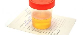
- general blood and urine analysis;
- biochemical blood test with determination of the concentration of total protein, glucose, bilirubin, glucose and alkaline phosphatase;
- coagulogram (APTT, INR, PTI, fibrinogen);
- Wasserman reaction, tests for the presence of antigens to HIV, as well as hepatitis types B and C;
- electrocardiogram.
- Gastroscopy
- Ultrasound of the abdominal organs
If the test results are within normal limits, the patient is allowed to undergo surgery. It is necessary to coordinate the use of any medications with the operating surgeon and anesthesiologist. On the day before surgery, it is recommended to finish eating by 6:00 pm and drinking liquids by 10:00 pm. After this, the patient is not allowed to take water or food until the start of the operation. No other preparation is required for laparoscopic gallbladder removal. Some clinics continue to perform cleansing enemas before surgery, but there is no point in this.
Diagnostics
Before making a final diagnosis, it is necessary to undergo a large number of tests, as well as visit a gastroenterologist: a specialist will conduct an examination and those studies, the results of which are necessary in a particular case. Gallbladder hurts
? Of course, if there is acute inflammation, the temperature immediately rises.
The development of inflammation of the gallbladder is indicated by certain symptoms, which can only be detected during an examination.
Laboratory and instrumental methods that are necessary to use to make a correct diagnosis:
- Blood analysis.
- Analysis of urine.
- Bile culture. This study is most important, as it allows us to fully assess the state of the microflora of the gallbladder.
- Ultrasound examination of the liver and gall bladder.
- CT.
- MRI.
When the gallbladder is inflamed, is there a fever?
? Since the inflammation continues beyond the gallbladder, the mobility of the abdominal wall on the right during breathing is limited. The temperature in mild forms increases to 37.5 - 38 degrees, in severe forms - up to 40 degrees. By the way, sometimes the pressure rises to 140 over 60.
Postoperative period after laparoscopy of the gallbladder
After the operation is completed, the anesthesiologist stops supplying the gas mixture and wakes the patient. On the day of surgery, he should remain in bed for 4 to 6 hours. After this time has passed, the patient is allowed to turn over in bed, sit down, and also get up and walk. He can perform simple actions.
From this time on, the patient is allowed to drink still water, and from the second day to eat soft, light food. The menu may consist of weak broth, fruits, low-fat cottage cheese, yogurt, boiled lean minced meat. Portions should be small, but you should eat 5 to 7 times a day. We should also not forget that throughout the second day after surgery the patient needs to drink a lot of water.
On the third day after surgery, you are allowed to eat regular meals. Those foods that cause the formation of large amounts of gas in the intestines should be excluded from the diet. These include brown bread and legumes. It is also not allowed to eat foods that stimulate the formation of bile (onions, garlic, spices and pickles). After laparoscopic cholecystectomy, from the third or fourth day, it is recommended to eat according to diet No. 5. Typically, a patient who has undergone laparoscopic removal of the gallbladder is discharged on the second day after surgery.
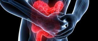
For the first 2 days after surgery, the patient may experience slight pain in the area of punctures on the skin, as well as in the right hypochondrium and above the collarbone. The pain will go away completely after 1 to 4 days. But in the case when the pain does not subside and its intensity increases, you should bring this to the doctor’s attention, as this may be a sign of a complication.
The postoperative period lasts from 7 to 10 days. During this time, patients are not recommended to lift heavy objects or perform work that involves physical exertion. During this period, they should wear soft underwear that does not irritate the puncture sites on the skin. On the 7th or 10th day after surgery, patients in the clinic have their stitches removed. This ends the postoperative period. Patients are given a certificate of temporary disability for the entire time they are in hospital and for another 10 or 12 days. If the patient develops any complications after surgery, then the period of disability is determined on an individual basis.
Treatment
A patient who suffers from an attack of cholecystitis necessarily requires hospitalization and treatment:
- Strict bed rest and constant rest.
- Drug treatment, the plan of which can only be drawn up by a specialist.
- Electrophoresis, paraffin, inductometry.
- Physiotherapy.
- Strict diet.
In the case when the disease is in the active phase, it is necessary to take fractional meals, the volume of which is less than usual. It is strictly forbidden to eat fried foods, smoked foods, egg yolks, fish and meat.
To treat inflammation of the gallbladder, medications such as:
- Antibiotic.
- Analgesic, antispasmodic.
- Enzyme-containing drugs.
- Sedative drugs.
- Maintenance therapy.
If a patient has a diagnosis of purulent gangrene, surgical treatment of cholecystitis is necessary: as a rule, the gallbladder is removed.
treatment of gallbladder inflammation
Rehabilitation after laparoscopic cholecystectomy
The rehabilitation period after laparoscopic removal of the gallbladder is short, in most cases it proceeds without complications. However, full recovery, physical and psychological, occurs after 5-6 months. But this does not mean that all this time a person feels unwell or is not able to work and live normally. Complete rehabilitation includes not only mental and physical recovery from surgical trauma and stress. This is also the accumulation of reserves that will allow a person to successfully withstand new stressful situations and life challenges without any harm to himself.
The person feels normal and is able to perform his usual work, not related to physical activity, within 10 or 15 days after surgery. From this point on, in order for rehabilitation to occur as quickly and fully as possible, patients should follow the following recommendations:
- organize proper nutrition that would help avoid constipation;
- sports training should begin with minimal loads one month after surgery;
- You should not engage in physical labor during the first month after surgery;
- during the first three months after laparoscopic surgery, you can lift no more than 3 kg of weight, and from the third to the sixth month - no more than 5 kg;
- From 3 to 5 months you should follow diet No. 5.
Diet after gallbladder removal
The purpose of the diet after laparoscopic gallbladder removal is to ensure normal liver function. Normally, this organ produces from 600 to 800 ml of bile per day. It should be released only after the bolus of food enters the duodenum. If bile secretion occurs voluntarily, this can create certain difficulties. To prevent this from happening, you should follow a diet that can minimize the consequences of the absence of a gallbladder.
So, on the 3rd and 4th days after laparoscopic surgery, the patient is allowed to eat pureed vegetables, low-fat cottage cheese, as well as boiled meat and low-fat fish. This menu is prepared for another 3 or 4 days, and then you can switch to diet No. 5.
It involves split, frequent meals in small portions. You should eat 5-6 times a day. All dishes must be warm, it is recommended to chop the products. You cannot eat cold or hot food. Gentle culinary technologies should be used to prepare dishes. Food can be boiled, baked or steamed. Under no circumstances should they be fried in fat.
Such foods should be excluded from the diet:
- salo;
- fatty meats and fish;
- high fat dairy products;
- canned fish, meat and vegetables;
- pickles and marinades;
- smoked meats;
- hot seasonings (horseradish, garlic, ginger, mustard, chili pepper);
- offal (kidneys, liver, brain, stomach);
- any mushrooms;
- raw vegetables, including green peas;
- fresh white and rye bread;
- baked goods and confectionery (pancakes, pies, pastries and cakes);
- alcohol;
- chocolate, black coffee and cocoa.
The menu of people who have had their gallbladder removed by laparoscopic access should consist of dishes prepared from the following products:
- lean meats (turkey, rabbit, chicken and veal);
- lean fish (pike, perch and pike perch);
- semi-liquid porridges from any cereals;
- soups with weak broth or water, seasoned with vegetables, pasta or cereals;
- steamed or stewed vegetables;
- low-fat or low-fat dairy products (kefir, milk, curdled milk, cheese);
- fresh non-acidic berries and fruits;
- compotes, mousses and jellies from the same berries;
- yesterday's white bread;
- honey, jam or jam.
These products are used to make up the diet and prepare various dishes. You can add 45 to 50 g of butter or 60 to 70 g of vegetable oil to them immediately before eating. This daily amount of fat should be distributed over several doses. You can eat no more than 200 g of bread and no more than 25 g of sugar per day. It is also recommended to drink one glass of low-fat kefir before going to bed.
It is recommended to drink weak tea, sweet juices diluted in half with water, compote, coffee with milk and rosehip infusion. The amount of drinking water consumed per day is set individually. It depends on the person’s well-being. So, for example, if bile is secreted quite often into the intestines, then it is recommended to reduce the amount of water consumed, and if not very often, then it can be increased. Starting from the fourth or fifth month of following diet No. 5, unchopped fish and meat, as well as raw vegetables, can be included in the diet. After this time, you can eat all your food little by little.
Gallstone disease and consequences of cholecystectomy: diagnosis, treatment and prevention
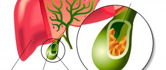
Gallstone disease (GSD) is a disease of the hepatobiliary system caused by impaired metabolism of cholesterol and/or bilirubin, characterized by the formation of stones in the gall bladder and/or bile ducts with the possible development of dangerous complications.
Ultrasound reveals gallstones in 10–15% of apparently healthy adults, the frequency of detection of which increases with age. Most gallstones form cholesterol by depositing in supersaturated bile, especially at night, when the concentration of bile in the gallbladder is at its highest.
Between meals, bile is concentrated in the gallbladder, which acts as a reservoir for bile acids (cholic and chenodeoxycholic).
Bile acids form the outer layer of the micelle, which contains fat-soluble cholesterol (cholesterol) in the center. Phospholipids concentrated in the center of the micelle increase its ability to keep cholesterol from crystallization.
Bile acids are necessary for the emulsification of fats, for their breakdown (hydrolysis) under the influence of lipase (mainly pancreatic) into triglycerides and fatty acids, the latter are absorbed (absorbed) together with bile acids mainly in the ileum.
A deficiency of bile acids involved in the enterohepatic circulation (eg, in terminal small intestinal disease) or an imbalance between the concentration of phospholipids and cholesterol (lithogenic bile) in the bile can lead to the precipitation of cholesterol crystals from the supersaturated bile, which then nucleate to form cholesterol bile stones
Factors predisposing to the formation of cholesterol gallstones include gender (women), obesity, diet (low dietary fiber), liver cirrhosis (30%), terminal ileal disease (Crohn's disease, etc.) or ileal resection , consumption of medications (oral contraceptives containing mainly estrogens, clofibrate).
Pigment stones consist of bilirubin with the formation of calcium-insoluble precipitates (calcium bilirubinate). Black, small, dense pigmented stones account for 70% of all radiopaque gallstones.
Brown pigment stones, also consisting mainly of calcium bilirubinate, are soft, predominantly intrahepatic and are found extremely rarely.
The following factors predispose to the formation of black pigment stones, consisting predominantly of calcium bilirubinate: chronic hemolysis (sickle-shaped and spherical red blood cells, for example, in sickle cell anemia, implanted artificial heart valves); cirrhosis of the liver; biliary tract infection (E. Coli, Clostridium Sp.). Infection of bile with microorganisms producing β-glucuronidase leads to an increase in the content of poorly soluble direct unconjugated bilirubin in the bile.
Brown pigment stones are usually formed in patients with sclerosing cholangitis and with biliary invasions (opisthorchiasis, clonorchiasis, giardiasis, etc.).
Along with cholesterol (single) and pigment (purely pigmented black and brown), mixed, usually multiple, gallstones are more common. It is extremely rare to find stones consisting of calcium carbonate and phosphorus.
Modern classifications provide for the identification of at least three stages of cholelithiasis. The first of them is physical and chemical. At this stage, the liver produces bile oversaturated with cholesterol, with a decrease in the content of bile acids and phospholipids (lithogenic bile).
Patients have no clinical symptoms of the disease; the diagnosis is based on the results of a study of gallbladder bile (portion B). A violation of the micellar properties of bile is revealed; cholesterol “flakes”, crystals and their precipitates are found in it. There are no gallstones.
The first stage of cholelithiasis can be asymptomatic for many years.
Therapeutic and preventive measures in this preclinical stage of cholelithiasis include a general hygienic regime, systematic physical activity, rational fractional nutrition with the exception of nutritional excesses (high-calorie and cholesterol-rich foods, especially in cases of obesity and hereditary predisposition).
Preventive measures also include adequate treatment of patients with gastrointestinal dysfunction (intestinal dysbiosis, colitis, etc.).
The second stage of cholelithiasis (latent asymptomatic stone carriage) is characterized by the same physicochemical changes in the composition of bile as the first stage, but with the presence of stones in the gall bladder. The process of stone formation at this stage is associated not only with physicochemical changes in bile, but also with the addition of gall bladder pathogenesis factors (stagnation of bile, damage to the mucous membrane, increasing the permeability of the bladder wall for bile acids, inflammation) and disturbances in the enterohepatic circulation of the bile acids
Most stones located at the bottom of the gallbladder do not manifest themselves in any way. The advancement of stones into the cystic duct and its blockage lead to the development of cholecystitis, which stops if the obstruction of the duct is eliminated, or progresses with the development of complications.
The third stage of cholelithiasis is clinical, complicated (calculous cholecystitis, acute, chronic, etc.). Clinical manifestations of cholelithiasis depend on the location of gallstones, their size, localization and activity of inflammation, the functional state of the biliary system, as well as damage to other digestive organs.
A stone lodged in the neck of the gallbladder obstructs its outlet, causing biliary (hepatic) colic. Subsequently, obstruction of the cervix may turn out to be temporary, and the stone returns to the gallbladder or penetrates the cystic duct, where it stops or passes into the common bile duct. If the size of the stone (up to 0.5 cm) allows, it can enter the duodenum and appear in the stool; the stone can also stop in the common bile duct, more often in its distal part, causing complete or intermittent obstruction (valve stone) with the corresponding clinic. In this case, the bile always becomes infected, and cholelithiasis is accompanied by inflammation (choledochitis, cholangitis).
Acute calculous cholecystitis (ACC) usually occurs when a stone enters the cystic duct, which leads to stagnation and infection of bile, swelling of the gallbladder wall with hemorrhages and ulceration of the gallbladder.
Signs of acute cholecystitis:
- Fever and constant pain in the right upper quadrant of the abdomen, in contrast to chronic cholecystitis.
- The main reason for the development of the disease is strangulation of a stone in the cystic duct (duct obstruction).
- Typical pain (with acute cholecystitis in less than 50% of patients).
- Pain that often occurs soon after eating and increases over the course of an hour, in contrast to attacks of pain with biliary colic, which are short-lived and disappear on their own.
- Fever joins the pain syndrome usually 12 hours after the onset of the attack and is associated with bacterial inflammation (invasion), as a result of which the pain becomes constant.
- Murphy's sign is usually positive, but it is not related to a specific test.
- An X-ray of the abdominal cavity taken in the supine position reveals calcified stones in the gall bladder and sometimes gas inside the biliary tree in some patients (15%) (the formation is associated with infection of the latter by Clostridium Welchii).
Complications of acute cholecystitis:
- chronicity (50%);
- cholangitis caused by the presence of stones in the bile ducts (10%);
- dropsy, empyema or gangrene of the gallbladder (1%);
- biliary peritonitis (0.5%) with mortality in 50% of cases.
Chronic calculous cholecystitis is usually characterized by recurrent attacks of biliary colic, less often by constant pain in the right upper quadrant of the abdomen. Patients show significant differences in the degree of thickening and fibrosis of the gallbladder wall and inflammatory infiltrate.
Biliary colic sometimes occurs suddenly, “for no reason” or after eating, and is combined with low-grade fever, nausea, and sometimes vomiting. The pain intensifies with movements and deep breathing. Severe pain usually disappears quickly.
An attack is provoked by fatty foods, spices, smoked foods, hot seasonings, severe physical stress, working in an inclined position, as well as infection. In women, colic sometimes coincides with menstruation or occurs after childbirth. The pain often radiates to the right scapula and subscapular region. Sometimes the pain radiates to the lumbar region, to the region of the heart, simulating an attack of angina. The pain varies in intensity: from strong cutting to relatively weak, aching. However, exacerbation of cholecystitis, especially non-calculous, is not always accompanied by typical attacks of biliary colic. The pain may be dull, constant or intermittent. Vomiting with cholecystitis does not bring relief.
The undoubted signs of calculous cholecystitis include:
- pain in the right upper quadrant of the abdomen - acute, episodic (less than 60 s) and cramping (from 1 to 72 hours);
- pain-free intervals (from several weeks to several months);
- intolerance to fatty and fried foods (often);
- flatulence - increased gas evolution (often);
- flatulence - bloating (often);
- positive palpation and percussion symptoms such as Murphy, Kera, etc. (sometimes absent even with exacerbation of chronic calculous cholecystitis);
- gallstones and a thickened wall of the gallbladder, always detected by ultrasound;
- non-functioning gallbladder, determined using oral cholecystography.
There is constant dull and variable pain in the right upper quadrant of the abdomen without irradiation. Cholecystolithiasis with a normal gallbladder wall, a functioning gallbladder even in the presence of dyspeptic disorders that occur in chronic calculous cholecystitis, are more characteristic of stone carriers rather than cholecystitis.
Along with calculous cholecystitis (acute, chronic), not stones are detected in the gallbladder, but sediment (sludge), associated with an increased content of mucin in it, on the matrix of which bile components crystallize. The formation of sediment in the gallbladder occurs when it is slowly or incompletely emptied. This condition is often associated with prolonged fasting or insufficient stimulation of gallbladder motility by cholecystokinin produced in the intestines. Although bile sludge is a reversible stage in the pathogenesis of gallstone disease, if appropriate therapy is prescribed, stones will inevitably form as it progresses, which leads to the appearance of corresponding symptoms.
Treatment includes: fractional hypocaloric nutrition aimed at reducing body weight if there is obesity; drug decontamination if a syndrome of excessive microbial contamination of the initial parts of the small intestine is detected; oral administration of bile acid preparations (chenodeoxycholic and ursodeoxycholic) for 1.5-2 months, endoscopic papilosphincterotomy, if a stenotic process is diagnosed at this level of the common bile duct.
Choledocholithiasis—stones in the common bile duct—is manifested by pain and jaundice. Choledocholithiasis occurs when a gallstone passes from the bladder into the common bile duct. Secondary stone formation in the common duct is possible, especially in the presence of stasis caused by duct obstruction.
The presence of stones in the common bile duct should be suspected in any patient with calculous cholecystitis whose serum bilirubin level exceeds 50 mmol/l and the alkaline phosphatase level is three normal levels. The level of aminotransferases may increase 2–10 times compared to normal, especially with acute obstruction. Once the obstruction is relieved, aminotransferase levels usually return to normal quickly, while bilirubin levels often remain elevated for 2 weeks, and elevated alkaline phosphatase levels persist even longer.
Symptoms, often intermittent, include colicky pain in the right hypochondrium, fever, chills and jaundice with a characteristic increase in alkaline phosphatase and transaminases in the blood serum. Choledocholithiasis, if it is not eliminated immediately, is almost always accompanied by ascending cholangitis, an infection of a confined space that can lead to sepsis.
Cholangitis is characterized by pain in the upper abdomen, often on the right, jaundice and fever, often accompanied by chills. Bacterial cholangitis is one of the most dangerous complications of cholelithiasis; it is usually associated with subhepatic cholestasis, which often occurs with calculous obstruction of the main bile duct. The severity of cholangitis depends on a number of factors, primarily on the duration of cholestasis and the level of cholemia. With a short-term, but repeatedly recurring violation of the outflow of bile, chronic cholangitis develops, in which (usually following a quickly passing attack of biliary colic) mild chills occur with an increase in temperature to subfebrile levels, the urine becomes dark in color and sometimes jaundice occurs. These symptoms usually last no more than 2–3 days. Blood tests in some cases reveal slight neutrophilic leukocytosis, a moderate increase in ESR, transient hyperbilirubinemia, and a short-term and slight increase in alkaline phosphatase levels.
Such exacerbations of cholangitis are more often associated with the passage of a stone through the common bile duct, less often due to the valve mechanism in choledocholithiasis and sometimes, possibly, papillitis (odditis). Between episodes of cholestasis, there may be no symptoms of cholangitis. This form of cholangitis is classified as chronic; its course is largely determined by the frequency of relapses and the duration of cholestasis, as well as the nature of the inflammatory process (catarrhal, purulent).
Diagnostic tests:
- The most effective non-invasive method for identifying gallstones is ultrasound. Often, asymptomatic stone carriers (silent stones) are diagnosed using this method. Occasionally, gallstones are not detected even with careful examination. In some patients, the gallbladder cannot be visualized due to intestinal gas, fibrosis of the gallbladder, or its unusual anatomical location. Ultrasound also turns out to be an informative method for detecting obstruction of the common bile duct, diagnosing acute and chronic cholecystitis and assessing gallbladder function, which is determined before and after the use of cholekinetics.
- CT has an advantage over ultrasound when it comes to identifying stones in the common bile duct.
Blood test:
- In chronic cholecystitis without exacerbation and asymptomatic cholecystolithiasis, the peripheral blood picture is normal.
- With choledocholithiasis, there is an increase in alkaline phosphatase (3 norms or more) and GGTP (3 norms or more).
Neutrophilic leukocytosis is characteristic of AC and cholangitis. The content of serum cholesterol is not important for the diagnosis of cholecystitis and cholangitis, but cholesterol naturally increases with primary biliary cirrhosis and sclerosing cholangitis.
Other studies:
- An abdominal x-ray has diagnostic value in the presence of acute abdominal pain. It may reveal calcifying stones in the gall bladder, bile ducts and intestinal lumen, an enlarged liver and the presence of air in the biliary tract (biliary enteric fistula, cholangitis caused by clostridia or occurring after surgery).
- To assess the function of the gallbladder in a patient with chronic cholecystitis who is undergoing conservative treatment, “oral” cholecystography can be performed.
- Isotope scanning with the Hida radiopharmaceutical has some diagnostic value only in acute illness, when it is possible to assess the function of the gallbladder, including determining its shutdown as a result of duct obstruction.
- ERCP is used for diagnosis of cholestatic syndrome and for the treatment of biliary obstruction (benign stricture in the area of the papilla of Vater, choledocholithiasis, etc.).
- Percutaneous transhepatic cholangiography is indicated in cases where there are dilated intrahepatic bile ducts identified by echohepatography, and it is not possible to perform ERCP or the condition of the intrahepatic bile ducts needs to be clarified (filling of the bile ducts with contrast can be blocked by a tumor or stricture that has arisen at one or another level of the biliary tract ).
- Carrying out intravenous cholangiography and a test with bromsulfalein to assess the state of the excretory function of the biliary system of the liver should be considered outdated methods.
Stasis of bile in the gallbladder (hypomotor dyskinesia of the gallbladder), caused by its hypokinesia, is considered typical for patients with cholelithiasis. It is also observed in a number of conditions that are risk factors for the development of cholelithiasis: during pregnancy, long-term use of anticholinergic and antispasmodics, after vagotomy, diabetes, obesity, etc.
Against the background of bile stasis in the gallbladder, ultrasound often reveals a sediment accumulating in it, known to surgeons as “putty,” found during cholecystectomy. On ultrasound, the sediment has the appearance of a “cloud” with multiple small echo-positive inclusions. Unlike gallstones, echogel is not detected, but a clear boundary between such a “cloud” and echo-negative bile can be determined. The “cloud” may include cholesterol crystals, bilirubinate particles, and calcium carbonate particles. Obviously, these patients may subsequently develop cholecystolithiasis. Along with microliths, changes on ultrasound of the gallbladder may be associated with the presence of polyps in it. True polyposis of the gallbladder can be differentiated from cholesterol parietal microlites by prescribing ursodeoxycholic acid (ursofalk) at a dose of 7.5 mg/kg for 3 months and performing dynamic ultrasound of the gallbladder. If, while taking the drug, the initially identified signs of the parietal “deposit” disappear, then polyposis is excluded and cholelithiasis is diagnosed at the stage of microlith formation.
When bile stasis in the gallbladder, one should keep in mind cholesterosis, characterized by the deposition of cholesterol in the CO of the bladder. Cholesterosis often develops in obese women with severe disorders of fat metabolism and high cholesterol levels in the blood.
In cholesterosis, lipids (mainly cholesterol) are found predominantly in the endothelial cells of the mucous membrane, from which then, if they are significantly deposited, a cholesterol polyp can develop. On ultrasound, this formation looks like a polyp, but histological examination does not determine the true structure of the polyp.
The presence of a correlation between cholesterosis, cholesterol levels in the blood and bile, and the development of atherosclerosis, including atherosclerosis of the aorta, coronary and cerebral arteries, has not been confirmed. The presence of malignant degeneration in cholesterosis has also not been established.
Stasis of bile in the gallbladder is usually clinically manifested by constant dull aching pain in the right hypochondrium, aggravated by shaking when driving, walking quickly, carrying a heavy weight in the right hand, or bending forward. Diagnosis is sometimes difficult, although duodenal probing reveals cholesterol crystals in portion B.
The choice of adequate treatment for cholelithiasis is often determined jointly by the therapist, surgeon and patient.
When determining the indications for cholecystectomy, we used the international recommendations presented in the table.
Treatment of the consequences of cholecystectomy
In patients who have undergone cholecystectomy, most often symptomatic manifestations are associated with dysfunction of the sphincter of Oddi of the pancreatic or biliary type. Preventive measures should be aimed at normalizing the chemical composition of bile, restoring the patency of the sphincter of Oddi, decontaminating the duodenal mucosa and restoring its motor-evacuation function.
As a symptomatic remedy, the drug Odeston (hymecromone) is prescribed, 200 mg 3 times a day before meals, the course of treatment is 1–2 weeks. The drug has a choleretic and antispasmodic effect on the sphincter of Oddi, but does not weaken intestinal motility.
For cholestasis and cholangitis in the absence of infection of the biliary tract and restoration of bile outflow, along with the elimination of stricture, we have accumulated positive experience in using the herbal preparation hepabene (1-2 capsules after dinner), and in case of infection of the biliary tract - an antibacterial drug from the macrolide group - clarithromycin (clacid, Klabaksa), for purulent infection - the drug Meronem.
Absolute indications for surgery:
- acute cholecystitis;
- chronic cholecystitis with a normal history (recurrent biliary colic) and non-functioning gallbladder (according to ultrasound or cholecystography);
- stones of the common bile duct: a) in persons under 70 years of age - ERCP; sphincterotomy; according to indications - cholecystectomy; b) in people over 70 years of age and in the presence of a high surgical risk, endoscopic sphincterotomy gives less mortality, but the risk of relapse of choledocholithiasis remains;
- gangrene of the gallbladder - urgent cholecystostomy (safer than cholecystectomy), cholecystectomy is possible in the future, but often you can limit yourself to spontaneous closure of the wound;
- intestinal obstruction caused by a gallstone is an operation to eliminate intestinal obstruction followed by cholecystectomy.
Relative indications for surgery: chronic calculous cholecystitis, if the symptomatic manifestations of the disease are associated with the presence of stones in the gall bladder. In this case, it is necessary to exclude gastric and duodenal ulcers, irritable bowel syndrome, chronic pancreatitis, and urinary tract diseases, which may have symptoms simulating chronic cholecystitis.
Currently, along with standard laparotomic cholecystectomy, laparoscopic cholecystectomy is being widely introduced into practice, the advantage of which is a short stay for the patient in the hospital (less than 48 hours) and earlier restoration of working capacity (after 5–7 days). Laparoscopic cholecystectomy, if performed by a highly qualified specialist and according to strict indications, allows you to quickly and much less traumaticly remove stones from the gallbladder even before the symptoms of inflammation increase. The advantage of cholecystectomy compared to conservative methods of treating cholecystolithiasis (lithotripsy, stone dissolution) is to eliminate the risk of recurrent stone formation.
Prevention of gallstone disease
The first stage of cholelithiasis can be diagnosed if appropriate biochemical studies of bile are carried out, mainly portion C. Lithogenic bile is characterized by oversaturation of bile with cholesterol, a decrease in the concentration of bile acids and phospholipids in it, as well as hypokinesia of the gallbladder and damage to the liver parenchyma. In order to prevent the progression of cholelithiasis (transition to the second stage - latent, asymptomatic stone carriage), it is recommended to change your diet and lifestyle. The effectiveness of preventive measures depends on their strict compliance by the patient.
In the second stage (latent stone carriage), the goal of prevention is to prevent the formation of complications of cholelithiasis and the consequences of cholecystectomy. Adequate tactics for patient management at this stage are determined jointly by the therapist and the surgeon.
For primary prevention of cholelithiasis it is recommended:
- In order to eliminate stagnation of bile and improve its quality, eat food at least 5 times a day (first breakfast at about 8 o'clock, second at 11 o'clock, lunch at 14 o'clock, afternoon snack at 17 o'clock and dinner at 21 o'clock). For patients predisposed to the development of cholelithiasis, as well as at the first stage of the disease, there are no prohibited foods and dishes. Leisurely eating at a strictly defined time stimulates the secretion of digestive juices (including bile) and the motor-evacuation function of hollow organs (including the gallbladder). The diet should include meat, fish, fats, vegetables, fruits and their juices. A decrease in working capacity and an increase in body weight (obesity, obesity) should not be allowed; a sufficient night's sleep, daily formed stools and sufficient and painless urination are necessary; It is not recommended to smoke and drink alcoholic beverages even in so-called “small” quantities.
- If there is a tendency to constipation or difficult, prolonged or painful defecation, it is necessary first of all to exclude organic pathology (hemorrhoids, peptic ulcer, diverticular colonic disease, polyposis of the rectum and colon, colorectal cancer, etc.), then make appropriate changes to the regimen nutrition and lifestyle (consume at least 0.5 kg of vegetables and fruits daily, increase fluid intake to 1.5-2 liters per day, eat only “dark” varieties of bread, increase daily physical activity (brisk walking, swimming, etc.) ...), after meals, take the drug allohol in the amount of 3-4 tablets per day. The so-called “morning block” is recommended: in the evening, soak 4 to 10 prunes in boiling water (prepare an infusion), and in the morning drink it and eat the fruits Then have breakfast with the obligatory consumption of a glass of any juice and a small amount of salad from fresh vegetables or fruits.
- For idiopathic functional constipation, along with compliance with the appropriate dietary and physical regimens, it is possible to prescribe laxatives for a short time, mainly mucofalk or laminarid (4 teaspoons of granules per day) or Forlax (2 sachets per day), or lactulose (30 ml of syrup or 20 g granules per day). Other laxatives are used less frequently.
- If the liver is involved in the process (fatty hepatosis, reactive hepatitis with weak activity, etc.), along with appropriate nutritional and physical regimens, drugs with a hepatoprotective and choleretic effect are prescribed for a long period (hepabene 2 capsules per day for a year).
- Lithogenicity of bile is successfully eliminated with long-term (many months) use of chenodeoxycholic acid (henosan, henofalk, henochol, etc.) in combination with ursodeoxycholic acid (ursofalk, ursosan, etc.) at the rate of 5-10 mg/kg body weight. For example, before going to bed, take 1-2 capsules of henochol and 1-2 capsules of ursofalk for 6 months.
- Prevention of the formation of brown and black pigment stones in the gallbladder and ducts includes treatment of patients with chronic hemolysis (splenectomy, sanitation of the biliary tract, etc.), adherence to an appropriate diet, and maintaining a healthy lifestyle. In some cases, it is possible to additionally prescribe choleretics and cholekinetics, which stop the process of stone formation.
- Considering the cholekinetic role of intestinal hormones formed in the mucous membrane of the duodenum (cholecystokinin, secretin, etc.), as well as the involvement in the inflammatory and, as a consequence, in the atrophic process of its mucosa due to excessive bacterial growth, it is advisable to conduct a course of antibacterial therapy (clarithromycin 500 mg 2 times a day + metronidazole or tinidazole 500 mg 2 times a day for 5 days). Antihelminthic and antiparasitic therapy is carried out according to indications.
- Cholelithiasis often occurs in the presence of a hereditary predisposition (burden), liver diseases (fatty hepatosis, hepatitis, cirrhosis, etc.), disorders of the digestive and absorption processes caused by pancreatitis, duodenitis, enteropathies, intestinal motor-evacuation disorders, including constipation. This disease often occurs in women. The occurrence of cholelithiasis is promoted by pregnancy, excess body weight, as well as a number of diseases and bad habits (alcohol abuse, smoking). In this regard, the prevention of cholelithiasis consists of normalizing the functional state of the liver and biliary system, including the gallbladder, duodenum, pancreas, and adequate treatment of existing diseases. Particular attention should be paid to the preservation of physical and mental activity, appetite, normalization of body mass index (20-25), relief of symptoms of certain diseases, including hemolytic anemia (for example, splenectomy for Minkowski-Schoff disease, etc.).
With the constant use of hepabene (a herbal preparation that has a hepatotropic and choleretic effect) 2 capsules after dinner, we, using dynamic ultrasound cholecystography, revealed the restoration of impaired contractility of the gallbladder, a decrease in the volume of residual bile and the time of contraction of the gallbladder. In almost 100% of cases, as a result of long-term use of hepabene, not only the contractility of the gallbladder was restored, but also the lithogenicity of bile disappeared. In the presence of sludge and polypous changes in the wall of the gallbladder, it is recommended to prescribe ursodeoxycholic acid (ursofalk, ursosan) for 3 months at the rate of 7.5 mg per 1 kg of body weight.
In the vast majority of patients at the first stage of cholelithiasis, such therapy stops the progression of the disease and stone formation.
Consequences of laparoscopic gallbladder removal
Unfortunately, after removal of the gallbladder, quite often patients are bothered by dyspeptic symptoms. They manifest themselves with the following symptoms:
- pain and rumbling in the stomach;
- nausea and vomiting;
- intestinal disorders (flatulence, diarrhea or heartburn);
- belching bitter;
- extremely rarely - increased body temperature and jaundice.
Dyspeptic symptoms can periodically bother a person, and it is not possible to get rid of them for life. Fortunately, patients rarely experience complaints and mostly after significant errors in diet. When these symptoms appear, it is necessary to strictly follow diet No. 5. If severe pain occurs, it is relieved with antispasmodics (No-spa, duspatalin). After consultation with a gastroenterologist, medications based on ursodeoxycholic acid (ursofalk, ursosan, etc.) are taken. It reduces the concentration of bile and thereby makes patients feel better