Infectious disease specialist
Sinitsyn
Olga Valentinovna
34 years of experience
Highest qualification category of infectious disease doctor
Make an appointment
The most common type of helminthiasis in the world is enterobiasis. This disease is caused by intestinal parasites - pinworms, which settle in the small intestine of a person, provoking a variety of intestinal and toxic-allergic disorders. In Russia, it accounts for up to 70% of all cases of human helminthiasis. A characteristic feature of this type of helminthiasis is the passage of the full cycle of development of the parasite in the host’s body.
Enterobiasis - what is it?
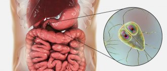
Enterobiasis is a disease caused by the parasite Enterobius vermicularis
, also called pinworms.
This is one of the most common parasitic infections in the world, manifested by disorders of the gastrointestinal tract. Humans are the only natural host of this infection. The disease most often affects children attending kindergartens and primary schools. Enterobius vermicularis
worms are tiny, thread-like, yellowish-white, so named because of the characteristic spike-like tail present at the rear of the female worms.
Treatment of pinworms
When pinworms are detected in the body, the doctor selects the optimal treatment based on the individual characteristics of the child, his age, body weight, as well as the volume and location of the lesion.
Therapy should be aimed at combating parasites of all phases of development (eggs, larvae, adults), eliminating the consequences of pinworm activity, as well as relieving itching and other unpleasant symptoms of the disease. For this purpose, anthelmintic drugs are first selected. Medicines can come in various forms (tablets, suspensions, suppositories), which allows them to be used to treat children of all ages. Specialists at the SM-Doctor clinic for children and adolescents strongly do not recommend choosing an anthelmintic drug without consulting a specialist. This may lead to intoxication and cause other side effects.
Taking an antiparasitic drug alone is not enough. To eliminate the consequences of pinworms in the body, the following groups of drugs are needed:
- Sorbents are prescribed to remove parasites and their metabolic products from the body.
- Probiotics – recommended in the presence of intestinal dysfunction caused by helminths.
- Antihistamines are prescribed if enterobiasis is accompanied by allergic reactions.
It should be remembered that to avoid re-infection, all members of the patient’s family should undergo treatment. A number of hygiene procedures are also required:
- Change of linen followed by washing and hot ironing.
- Heat treatment of toys and other objects with which the child has been in contact.
- Regular wet cleaning of the premises.
To confirm the fact of recovery, the child must be re-examined for enterobiasis 2 weeks after the therapy.
Causes of enterobiasis (etiology)
Enterobiasis most often occurs in children, but people of any age can get sick. Infection is caused by pinworm eggs entering the body. Transmission most often occurs by the fecal-oral route, but can also occur through contact with contaminated clothing, bedding, personal care items, and furniture. Risk factors for infection include:
- insufficient hygiene,
- eating after touching contaminated objects,
- living with an infected person.
It is believed that more than a billion people worldwide are infected with pinworms and suffer from enterobiasis. According to various estimates, the prevalence of enterobiasis among students in kindergartens and primary schools in Europe reaches 20%.
Causes of infection
Most often, doctors detect enterobiasis in children, since they are much more likely than adults to:
- do not follow hygiene rules: do not wash their hands after contact with potentially contaminated surfaces, soil, visiting a public bathroom, before eating, etc.;
- eat on the street;
- lick or suck fingers, toys and other objects;
- They bite their fingernails.
The same actions cause helminthic infection in adults: enterobiasis is often called the disease of dirty hands.
Pathogenesis
Enterobius vermicularis
is a parasite that primarily lives in the ileum and cecum. Once E. vermicularis eggs are ingested, they take 1 to 2 months to develop into adult worms in the small intestine. During this period, the infection does not manifest itself with any symptoms. Adult females migrate to the anal area mainly at night and lay eggs in the perianal area, causing itchy skin. Children try to relieve itching by scratching the perianal area with their fingers. Scratching the perianal area leads to a massive release and spread of eggs into underwear and the environment, including landing on the fingers, and then in the face, after which they enter the body again as a result of ingestion (autoinfection and resumption of the worm’s life cycle). Sometimes the larvae migrate back to the rectum and small intestine and begin their life cycle (retroinfection). The time interval between the ingestion of infectious eggs and the laying of eggs by an adult female pinworm is 2-6 weeks.
Structure and physiology of pinworms
The human pinworm is the causative agent of enterobiasis. These helminths parasitize the human intestines, but the eggs need to be released into the environment to fully mature. Pinworms have the same structure as other roundworms.
The body of the helminth is round in cross-section and elongated in shape. Adults vary in sex. The rear end of the female is pointed, while that of the male is curled on the ventral side. That's why these parasites got their name. On the sides of the caudal end there are growths of the cuticle, which are called caudal wings. Females are usually larger than males and measure between nine and twelve millimeters. Males are only two to five millimeters in size (Figure 1).
Figure 1 - Male pinworm under formaldehyde
At one end of the body there is an opening for the mouth, framed by three folds resembling lips (one of them is located on the side of the “belly”, the other two are on the back). Behind them is a special swelling of the cuticle, which is called a vesicle. Thanks to it, pinworms are retained in the intestines of the host. The anus is located at the end opposite the opening of the mouth and is located on the abdomen.
The helminth is covered with a special skin-muscular sac. This is a complex cellular formation, the first layer of which is the cuticle, the second is the muscles, which are represented by longitudinal ribbons, and the third is the hypodermis, which forms ridges between the muscles. The cuticle has a complex biochemical structure. Its main function is to provide a barrier with the environment; it prevents the penetration of various substances into the helminth, ensuring its internal constancy of the environment (homeostasis). Also, the cuticle is a specific skeleton, since muscle fibers are attached to its cells.
The hypodermis lies under the cuticle. It looks like compacted ridges located throughout the body of the pinworm. The hypodermis takes part in the barrier function and in the formation of the cuticle, and many useful microelements also accumulate in it.
Figure 2 - Structure of pinworms
Immediately below the hypodermis there is a layer of longitudinally arranged muscle fibers. The space under the skin-muscle sac is a cavity filled with a toxic liquid that has a complex biochemical composition. It is a hydroskeleton that functions as a support.
The digestive system is well developed and is represented by a straight tube stretching from the opening of the mouth to the anus. Behind the mouth opening is the esophagus. Its initial compartment is separated from the final one by a deep constriction. The esophagus works like a pump. It is followed by another extension, the so-called bulbus. It is also involved in parasite attachment. Thus, food moves through the helminth’s intestines in one direction and is better absorbed.
The excretory system consists of cervical glands, from which long canals extend. They are located inside the hypodermal ridges. The channels of the right and left halves of the body merge into one duct that opens on the abdomen. The function of the excretion system is to excrete waste as well as maintain internal pressure.
The female genital organs are double. They are represented by two ovaries, which flow into two oviducts, followed by double uteruses. They merge and form a short vagina, which opens with a slit in the anterior quarter of the body, located transversely. The male genital organs are always unpaired. First comes the testis, after it is the vas deferens, which turns into the ejaculatory canal. Behind it a complex copulatory organ is formed. The circulatory and respiratory systems are not developed. Breathing is carried out using the integument, and sometimes only through chemical processes.
Classification of the disease
Enterobiasis can occur in mild, moderate and severe forms.
- With a mild course of the disease, there are usually no symptoms other than mild nighttime itching in the perianal area.
- In the moderate form, the itching becomes more intrusive, and symptoms of a functional disorder of the gastrointestinal tract and astheno-neurotic symptoms often appear.
- The onset of a severe form is accompanied by severe itching, sleep disturbances, inflammation, digestive disorders, abdominal pain, and the possible development of appendicitis and other inflammatory processes dangerous to health.
Diagnostic methods
Of no small importance for diagnosis is asking the patient about the place of work (preschool and school institutions), about contact with children with enterobiasis, about the nature of the excreted feces (sometimes mature helminths can be seen on the surface of the feces), as well as about the detection of helminths on the surface of the body during active crawling. It is worth keeping in mind that children and food industry workers may not be aware of the latest facts.
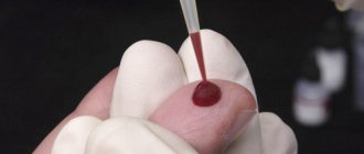
The main laboratory method for making a diagnosis is a microscopic examination of a scraping, which is obtained from the patient’s anal area using tampons, spatulas, transparent adhesive tape, a glass eye stick with an adhesive layer according to Rabinovich. Level of evidence – A.
Other diagnostic methods are used less frequently:
- 1 stool PCR (level of evidence – A);
- 2microscopic examination of stool for pinworm eggs and scatological method (level of evidence – B);
- 3 a general blood test to detect eosinophilia (level of evidence – C);
- 4 histological examination of specimens obtained from a biopsy of the intestinal mucosa (level of evidence – D).
Instrumental diagnostic methods can be divided according to levels of evidence:
- Level 1 evidence - B - endoscopic examination of the gastrointestinal tract, vulvoscopy, vaginoscopy, hysteroscopy;
- Level 2 evidence - C - comprehensive ultrasound examination of the abdominal organs, laparoscopy.
Symptoms of enterobiasis
About a third of patients with enterobiasis are asymptomatic. The most common symptom associated with pinworm infection is perianal itching. Perianal erythema may occur due to itching and scratching. Sometimes a superficial bacterial infection can occur in areas where scratching occurs, leading to redness and inflammation. Constant itching can cause sleep disturbances and lead to insomnia. Children often experience teeth grinding, enuresis, anorexia, and irritability. Sometimes there is the addition of genitourinary infections (invasion of the female genital tract with vulvovaginitis and granulomas of the pelvis or peritoneum), watery diarrhea, and abdominal pain. Sometimes tiny thread-like worms may be visible to the naked eye in the perianal area.
How to avoid infection
The main measures to prevent enterobiasis are to comply with hygiene requirements:
- mandatory hand washing after visiting the toilet, walking, visiting public places, contacting animals, etc.;
- nail care, getting rid of the habit of biting nails;
- frequent replacement of bed linen, towels, hygiene items;
- washing underwear and bed linen in hot water, followed by ironing and steaming;
- wet cleaning with disinfectants every two to three days.
Enterobiasis is a helminthiasis from the group of nematodes, caused by worms from the genus pinworms. The causative agent of enterobiasis is one of the most common helminthiases. The length of the female reaches 12 mm, and the male - up to 3 mm. Pinworms are localized in the lower part of the small intestine and the upper part of the large intestine.
In some cases, after removal of the appendix they can also be found in the appendix. The most susceptible population to helminths are children aged 2-7 years. Infection occurs through the fecal-oral route (through dirty hands, toys, household items, on playgrounds).
Enterobiasis is a contact helminthiasis, one of two cases when the whole family must be treated. This is determined by the peculiarities of the biology of the parasite: mature, egg-filled female pinworms actively crawl out of the anus, most often at night, and lay eggs on the skin of the perianal area, buttocks, and sometimes on bed linen.
The crawling worms secrete isovaleric acid, which causes severe itching. A person in a dream involuntarily scratches himself and collects eggs on his fingers and under his nails. From there they enter the external environment and become a component of house dust.
Man is the only owner of pinworms, i.e. The entire life cycle takes place in the human body.
The prevalence is very wide; representatives of all socio-economic groups are affected. It is most common in children 5-10 years old, least rare in children under 2 years of age who do not attend kindergarten.
Infection occurs by ingestion of eggs. In the small intestine, larvae hatch from the eggs, and pinworms attach with their mouth opening to the mucous membrane, irritating it.
Sexually mature female pinworms, filled with eggs, descend into the large intestine, then into the rectum. Eggs are laid in the anal area (in the so-called perianal folds), sometimes on the skin of the buttocks or nightwear/bed linen. The lifespan of pinworms is only 20 days, some champions live up to 40.
Eggs remain invasive (infectious) depending on external conditions from several hours to several months. They are resistant to most environmental influences and chlorination, but quickly die under high insolation. Therefore, the prevalence of enterobiasis is significantly lower in countries with hot, dry climates.
Forms of enterobiasis
There are acute and chronic forms of enterobiasis. In the absence of hygiene measures and preventive therapy, a chronic form occurs, which leads to constant intoxication of the body and the formation of complications.
Causes of enterobiasis
According to epidemic sources, enterobiasis is found in 20% of preschool children.
Children very often bite their nails, are in close contact with children in organized institutions (kindergartens, schools, baby clubs, development centers), and do not always observe hygiene measures.
Provoking factors for pinworm infection are: the presence of allergic dermatoses in children or other skin diseases, sleep and appetite disturbances, frequently ill children, children with obsessive, neurosis-like conditions.
Symptoms and course of enterobiasis
The main symptom of pinworm infection is itching in the anal area, especially at night. The itching is caused by an allergic reaction to egg laying and the presence of an adult in the perianal area.
Scratching leads to eggs getting on the hands, followed by self-infection and the spread of eggs into the environment. Intense scratching leads to the appearance of inflammatory areas and their secondary bacterial infection.
In rare cases, with a large number of worms in the host’s body, abdominal pain, nausea, and vomiting may develop. Pinworms crawling into the caecum can lead to appendicitis.
Complications of enterobiasis
Complications are associated not only with intestinal damage, but also with pinworms entering atypical habitats. The following possible complications are described:
- Eosinophilic enterocolitis as a special, individual reaction with increased sensitivity to pinworm antigens or the result of a very severe infection.
- Vulvovaginitis and secondary urinary tract infection caused by it. This problem is common in little girls. Therefore, in case of vulvovaginitis, it is necessary to perform an examination for enterobiasis.
- Salpingitis and oophoritis develop if pinworms penetrate the pelvic organs through the female genital tract.
- In rare cases, pinworms can pierce the intestinal wall and exit into the abdominal cavity. This can lead to peritonitis.
Diagnosis of enterobiasis
Diagnosis is not difficult; scraping or imprinting with adhesive tape from the skin of the perianal area reveals pinworm eggs. The effectiveness of the test is maximum in the morning before going to the toilet.
A clinical blood test reveals a persistent increase in eosinophils.
Stool analysis does not reveal pinworm eggs.
Treatment of enterobiasis
The effectiveness of treatment of enterobiasis lies in compliance with general hygiene measures:
- frequent change of bedding, underwear,
- thoroughly washing children before and after sleep,
- cutting nails, washing hands frequently, especially after going outside, going to the toilet or playing active games outside.
Specific therapy includes pyrantel, vermox, and nemozol. The dose of the medicine depends on the child’s age and body weight.
Preventive therapy is prescribed to all family members, even if they do not have symptoms of the disease. Treatment is carried out simultaneously.
Some medications are excellent for treating newborns and infants. A repeat course is carried out after 12-18 days.
In addition, children are prescribed diet therapy. Products that help eliminate the pathogen from the body include: fresh carrots, carrot juice, walnuts, wild strawberries, garlic, green lovage, St. John's wort, elecampane. Bificol, lactic acid bacteria, and lactulose also provide protection to the body from pinworms.
Sick children are monitored - after completing the course of treatment, 2, 3 and 4 weeks after the treatment.
Prevention
From an early age, a child must be instilled with the habit of observing the rules of hygiene, which is important not only in the prevention of helminthic infestation, but also in general for maintaining social status.
A fairly important preventive measure is the mechanical removal of pinworms from the perianal folds daily by taking a shower or bath, using hygiene products. It is also important to change bed and underwear daily; Linen should be washed at high temperatures of at least 60 degrees, wet cleaning of the premises, with frequent changes of water and rinsing of rags. Low temperatures have a negative effect on pinworms, so soft toys and other household items can be left in the cold for 2-3 hours.
In children's groups, preventive deworming is carried out among frequently ill children, children with obsessive conditions (biting nails, sucking fingers, etc.), since this contingent of children may pose a threat to the spread of enterobiasis.
Read also:
- Helminthiasis (in common parlance - “worms”) - damage to the body by parasitic worms
- Prevention of helminthiases transmitted through meat and meat products
Enterobiasis: how to repel the attack of pinworms
Along with roundworms, pinworms are the most common parasite in the children's (and, let's be honest, in adults) digestive tract. They primarily affect the upper parts of the large intestine. Why enterobiasis? This is a derivative of the Latin name for pinworms - Enterobius vermicularis. They are transmitted only from person to person; pets and other animals are beyond suspicion here. So, to paraphrase a well-known saying, in relation to enterobiasis, man is not a friend to another, much less a brother, but a source of infection. Perhaps the most inquisitive readers of our site have already read the articles about ascariasis and toxocariasis, and they have a question: what is the difference, in general? Worms are worms, so why go into so much detail. This is a fairly common mistake of young mothers. It is erroneous, because parasitic organisms even within the same species group (in our case, Nematodes) may differ, which requires different approaches to the diagnosis and treatment of helminthic infestations.