© Author: Soldatenkov Ilya Vitalievich, doctor of the therapeutic department, especially for SosudInfo.ru (about the authors)
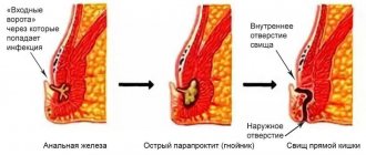
Splenic rupture is a pathology in which the structure and functions of the organ are disrupted. The cause of this emergency condition is traumatic injury - a fall from a height, compression of the abdomen, wounds. The force from the impact falls on the left hypochondrium. The victims require hospitalization in a trauma hospital. Only urgent surgical intervention can save a patient from death.
The spleen is an organ of the lymphatic system located on the left side of the abdomen behind the stomach and performs a number of vital functions. It is involved in protein metabolism, blood cell synthesis, and the fight against infectious agents. The organ destroys old blood cells, produces antibodies, and ensures the balance of blood and lymph. The spleen is richly supplied with blood: due to the large number of blood vessels, it has a purple color. The organ is reliably protected from negative external influences by a dense tissue membrane and a frame of ribs. But despite this, he is easily injured.
Splenic rupture can be isolated or combined. Usually it is part of polytrauma along with damage to other structures of the body. The pathology often leads to heavy internal bleeding and shock. The spleen becomes damaged and ruptured as a result of certain diseases. The organ capsule becomes thinner, and the parenchyma swells. Moreover, any, even the most minimal, impact on the stomach can injure it. In the absence of emergency medical care, victims quickly die.
example of 2nd degree splenic rupture
Clinical manifestations of splenic rupture: pain, symptoms of internal bleeding, anemia and peritoneal irritation. This dangerous condition occurs relatively often, especially among physically active people who are most at risk of getting into an extreme situation. But despite this, an organ injury can be sustained by a person of any age, gender and lifestyle - an elderly person, a teenager, a child. Diagnosis of pathology is based on the main clinical manifestations, the results of laparoscopic and other laboratory and instrumental studies. Treatment of the pathology is surgical, consisting of suturing the organ or removing it. The operation is performed by specialists in the field of traumatology or abdominal surgery.
Causes
High-energy injuries are the immediate cause of splenic rupture. The organ is damaged by a fall, injury or blow to the stomach, as a result of a car, household or industrial accident. Injuries to the spleen can be professional or criminal in nature. In children, it is damaged during fights or outdoor games. The child falls or is hit in the left chest and abdomen.
Factors contributing to organ rupture:
- Thinned capsule, full-blooded and loose parenchyma,
- Immobility of the spleen
- Dysfunction of the hematopoietic system,
- Increased load on the spleen during prolonged and severe infectious processes,
- Excessive physical stress
- Difficult birth
- Colonoscopy and other invasive diagnostic and treatment procedures,
- Weakness of the abdominal muscles.
Diseases in which splenomegaly develops often lead to organ rupture. Non-traumatic causes of injury include:
- Infectious mononucleosis,
- Enlarged spleen of toxic, autoimmune or infectious origin,
- Hematological diseases,
- Oncological pathologies,
- Abscess of the spleen,
- Malarial infection
- Metabolic disorders
- Congenital anomalies of the spleen,
- Inflammation of nearby organs - liver, kidneys.
If a patient has the listed diseases, the lining of the spleen gradually becomes thinner, and sensitivity increases. The spleen can rupture regardless of a person's general well-being. The ideal functioning of internal organs minimizes the risk of such an event occurring. If the functioning of the bone marrow, endocrine glands and immune system is impaired, the likelihood of rupture increases significantly.
There is also spontaneous splenic rupture, which occurs for no apparent reason. This name is considered conditional, since in most cases, organ damage is preceded by minor trauma. In the presence of normal parenchyma, it does not lead to a violation of integrity. For spontaneous rupture of a pathologically altered spleen to occur, it is enough to make an awkward movement or sneeze.
Pathogenesis
There are two ways to rupture the spleen - two-stage and simultaneous. In the first case, under the influence of traumatic factors, only the parenchyma of the organ is damaged. Blood does not exit into the abdominal cavity, but accumulates under the capsule. This forms a hematoma containing many clots. Any, even minor physical stress can suddenly cause the capsule to rupture, which will lead to bleeding. The time interval between the traumatic impact and the direct release of blood from the organ varies from several hours to weeks and months.
degree of splenic rupture according to international grading
Another option for two-stage damage to the spleen is a rare imaginary rupture, in which independent tamponade of the organ occurs. A large blood clot covers the capsule rupture. This allows you to delay bleeding for some time. A similar process is observed with arterial hypotension or spasm of the vessels of the spleen. With physical activity, sneezing, coughing, defecation, turning in bed, the blood clot comes away. Suddenly, internal bleeding occurs. Usually it is profuse, accompanied by a sharp increase in symptoms and deterioration of the patient’s condition.
More often than others, a simultaneous rupture occurs, in which all structures of the organ are damaged. The blood immediately exits into the abdominal cavity. Internal bleeding is very dangerous for the functioning of the body and the life of the patient. It was recently impossible to restore systemic circulation after a ruptured spleen. The damaged organ was completely removed. Modern doctors successfully perform conservation operations.
Small ruptures of the spleen usually occur. They are characterized by an erased clinic and are not detected immediately. A few hours after the organ damage, the patient’s condition worsens. The reason for the appearance of typical signs of pathology, allowing a diagnosis to be made, is a large loss of blood and its accumulation in the abdominal cavity.
Features of treatment
Since bleeding that occurs when the spleen ruptures almost never stops on its own, surgery should be performed in the early stages of the development of the pathology.
The prognosis is significantly complicated by increasing blood loss. Therefore, doctors should act as quickly as possible.
The classic traumatological technique used for organ rupture is removal of the spleen. However, at the moment it is also possible to carry out organ-preserving procedures. One of these operations involves suturing wounds of the spleen.
Complete removal of the organ is recommended in the presence of extensive ruptures, crush injuries, through and lacerations, as well as if it is impossible to perform the wound suturing procedure. However, experts are trying to do everything possible to preserve the organ, because the spleen plays a number of important functions in the human body.
Manifestations
The symptoms of the pathology are polymorphic. Its severity is determined by the severity of the injury, the presence of concomitant disorders and the time that has passed since the injury.
Pain syndrome is the first and main manifestation of splenic rupture. The victims experience unbearable, intolerable pain in the left hypochondrium and epigastrium, radiating to the left shoulder and limb. Acute pain from a subcapsular rupture forces patients to take a gentle position - lying down with their knees tucked in. The person breathes through the chest, because every breath is accompanied by sharp, excruciating pain. The victim instantly loses strength, becomes weak and apathetic. He develops dyspeptic symptoms - nausea and vomiting, as well as bloating, rumbling, and stool retention. These are signs of intestinal paresis, which develops several hours after the injury. Percussion reveals a dull sound, and auscultation reveals the disappearance of peristaltic sounds.
Local manifestations of the lesion are accompanied by a deterioration in the patient’s general condition, symptoms of acute blood loss, and peritoneal signs:
- Pale skin
- Sticky and cold sweat,
- Hypotension,
- Frequent heartbeat
- dizzy
- Cephalgia,
- dyspnea,
- Noise in the ears
- With blurred vision,
- Psychomotor overexcitation,
- Brad,
- Anxiety, anxiety,
- Loss of orientation and consciousness.
Symptoms of hypovolemia are nonspecific. They occur with any serious damage and do not allow a diagnosis to be made. But despite this, these manifestations of the disease cannot be ignored. They indicate the need for immediate hospitalization and surgical intervention. Otherwise, extremely serious consequences may develop - anemia, shock.
All symptoms of splenic rupture must be considered together. Only in this way will specialists be able to diagnose pathology in the shortest possible time and prevent the development of dangerous complications. Timely and adequate assistance to the victim makes the prognosis for recovery favorable. Self-diagnosis and self-medication are one of the main causes of death of a patient due to damage to internal organs.
Diagnosis
A splenic rupture is accompanied by intense pain that cannot be ignored. It is this that forces a person to see a doctor. The specialist takes into account not only the victim’s complaints, but the physical examination data, the results of measuring pulse and pressure. When examining a patient, doctors pay attention to the abdominal wall that is swollen and hard to the touch, to the presence of abrasions, hematomas and other signs of injury. However, making a diagnosis based on symptoms alone is quite difficult. Similar manifestations occur when other internal organs are affected.
At the initial stages, the diagnosis of pathology is carried out taking into account clinical symptoms, the results of radiography, laparoscopy and other methods.
splenic rupture, subphrenic hematomaOn the radiograph on the left, under the diaphragm, a uniform darkening is visualized, displacing the dilated stomach, kidney and part of the intestine to the right and down. In the absence of obvious symptoms, X-ray data are considered nonspecific - not allowing a diagnosis.
- Angiography is a radiopaque study that can detect vascular damage and determine the extent of bleeding. This method requires a lot of time, expensive equipment and specially trained personnel. It is used extremely rarely.
- Ultrasonography - assessment of the condition of the damaged organ, detection of free fluid in the abdominal cavity and immobility of the diaphragm, determination of the symptom of “floating loops” and decreased echogenicity of the spleen.
- Laparoscopy is a diagnostically significant, informative and widely used method for detecting damage. It allows you to confirm the suspected diagnosis and accurately determine the location of the source of bleeding. Specialists on the surgical table puncture the abdominal cavity and insert an endoscope inside. It is used to determine the presence of injury.
- Laparocentesis is performed in the absence of endoscopes or if the patient has contraindications to laparoscopy. This is an alternative technique that consists of piercing the anterior abdominal wall with a trocar, inserting a catheter through it and removing contents from the abdominal cavity. Laparocentesis confirms the presence of bleeding, but does not determine its source.
- Blood tests may initially give normal results. Their lack of information is due to the compensatory restoration of the composition of peripheral blood. Subsequently, signs of anemia, hypovolemia, and endotoxemia increase.
Detecting a splenic rupture is not at all easy. Diagnosis requires a thorough examination of the victim, study of the clinical picture, and correct interpretation of the results of hardware studies. Only during surgery can specialists confirm with 100% certainty that the spleen has ruptured.
In the 21st century, trauma has become a pandemic. A systematic review of the literature [63] found that 5.8 million people die from injury each year, and by 2022 this figure could rise to 8.4 million. Road traffic accidents are the most common cause of death in older people from 15 to 44 years old.
Among closed injuries of abdominal organs, splenic injury, according to various authors, ranges from 16 to 50%, ranking 1st-2nd in frequency [11, 14, 27, 57, 77].
Surgery for spleen injuries has gone through several stages. Splenectomy was performed in China in the second century AD [64], but was first described in the medical literature in 1549 [43]. In 1581, Viard removed part of the spleen that had prolapsed through a stab wound in the abdominal wall [24]. In both cases there was recovery. Until the 1980s, splenectomy was the only operation for splenic injury. Thus, in the manual on operative surgery it is written: “In connection with this circumstance characterizing the parenchymal organ, except for splenectomy, that is, complete removal of the spleen, no other interventions on it are recommended” [15]. Increased knowledge about the numerous functions of the spleen [18], including the immune function [7, 16, 17], led to the development of organ-sparing operations [2, 8, 10] and autotransplantation of spleen tissue [1, 19]. After the successful use of conservative therapy for solid organ injuries in children, this tactic was extended to adults [68].
Morphological aspects of splenic injury, including two-stage rupture, were studied in the works of A.P. Vilka et al., M.A. Sapozhnikova and I. Riezzo et al. [9, 20, 69].
To damage the spleen, especially a pathologically altered one, great force is not required. Cases of its spontaneous rupture in various diseases are well known [25, 42, 46, 51, 74, 87]. The tensile strength of the spleen is 4/5 provided by the intact capsule [73].
Currently, the generally accepted classification of injuries (Organ Injury Scaling), The American Association for the Surgery of Trauma [58]. The section on splenic injuries was published in 1989 and revised in 1994 ( see table
).
Damage to the spleen can be assumed based on the mechanism of injury (a blow to the left half of the body), in the presence of fractures of the VIII-XII ribs on the left. K. Boris et al. [30] showed that the likelihood of splenic injury in victims with fractures of five or more ribs on the left side exceeds that in victims with fractures of one to four ribs, but the severity of splenic injury does not correlate with the number of broken ribs. The clinical picture of splenic rupture is characterized by signs of intra-abdominal bleeding. The symptoms of Kehr (phrenicus symptom, i.e. pain in the area of the left shoulder girdle and left shoulder joint) and Vanka-vstanka (increased pain in the horizontal position of the patient) are considered pathognomonic for spleen injury, although they are very rare [3]. It should be remembered that in a victim with impaired consciousness and with a combined injury with dominant injuries in other areas of the body, the clinical picture may be veiled [79]. Therefore, any patient with abdominal trauma is advised to undergo instrumental examination and clinical observation.
The capabilities and limitations of various diagnostic methods (ultrasound, CT, laparocentesis and laparoscopy) were discussed in the previous lecture. According to a survey, more than 80% of Swiss surgeons begin instrumental examination for abdominal trauma with ultrasound [72], and the author of the lecture supports this point of view. Since the FAST technique has low sensitivity in detecting splenic injury [35], in hemodynamically stable victims, ultrasound should examine the abdominal and retroperitoneal organs. Suspicion of abdominal trauma based on ultrasound findings (including fluid in the abdominal cavity and retroperitoneal hemorrhage) is an indication for CT scanning.
CT is the most accurate diagnostic tool to determine the severity of splenic injury and detect signs of ongoing bleeding [70]. The use of CT in trauma, according to F. Swaid et al. [77], contributed to a significant reduction in the number of diagnostic laparotomies. Intravenous contrast enhancement and scanning in the arterial, portal venous and delayed phases are mandatory [31]. In the arterial phase, arterial damage is better identified [80], while in the venous and delayed phase, ongoing bleeding and parenchymal rupture are detected. A false arterial aneurysm, the presence of a zone of increased accumulation of contrast agent in the parenchyma (blush), and a large amount of fluid in the abdominal cavity are signs of ongoing bleeding [32]. Considering the risk of rebleeding in patients treated conservatively, many researchers recommend performing a repeat CT scan within 48 hours [55] to 7 days [60] after injury.
Laparocentesis and diagnostic peritoneal lavage have been used less frequently in recent years than non-invasive diagnostic methods. The indication for the use of laparocentesis and peritoneal lavage is the uninformativeness of ultrasound and the inability to perform CT. We consider it necessary to remind you that laparocentesis and laparoscopy do not allow diagnosing central splenic hematoma and, therefore, carrying out therapy adequate for this condition. In such situations, intense intra-abdominal bleeding with a two-stage rupture of the spleen comes as a complete surprise to both the victim and the surgeon.
All researchers consider hemodynamic parameters to be the main factor influencing the choice of tactics for splenic injury [48, 49, 53, 86, 89].
Victims with unstable hemodynamics and a large amount of fluid in the abdominal cavity, according to ultrasound or a positive laparocentesis result, require immediate laparotomy [75].
Surgery for intense intra-abdominal bleeding is aimed at saving lives, and therefore should be performed as quickly as possible. The author of the lecture, who has 25 years of experience in emergency surgery, mainly in trauma surgery, considers it necessary to recall the strict sequence of actions during laparotomy in a patient with abdominal trauma. As already mentioned in the previous lecture, the universal access for abdominal trauma is the upper mid-median laparotomy.
Laparotomy, removing the tamponing effect, can lead to hypotension. If the decrease in pressure is pronounced or critical, and inspection of the abdominal organs is difficult or impossible due to large hemoperitoneum, it is advisable to press the aorta with a fist to the spine immediately below the diaphragm. This technique allows you to achieve some hemodynamic stabilization, provides additional time for infusion therapy and blood aspiration, which is advisable to perform using reinfusion devices.
After evacuation of blood from the abdominal cavity, a full and rapid inspection is performed. If necessary, a nasogastric tube is inserted to empty the stomach. The small intestine is ventrated from the abdominal cavity, and the transverse colon is pulled caudally - this improves the view of the upper abdominal cavity. The abdominal organs are examined and palpated in the following order: the diaphragmatic surface of the right lobe of the liver and the right half of the diaphragm; visceral surface of the right lobe of the liver and gallbladder; diaphragmatic and visceral surfaces of the left lobe of the liver; left subphrenic space (spleen and left half of the diaphragm). Each stage of the examination ends with tamponade of the right subdiaphragmatic, subhepatic and left subdiaphragmatic spaces. Then the abdominal part of the esophagus, the anterior wall of the stomach with the lesser omentum, and the part of the duodenum visible through the peritoneum are examined. After this, the colon is inspected in the direction from the cecum to the sigmoid with simultaneous examination of the peritoneum of the lateral canals and palpation of the kidneys. Having moved the transverse colon caudally, sequentially, from the ligament of Treitz to the ileocecal junction, the small intestine, its mesentery and both mesenteric sinuses are examined. Billroth or vascular clamps are applied to the damaged arteries - depending on the need for reconstructive vascular surgery; through ruptures of the gastrointestinal tract are isolated with napkins moistened with an antiseptic.
After the revision of the abdominal cavity is completed, the tampons are removed. Blotting the swab with blood indicates ongoing bleeding and indicates its possible source. It should be emphasized that a full revision of the abdominal organs precedes the restorative stage of the operation. A typical mistake of young surgeons is that they first suture the damage that first caught their eye, then continue the inspection and interrupt it again to repair another discovered damage, etc. With such an incorrect sequence of actions, injury to the aorta or inferior vena cava will be the last to be detected with all the ensuing consequences. Indications for revision of the retroperitoneal space and methods for its implementation will be discussed in one of the following lectures.
So, a spleen injury was discovered. What to do? The key to any operation on the spleen is to mobilize it and bring it to the level of the laparotomy wound. Unlike patients with hematological diseases, this can be done in the majority of victims. Attempts to perform surgery on the spleen deep in the left subphrenic space are fraught with additional blood loss, poor hemostasis, and injury to the tail of the pancreas. Two methods of mobilization of the spleen have been proposed; they can be conventionally called “anterior” and “posterior”.
In the first method, the gastrosplenic ligament is dissected in the avascular zone, then the ligament incision is expanded with mandatory hemostasis from short gastric arteries and veins, providing sufficient access to the omental bursa. Complete dissection of the gastrosplenic ligament is not required at this stage of the operation. After visualization of the splenic artery along the upper edge of the pancreas, the parietal peritoneum above it is dissected over a short distance. The artery is carefully “bypassed” from all sides (for this, a long dissector with short sharp tips bent at a right angle is most convenient), after which a non-absorbable ligature of USP size 0 or 1 is placed under it. After tying the ligature, the blood flow to the spleen is significantly reduced and its size, blood loss is reduced. The second stage completes the division of the gastrosplenic ligament, ligating and crossing the branches of the left gastroepiploic artery and vein in the caudal direction, and the short gastric vessels in the cranial direction. The distance between the stomach and the spleen is minimal in the area of the upper pole of the spleen; the ligatures applied here can slip off during postoperative dilatation of the stomach, so ligation of the short gastric vessels in this place is best done by suturing the serous and muscular membranes of the stomach wall with 8-shaped sutures. Then the surgeon with his left hand covers the spleen from the diaphragmatic side and moves it towards the laparotomy wound. This technique allows you to stretch the splenorenal ligament, which does not contain blood vessels. The splenorenal ligament is cut with scissors and the spleen is brought out into the laparotomy wound. The next step is to cross and ligate the splenocolic ligament between the clamps. The author draws the attention of readers to the fact that in the currently used international anatomical nomenclature, the spleen has only two ligaments - the gastrosplenic and splenorenal, which was previously called the diaphragmatic-splenic [13]. Surgeons also distinguish the splenocolic and pancreasplenic ligaments [15, 90]. The authors of a well-known anatomy textbook [21] call the gastrosplenic and splenorenal ligaments large, and the rest small.
The “posterior” method is used for a mobile spleen; it is easier to perform in thin patients and in the presence of a wide costal angle. Mobilization begins with displacement of the spleen towards the laparotomy wound and intersection of the splenorenal ligament. Having penetrated the fatty tissue with the index finger of the right hand and then the hand, the surgeon dissects it, thereby mobilizing the spleen with the tail of the pancreas [5]. It should be emphasized that during this maneuver it is necessary to feel the anterior surface of the kidney with the back surface of the finger or hand; this indicates that it is “in the layer”, which eliminates additional injury to the spleen and pancreas. Rotation of the spleen and its displacement towards the laparotomy wound make it possible to quickly apply clamps and stop bleeding. Since with this method of mobilization, clamps are often applied simultaneously to the “leg” of the spleen and the gastrosplenic ligament, you need to be very careful not to catch the tail of the pancreas and the greater curvature of the stomach in the clamp. The operation is completed by ligating and cutting the splenocolic and gastrosplenic ligaments (if this was not performed at the previous stage).
Some surgeons [4, 22] believe that ligation of the splenic artery can be combined with organ-sparing surgery on the spleen and this does not lead to an increase in postoperative complications such as pancreatitis and splenic ischemia. Other authors [86] write: “...those spleens that we could previously save during surgery, now we treat without surgery,” so if surgery is indicated for a patient with a splenic injury, it is always a splenectomy [90]. This is also supported by the fact that with a closed injury, unlike a stab wound, the volume of intraorgan damage is unknown and is most often greater than its external manifestations.
In our practical activities we act as follows. If, based on the results of the examination, a decision is made about splenectomy, the bleeding is not intense, and the anatomical conditions are complex, we perform mobilization using the “anterior” method. If the bleeding is intense and access to the spleen is technically simple, we choose the “posterior” method. If an organ-preserving operation is planned, then we do not ligate the splenic artery, but put it on a tourniquet, which we remove after completing the surgical procedure on the spleen.
For victims with stable hemodynamics and signs of injury to the abdominal organs according to ultrasound, a CT scan is performed and the possibility of conservative therapy is clarified. In first-level trauma centers, in 50-75% of cases with splenic injury, conservative therapy is started [55, 61, 66]. Necessary organizational conditions for conservative therapy are repeated examinations, laboratory and instrumental examination methods, constant availability of the operating room and anesthesiology service [76].
Endovascular embolization is an important component of conservative therapy [47, 59, 65, 75], although it is used in less than 10% of cases [38]. Indications for angiography and its effectiveness remain the subject of debate. Most authors believe that angiography is indicated for grade IV-V splenic damage, ongoing bleeding (blush) [29], and vascular damage [56]. Some authors consider hypotension that cannot be corrected by infusion therapy and the need for repeated blood transfusions to be additional indications for angiography [34].
The indication for endovascular surgery is the detection during angiography of extravasation of a contrast agent (into the abdominal cavity or intraparenchymal), a false arterial or arteriovenous aneurysm. Indirect indications are “breakage”, spasm or thrombosis of a branch of the splenic artery. There are three embolization methods. During proximal embolization, the trunk of the splenic artery is occluded after its branches branch off to the pancreas (dorsal pancreatic artery). Distal (superselective) embolization involves identification and occlusion of only the damaged vessel. The combination of two methods is called combined embolization. Each method has its own indications, but the format of the lecture for surgeons does not allow discussing this issue. Proximal embolization is more reliable and faster, but complications occur more often after it, although J. Frandon et al. [44], B. Schnüriger et al. [71] disagree with this. “Major” complications of embolization include rebleeding [88], abscess [78] of the spleen and sepsis, pancreatitis [62], total splenic infarction, and “minor” complications include infarction of part of the spleen [50]. General complications of angiography (vessel damage, nephropathy, etc.) are also not discussed in this lecture.
According to a meta-analysis by J. Requarth et al. [66], who included 10,157 victims, conservative therapy was successful in 91.7% of cases. Obviously, the stricter the criteria for selecting victims for conservative therapy, the better its results. Conservative treatment of splenic injuries is more successful in first-level trauma centers, university clinics, and hospitals that regularly use angiography [28].
Predictors of failure of conservative therapy F. Carvalho et al. [36] consider the overall severity of the injury and the degree of damage to the spleen. In 2000, G. Velmahos et al. [81, 82] noted that conservative therapy for splenic injury of grade III or more and the need for transfusion of 1 liter of blood is less successful; by 2010, they somewhat changed their opinion [83] and called such factors already grade V injury to the spleen and cranial -brain injury.
How long to observe a patient with a splenic injury who is treated without surgery? There are known cases of late splenic rupture [38, 67]. In a retrospective study by R. Crawford et al. [39] found that of 691 patients with splenic injury, 499 (72%) received conservative therapy, which was unsuccessful in 36 (7%). In 26 victims, bleeding occurred in the first 3 days after admission, in 7 - on the 4-8th day, in 3 - on the 12-22nd day. In all patients with late bleeding, except one, it occurred during their hospital stay, the cause of which was other injuries. There were no deaths. The authors conclude that observation of a victim with an isolated splenic injury in a hospital for more than 3 days is impractical.
What to do if conservative therapy fails? If the clinical picture of bleeding is obvious, there is no alternative to open surgery. If ongoing bleeding is manifested by an increase in the amount of fluid in the abdominal cavity according to ultrasound, and hemodynamics continue to remain stable, then it is advisable to perform laparoscopic splenectomy [23, 26].
Stopping bleeding from a splenic rupture is carried out using sutures, chemical (Tachocomb, SURGICEL, 3% sodium tetradecyl sulfate solution) or physical (ultrasonic, electrical, argon plasma) effects, however, due to the extreme rarity of such observations, we do not include them in this lecture We are considering.
Autotransplantation of fragments [19, 45] or tissue [6, 12, 54] of the spleen, quite common in the 90s of the 20th century, is now used much less frequently, since its practical benefit has not been proven.
Postoperative complications and complications during conservative treatment of a victim with splenic injury include re-bleeding, traumatic pancreatitis, infiltration and abscess of the left subdiaphragmatic space. Iatrogenic trauma to the greater curvature of the stomach can lead to necrosis of its wall, the formation of an abscess and gastric fistula. Treatment of these complications is carried out according to the general principles of surgery. The author of the lecture is convinced that careful adherence to all stages of the operation reduces the occurrence of such complications to a minimum.
A specific complication of splenectomy, overwhelming postsplenectomy infection (OPSI), or overwhelming postsplenectomy infection, was described by H. King and H. Shumaker [52] in children operated on for various hematological diseases. The overwhelming postsplenectomy infection is caused by encapsulated microorganisms (pneumococci and meningococci), the infection develops very quickly and is fatal in 50% of cases. The overwhelming postsplenectomy infection in adult patients operated on for trauma, in contrast to the pediatric population, is a casuistry [37]. J. Wang et al. [85] conducted a telephone survey of 889 patients who had undergone splenectomy and found that overwhelming postsplenectomy infection occurred in one of them, accounting for 0.1%.
Many countries have recommendations for patients who have undergone splenectomy or have a nonfunctioning spleen, which include mandatory vaccinations, patient education and education, and antibiotic therapy, either on demand or ongoing [40], but audits show that these recommendations are poorly adhered to by both doctors and patients [ 33, 41, 84].
To summarize, we consider it necessary to say that careful adherence to the stated principles of diagnosis and treatment of victims with closed splenic trauma allows us to reduce mortality and the number of complications to a minimum. So, over the past 25 years at the Research Institute of Emergency Medicine named after. N.V. Sklifosovsky, the death of a patient with a closed isolated injury to the spleen is casuistry.
Therapeutic measures
Persons with a ruptured spleen are subject to mandatory hospitalization and emergency surgery. If you experience sharp pain in the left hypochondrium and a so-called “push” in the projection of the organ, you must call an ambulance. Before doctors arrive, the victim should be given proper first aid. Pre-medical general therapeutic measures alleviate the condition of patients and shorten the duration of the rehabilitation period after surgery. To do this, the victim is placed on a hard surface, a cold compress is placed on the stomach and periodically pressed with fingers on the chest to stop the bleeding.
Operation
Patients are hospitalized in a surgical hospital, where surgery is performed. Its purpose is to stop internal bleeding, which can lead to death. Before proceeding with the operation, it is necessary to restore hemodynamic parameters. Patients are given blood substitutes or plasma and their blood pressure is normalized. If in this way it is not possible to achieve stabilization of the general condition, the patient is connected to special devices and an operation is performed.
In emergency cases, surgical and infusion treatment are carried out simultaneously. The affected organ is removed completely or its damaged parts are sutured. Currently, surgeons are increasingly performing organ-saving operations. Upper midline laparotomy is the most convenient access for a wide examination of the abdominal organs, which is usually supplemented with a transverse incision. Bleeding is stopped with an electrocoagulator or hemostatic sponge. After the operation, the organ bed is sutured with catgut, and the resulting space is drained with a tube for three days. Drainage allows you to remove serous effusion and not miss a possible recurrence of bleeding.
When it becomes very difficult to cope with extensive damage to the spleen, it is removed. Splenectomy is indicated if the organ moves away from the pedicle, sutures are cut, there are wounds and cracks affecting the hilum of the spleen. Lacerations and gunshot wounds are absolute indications for removal. Splenic tissue is implanted into the greater omentum to preserve hematopoietic and immune function. Several organ fragments are used for autotransplantation.
Recovery
The rehabilitation period lasts 2-3 months and is relatively difficult. Patients are advised to maintain strict bed rest, avoid hypothermia, take painkillers, antibiotics, immunostimulating drugs and multivitamin complexes.
Diet is of great importance in the postoperative period. Patients are recommended to use weak fish, meat and vegetable broths; buckwheat, pearl barley and oatmeal; steamed or boiled sea fish; vegetable stew. Sweets, fatty meats, fried foods, baked goods, canned food, and smoked foods should be excluded from the diet. The total caloric intake of the diet should be 2000-2500 calories. This is enough for the weakened body to gradually recover. Persons who have undergone surgery are prohibited from smoking and drinking during rehabilitation.
If you follow all medical recommendations and prescriptions, the pain syndrome will gradually fade and completely disappear in 2-3 weeks.
Methods of treatment and rehabilitation
The main treatment for splenic rupture is surgery. In this case, the therapeutic process involves the complete removal of the damaged organ, or suturing its tissues while preserving its physiological functions.
In the first case, excision of the spleen is indicated in the presence of severe injuries with signs of crushing and detachment of its individual fragments. The decision to suture organ tissue is made by a surgeon if the injuries to the parenchyma are single, shallow and allow for organ-preserving surgery.
In parallel with the surgical intervention, emergency resuscitation measures are carried out aimed at restoring stable hemodynamics.
The attending physician assesses the severity of the consequences caused by internal bleeding.
The patient is prescribed a blood transfusion and administered blood substitutes. This surgical operation is performed under general anesthesia, and its average duration ranges from 1.5-2 hours, depending on the severity of the clinical case, as well as the presence of concomitant injuries to the abdominal organs.
In a public hospital, this type of surgery falls under the category of emergency and urgent care, for which no fee is charged. In a private clinic, the average cost of removing the spleen will be from 10 to 15 thousand rubles.
Rehabilitation after complete removal of the spleen involves the following steps:
- wearing a special bandage that supports the muscles of the anterior abdominal cavity;
- compliance with postoperative dietary nutrition;
- walking slowly in the first days after surgery to prevent the development of adhesions;
- exclusion of physical activity.
In the first 3-5 days after surgery, you may need to take antibacterial and painkillers, the type and dosage of which is determined by the attending surgeon. The average duration of postoperative recovery of the body is 3 weeks.
Consequences and prognosis
A ruptured spleen has dangerous consequences. The risk of death depends on the general condition of the victim, the extent of the injury and the correctness of medical care. But even timely surgical treatment cannot completely return a person to their usual way of life. The operation only stops the bleeding and partially preserves the functions of the damaged organ. At the same time, the patient’s immune defense decreases, the number of platelets increases, and he begins to get sick more often and longer. Severe infections are complicated by serious processes. In the early postoperative period, secondary bleeding, peritonitis, pancreatic necrosis, and subphrenic abscess often occur. A little later, thrombocytosis, fulminant sepsis, decreased resistance to infection and oncopathology, and the development of asthenia develop. The body of newborns and elderly people is often unable to cope with severe pathology.
Damage in which the integrity of the outer shell of the organ and its parenchyma is disrupted is called splenic rupture. The main sign of the process is the dissociation and separation of the organ structure with the formation of a slit-like space. Closed injuries are diagnosed more often than open ones. A ruptured spleen is a medical emergency. Otherwise, the victim will die from massive bleeding and shock. You can live without a spleen. If the organ is completely removed, the liver takes over its functions. When an organ ruptures, the entire body suffers.