Small intestine
Small intestine
(lat.
intestinum tenue
) - a section of the gastrointestinal tract located between the stomach and large intestine. Together with the large intestine, they make up the intestines. The name of the small intestine is due to the fact that its walls are less thick and durable, and the internal diameter of its lumen is smaller than that of the large intestine.
Anatomy of the small intestine
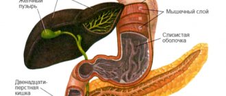
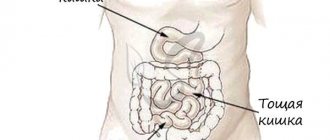
The small intestine has three sections: duodenum (lat. duodenum
), jejunum (lat.
jejunum
) and ileum (lat.
ileum
). The jejunum and ileum do not have a clear boundary between each other. Typically, the first 2/5 of the total length is allocated to the jejunum, and the remaining 3/5 to the ileum. At the same time, the ileum has a larger diameter, its wall is thicker, and it is richer in blood vessels. in relation to the midline, the loops of the jejunum lie mainly on the left, the loops of the ileum on the right. The small intestine is separated from the upper parts of the digestive tract by the pylorus, which acts as a valve, and from the colon by the ileocecal valve.
The thickness of the wall of the small intestine is 2–3 mm, and during contraction it is 4–5 mm. The diameter of the small intestine is not uniform. In the proximal part of the small intestine it is 4–6 cm, in the distal part it is 2.5–3 cm. The small intestine is the longest part of the digestive tract, its length is 5–6 m. The weight of the small intestine of a “conditional person” (with body weight 70 kg) normal - 640 g.
The small intestine occupies almost the entire lower floor of the abdominal cavity and partially the pelvic cavity. The beginning and end of the small intestine are fixed by the root of the mesentery to the posterior wall of the abdominal cavity. The rest of the mesentery ensures its mobility and position in the form of loops. They are bordered on three sides by the colon. Above is the transverse colon, on the right is the ascending colon, on the left is the descending colon. The intestinal loops in the abdominal cavity are located in several layers, the superficial layer is in contact with the greater omentum and the anterior abdominal wall, the deep layer is adjacent to the posterior wall. The jejunum and ileum are covered on all sides by peritoneum.
The structure of the wall of the small intestine
The wall of the small intestine consists of four membranes (often the submucosa is referred to as the mucous membrane and then the small intestine is said to have three membranes):
- mucous membrane, divided into three layers:
- epithelial
- lamina propria, which has depressions - Lieberkühn's glands (intestinal crypts)
- muscle plate
The mucous membrane of the small intestine has a large number of circular folds, most clearly observed in the duodenum. The folds increase the absorptive surface of the small intestine approximately threefold. In the mucous membrane there are lymphoid formations in the form of lymphoid nodules. If in the duodenum and jejunum they are found only in a single form, then in the ileum they can form group lymphoid nodules - follicles. The total number of such follicles is approximately 20–30.
Functions of the small intestine
The most important stages of digestion occur in the small intestine.
The mucous membrane of the small intestine produces a large number of digestive enzymes. Partially digested food, chyme, coming from the stomach, is exposed in the small intestine to intestinal and pancreatic enzymes, as well as other components of intestinal and pancreatic juices, bile. In the small intestine, the main absorption of food digestion products into the blood and lymphatic capillaries occurs. The small intestine is also where most orally administered drugs, poisons and toxins are absorbed.
The normal residence time of the contents (chyme) in the small intestine is about 4 hours.
Functions of various parts of the small intestine (Sablin O.A. et al.):
| Duodenum | Jejunum | Ileum |
| Release of enzymes, hydrolysis of proteins, fats, carbohydrates, enrichment of chyme with bile, change in acidity of the medium, mixing of contents and its transport, absorption | Hydrolysis of polymers, endocrine, absorption, motor, evacuation, hormonal | Absorption of hydrolysis products, bile acids, immune, endocrine, motor-evacuation |
Endocrine cells and hormone content in the small intestine
The small intestine is the most important part of the gastroenteropancreatic endocrine system.
It produces a number of hormones that regulate the digestive and motor activity of the gastrointestinal tract. The proximal parts of the small intestine contain the largest set of endocrine cells among other organs of the gastrointestinal tract: I-cells producing cholecystokinin, S-cells - secretin, K-cells - glucose-dependent insulinotropic polypeptide (GIP), M-cells - motilin, D -cells - somatostatin, G-cells - gastrin and others. The liberkühn glands of the duodenum and jejunum contain the vast majority of all I cells, S cells and K cells in the body. Some of the listed endocrine cells are also located in the proximal part of the jejunum and an even smaller part in the distal part of the jejunum and in the ileum. In the distal part of the ileum there are, in addition, L-cells that produce the peptide hormones enteroglucagon (glucagon-like peptide-1) and peptide YY. The content of hormones (in pmol/g) and the number of cells of their producers per 1 mm2 in the sections of the small intestine (SRBloom, JMPolak; G.F. Korotko):
| Sections of the small intestine | ||||
| Hormone | duodenum | skinny | ileum | |
| gastrin | gastrin content | 1397±192 | 190±17 | 62±15 |
| number of producing cells | 11–30 | 1–10 | 0 | |
| secretin | secretin content | 73±7 | 32±0,4 | 5±0,5 |
| number of producing cells | 11–30 | 1–10 | 0 | |
| cholecystokinin | cholecystokinin content | 26,5±8 | 26±5 | 3±0,7 |
| number of producing cells | 11–30 | 1–10 | 0 | |
| pancreatic polypeptide (PP) | PP content | 71±8 | 0,8±0,5 | 0,6±0,4 |
| number of producing cells | 11–30 | 0 | 0 | |
| GUI | GUI content | 2,1±0,3 | 62±7 | 24±3 |
| number of producing cells | 1–10 | 11–30 | 0 | |
| motilin | motilin content | 165,7±15,9 | 37,5±2,8 | 0,1 |
| number of producing cells | 11–30 | 11–30 | 0 | |
| enteroglucagon (GLP-1) | GLP-1 content | 10±75 | 45,7±9 | 220±23 |
| number of producing cells | 11–30 | 1–10 | 31 | |
| somatostatin | somatostatin content | 210 | 11 | 40 |
| number of producing cells | 1–10 | 1–10 | 0 | |
| VIP | VIP content | 106±26 | 61±17 | 78±22 |
| number of producing cells | 11–30 | 1–17 | 1–10 | |
| neurotensin | neurotensin content | 0,2±0,1 | 20 | 16±0,4 |
| number of producing cells | 0 | 1–10 | 31 | |
Small intestine in children
The small intestine in children occupies a variable position, which depends on the degree of its filling, body position, intestinal tone and peritoneal muscles. Compared to adults, it is relatively long, and the intestinal loops lie more compactly due to the relatively large liver and underdevelopment of the pelvis. After the first year of life, as the pelvis develops, the location of the loops of the small intestine becomes more constant. The small intestine of an infant contains relatively a lot of gases, which gradually decrease in volume and disappear by the age of 7 (adults normally do not have gases in the small intestine). Other features of the small intestine in infants and young children include: greater permeability of the intestinal epithelium; poor development of the muscle layer and elastic fibers of the intestinal wall; tenderness of the mucous membrane and a high content of blood vessels in it; good development of villi and folding of the mucous membrane with insufficiency of the secretory apparatus and incomplete development of the nerve pathways. This contributes to the easy occurrence of functional disorders and facilitates the penetration into the blood of undigested food components, toxic-allergic substances and microorganisms. After 5–7 years, the histological structure of the mucous membrane no longer differs from its structure in adults (Bokonbaeva S.D. et al.).
Small intestine in newborns
A newborn has a relatively large length of the small intestine: 1 m per 1 kg of body weight, and 10 cm in adults. Due to the relatively large size of the liver and underdevelopment of the pelvis, the intestinal loops lie more compactly than in adults.
The small intestine is where most of the digestion and absorption of food occurs. The small intestine of an infant contains a relatively large amount of gases, the volume of which gradually decreases until it completely disappears by the age of 7 (adults normally do not have gases in the small intestine).
The mucous membrane is thin, richly supplied with blood vessels and has increased permeability (especially in children of the first year of life). In newborns, single and group lymphoid follicles are present in the thickness of the mucous membrane. Initially, they are scattered throughout the intestine, and subsequently are grouped mainly in the ileum in the form of group lymphatic follicles (Peyer's patches). Lymphatic vessels are numerous and have a wider lumen than in adults. Lymph flowing from the small intestine does not pass through the liver, and absorption products enter directly into the blood (Geppe N.A., Podchernyaeva N.S.).
Still from the video lecture “Small intestine in newborns” by E.V. Gostishcheva “Anatomical and physiological features of the digestive organs in children. Methods and methods for studying the digestive system in children" for students of the Medical Academy named after S.I. Georgievsky Crimean Federal University named after V.I. Vernadsky.
Some diseases of the small intestine
Some diseases of the small intestine and syndromes (see):
| Inflammatory process in the small intestine |
- enteritis
- small bowel cancer
- duodenitis
- gastroduodenitis
- gurgles
- duodenal ulcer
- functional dyspepsia, including dyspepsia in children
- helminthiasis:
- hookworm (hookworm, necatoriasis)
- strongyloidiasis
- taeniasis and teniarinhoz
- diphyllobothriasis
Duodenal motility disorders:
- duodenal hypertension
- duodenal obstruction
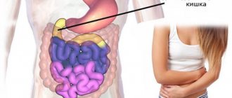
Some symptoms that may be associated with diseases of the small intestine:
- stomach ache
- nausea
- vomit
- flatulence
- diarrhea (diarrhea), including diarrhea (diarrhea) in children
- melena
On the website in the “Literature” section there is a subsection “Diseases of the small intestine”, containing publications for healthcare professionals, including issues of diagnosis and treatment of diseases of the small intestine.
Patient Materials
The GastroScan.ru website contains materials for patients on various aspects of gastroenterology:
- “Advice from doctors” in the “Patients” section of the site
- “Popular gastroenterology” in the “Literature” section
- “Popular gastroenterology” in the “Video” section
Back to section
Histology.RU
Material taken from the site www.hystology.ru
The large intestine, which combines the cecum, colon, and rectum, consists of mucous, muscular and serous membranes (Fig. 272).
The mucous membrane has a folded surface, does not form villi and consists of an epithelial layer, a main lamina, a muscular lamina, and a submucosa.
Rice. 272. Large intestine:
1 - mucous membrane; 2 - muscular layer; 3 - serous membrane; 4 - submucosa; 5 - single-layer intestinal epithelium; 6 - goblet cells; 7 - crypts; 8 — own plate; 9 - muscular plate; 10 - submucosal nerve plexus; 11 - lymphatic follicle; 12 - blood vessels; 13 - annular layer of the muscular layer; 14 - longitudinal layer of the muscularis propria; 13 - mesothelium of the serous membrane.
The epithelial layer is represented by a single-layer columnar bordered epithelium. The epithelium sinks into the underlying lamina and forms crypts (7). The epithelial layer covering the surface of the mucosa and crypts is represented by epithelial cells with a striated border, epithelial cells without a border and goblet cells. Bordered enterocytes (as in the small intestine) are columnar in shape, with pronounced polar differentiation and a thinner border. Since this layer is involved in the formation of feces, it is characterized by an abundance of goblet cells. Epithelial cells without a striated border are characterized by high mitotic activity. Due to their divisions, the integumentary and glandular (goblet) cells are restored. This zone is usually located in the bottom part of the crypts, where, as a rule, chromaffin and Paneth cells are absent.
Layers of loose connective tissue lying between the crypts continue into the tissue of the main plate. The latter is built from loose connective tissue containing a significant amount of reticular tissue, accumulations of lymphocytes that form lymph nodes with reproduction centers. From here, lymphocytes can migrate to all layers of the mucous membrane.
The muscle plate is intensively developed and is built from two layers of smooth muscle tissue - internal (circular) and external (longitudinal).
The submucosa consists of loose fibrous connective tissue. The choroid and submucosal nerve plexuses are located here; lymph nodes are more developed than in the small intestine. They can connect to each other.
The muscular layer is formed by smooth muscle tissue, forming two layers. The bundles of the inner layer are arranged circularly, the outer ones - longitudinally. As in all organs of the digestive tube, between the layers of the muscular layer is the intermuscular nerve plexus. Its cytoarchitecture is described in the chapter “Nervous System”.
The serous membrane covering the outside of the large intestine has an intensively developed connective tissue layer covered with mesothelium.
The wall of the rectum is built from the same membranes. In its most caudal part, the single-layer columnar epithelium is replaced by stratified squamous epithelium, and clusters of lymphoid nodules reach their greatest development. In some farm animals, mucous glands are located in the wall, and in carnivorous animals, there are peri-anal glands, which have a significant resemblance to the sebaceous glands of the skin.
Reviews (0)
Add a review
The answers are not ours / gista_ / ticket 21 / LARGE INTESTINE
COLON
Functions:
1 suction
water and electrolytes from chyme and fecal formation;
2 suction
compounds formed as a result of the activity of intestinal microflora: vitamins K and B, products of fiber hydrolysis;
3 mechanical -
pushing the contents of the intestine (feces) in the distal direction and removing them from the body;
4 endocrine -
due to the presence of DES cells in the intestinal epithelium, which produce hormones that have local and systemic effects;
5 immune -
is provided by diffuse lymphoid tissue in the intestinal wall, as well as by special structures - single lymph nodes and their accumulation in the appendix.
Colon
consists of
four
sections:
the cecum with the appendix, the colon (ascending, transverse and descending), the sigmoid and rectum.
Although it is significantly shorter in length than the small intestine, amounting to only about 1.5 m, the duration of passage of undigested food debris through it reaches
90%
of the total duration of residence of nutrients in the intestine (2-3 days).
The wall of the colon is formed by three
membranes:
mucous, muscular
and
serous
1. Mucosa
consists of
four
layers:
epithelium, lamina propria, muscularis lamina and submucosa.
Its surface is increased due to permanent
semilunar folds.
There are no villi,
intestinal crypts (glands) are
deeper than in the small intestine, are located more often, have a wider lumen and contain cambial elements of the epithelium.
a) epithelium - single-layer prismatic,
contains
four
types of cells:
(1) prismatic, (2) goblet, (3) undifferentiated (poorly differentiated), (4) endocrine.
(1) prismatic cells -
located on the surface of the mucous membrane and in the crypts: tall, narrow, similar to the border cells of the small intestine, but their brush border is much less developed.
They are formed in the depths of the crypt, migrating through which they produce and secrete glycoproteins that accumulate in vesicles in the apical part of the cytoplasm; As they approach the mouth of the crypt, the vesicles disappear, and the microvilli become more numerous and elongate. Provide absorption processes.
(2) goblet cells -
are located in the crypts and (in smaller numbers) on the surface of the mucous membrane.
They are formed in the depths of the crypts from undifferentiated cells, filled with mucous granules. Their number increases in the direction of the rectum. They produce mucus,
which prevents damage to the mucous membrane and facilitates the movement and removal of feces.
(3) undifferentiated cells -
lie deep in the crypts, are cambial
intestinal epithelium;
As they migrate toward the mouth, the crypts differentiate into goblet
or
prismatic cells
Renewal of the epithelium in the large intestine is slower than in the small intestine and takes about 6 days. The desquamation of differentiated cells from the surface of the mucous membrane occurs in the middle between the crypts.
(4) endocrine cells -
located in the bottom of the crypts, they belong mainly to
EC and ECL cells
(see table).
b) own record -
consists of
loose fibrous tissue
in which fibroblasts, lymphocytes, eosinophils, macrophages, mast and plasma cells are found.
Contains capillaries intertwining crypts and nerve fibers. Reticular fibers look like a dense network. It contains single lymph nodes
(with a total number of more than 20 thousand), which often penetrate through the muscular plate into the submucosa.
c) muscle plate
mucous membrane - consists of
two
layers
of smooth muscle cells
(inner
circular
and outer
longitudinal);
d) submucosa -
formed
by loose fibrous connective tissue with
a large number of elastic fibers, often contains adipose tissue.
It contains lymph nodes
(not permanently), elements
of the submucosal nervous, venous and lymphatic plexuses.
2.
Muscular membrane -
formed
by two
layers
of smooth muscle tissue:
internal
circular
and external
longitudinal.
The latter has the appearance of three
ribbons,
between which the muscle tissue is poorly developed.
These ribbons are shorter than the intestine itself, as a result of which it forms multiple sac-like protrusions - haustra coli .
Between the layers of the muscular layer there are layers of connective tissue and elements
of the intermuscular nerve plexus.
3.
Serous membrane -
covers some parts of the colon completely, others - partially, where it is replaced by adventitia.
Forms protrusions in the form of processes containing adipose tissue ( appendices epiploicae ).
Appendix
- a finger-shaped outgrowth of the cecum with a narrow stellate or irregularly shaped lumen, which contains cellular detritus and can become obliterated. The wall of the process is relatively thick due to the high content of lymphoid tissue in it.
1. Mucosa
~
contains the same layers as other parts of the colon.
a) epithelium -
includes
prismatic and goblet cells,
and in the crypts also
poorly differentiated cells, individual Paneth cells
and numerous
endocrine cells.
The areas where lymphatic follicles are located
(domes)
are covered with epithelium containing M
cells.
b) own record
contains short
crypts,
the size and number of which decrease with age, as well as numerous
secondary lymph nodes (B-dependent zone)
and
interfollicular accumulations of lymphoid tissue (T-dependent zone).
The connective tissue contains a large number of diffusely scattered lymphocytes, plasma cells, and eosinophils.
c) muscle plate
is poorly developed and consists of an inner
circular
and outer
longitudinal
layer
of smooth muscle cells, and
is interrupted in places.
d) submucosa
represented by
loose fibrous connective tissue
with a high content of elastic fibers;
Lymatic nodules
are partially located in it 2.
The muscular layer
is formed by the inner
circular
and outer
longitudinal
(solid) layers
of smooth muscle tissue.
3.
The serous membrane
completely covers the appendix.
The vermiform appendix performs a protective function
and is, along with Peyer's patch,
a peripheral organ of the immune system,
part of the KALT.
It ensures the absorption of antigenic material
from the lumen of the colon, its
presentation
to immunocompetent cells with
the development of immune reactions;
contains effector B and T cells.
Inflammation of the appendix (appendicitis),
which can result in the destruction and rupture of its wall with the subsequent development of inflammation of the peritoneum
(peritonitis) -
a common disease requiring surgical treatment. It probably occurs due to the activation of the microbial flora located in the lumen of the appendix. An excessively strong (hyperergic) reaction of lymphoid tissue to incoming antigens may play a certain role.
Rectum
- the distal portion of the large intestine, ending in the anal
Above the expanded lower part
(rectal ampulla)
there are 2-3 transverse folds of the mucous membrane. The ampoule is dominated by mucous cells; crypts are long.
The anal canal
is
a continuation of the tapering lower part of the ampulla;
the mucous membrane forms 5-10 longitudinal folds - anal or rectal columns (Morgagni),
which are connected at the bottom by transverse folds
(anal valves).
Between the columns there are depressions in the form of pockets -
anal sinuses.
The crypts in the distal direction shorten and disappear, and
the single-layer prismatic epithelium
along
the dentate (anorectal) line
is replaced
by multilayered squamous non-keratinizing epithelium.
Often in the area where the epi-telnev changes there is
a transition zone
containing
multilayered prismatic or cuboidal epithelium
that secretes mucus.
The stratified non-keratinizing epithelium is then replaced by keratinizing skin epithelium,
sebaceous and apocrine sweat glands and hair appear.
Anal glands
- rudimentary formations located in
the submucosa of the anal canal
(in some places they penetrate into the muscular layer) and open into the anal sinuses.
MUCOSA
Mucous membrane
[
tunica mucosa
(PNA, BNA)] - the inner lining of hollow organs communicating with the external environment (respiratory organs, urinary, reproductive and digestive systems).
The mucous membrane of various organs consists of epithelium (epithelium), lamina propria (lamina propria) and muscular plate (lamina muscularis), the edges are adjacent to the submucosa (tela submucosa), separating the S. o. from other layers of the organ wall (for example, from the muscular layer). The figure shows the schematic structure of the S. o. The surface of the mucous membrane is always covered with mucus (mucus).
Schematic representation of the structure of the mucous membrane: I - epithelium; II—lamina propria; III - muscular plate; IV - submucosa; 1 - nerve trunk; 2 - nerve plexus; 3 - complex (alveolar-tubular) glands; 4 - simple tubular glands; 5 - lymphoid follicle; 6 - venous vessel; 7 - lymphatic vessel; 8 - arterial vessel.
The mucous membranes of various vertebrates have a number of structural and histochemical features. Thus, in amphibians and reptiles S. o. The esophagus is lined with multilayered ciliated epithelium, and in mammals (including humans) with multilayered squamous epithelium. There are differences (depending on the type of nutrition) in the histochemical characteristics of the epithelium of the lake. stomach of mammals. Thus, in carnivores it includes neutral glycoproteins, sialoglycoproteins, sulfoglycoproteins, and in plants it contains only neutral glycoproteins.
Tissue components of S. o. originate from various embryonic rudiments. The epithelium develops from all three germ layers (see): stratified squamous non-keratinizing epithelium S. o. oral cavity, anal rectum, vagina and cervix - from the ectodermal germ layer; single-layer epithelium of S. o. stomach, intestines, gall bladder - from endodermal; single-layer epithelium of S. o. genital tract - from the mesodermal; blood and lymph, vessels, lamina propria and muscle of the S. o. - from mesenchyme.
Epithelium S. o. protects the body from the penetration of harmful substances from the outside and ensures the metabolism of the body with the environment. The structure of the epithelium of various organs varies depending on the functional specificity of S. o. The mucous membrane, which performs the function of protecting the organ wall from rough, traumatic components, is covered with non-keratinizing stratified squamous epithelium (oral cavity, pharynx, esophagus, anus of the rectum); the mucous membrane, which ensures the transport of substances from the environment into the body or their elimination, is single-layered (stomach, intestines).
Own record of S. o. located directly under the epithelium and consists mainly of loose fibrous connective tissue (see). In areas exposed to rough mechanical stress (gums, hard palate, epiglottis), it contains powerful intertwining bundles of collagen fibers. In the lamina propria of the mucous membrane, reticular tissue (see) is abundantly developed, in which single, and in the mucous membrane of the ileum - group, or generalized, lymph, follicles (Peyer's patches), as well as lymphocytes are localized. Lymphocytes play a major role in protecting the body from bacteria, viruses, and other antigenic factors that enter the body through the covering layer of the lake. epithelium. It has been shown that in response to an antigenic stimulus, lymphocytes turn into plasma cells (see), producing immunoglobulin A (see Immunoglobulins), called secretory (SIg A), because it is actively transported to the surface of the epithelium by its secretory cells.
Muscular plate S. o. formed by smooth, unstriated muscle tissue (see). Unlike the epithelium and the lamina propria, the muscular plate is well expressed only in the S. o. organs of the digestive system (see) and bronchi (see).
In other organs it is either absent or represented by separate bundles of smooth myocytes.
The submucosa consists of loose fibrous connective tissue, which provides the possibility of displacement of the lake. and the formation of folds.
The mucous membrane has its own glandular apparatus. The most common are unicellular mucous glands (see), localized in the epithelium itself (endothelial). They are usually located mosaically, interspersed with other epithelial cells (goblet cells, endocrinocytes), less often in groups (Paneth cells of the intestine) or in the form of glandular fields (in the epithelium of the stomach and uterus). The mucous glands of the lamina propria are multicellular (simple, tubular, less often branched).
The vast majority of the glands of S. o. They are exocrine (see Mucous glands) and produce mucous (less often protein) secretion. Released onto the surface of the epithelium, mucus moisturizes it, protects it from damage, and in some organs adsorbs foreign particles, facilitating their removal. Endocrinocytes, Paneth cells of the intestine, and tubular glands of the stomach have more specific functions. Endocrinocytes are found in the epithelium of almost all S. lakes, where they form the local endocrine apparatus. As part of the epithelium of S. o. The olfactory region of the nasal cavity contains cells specialized in the perception of odors, and in the epithelium of the papillae of the tongue there are taste buds that perform the reception of flavoring agents. In addition to its own unicellular glands, on the surface of S. o. the oral cavity, esophagus, duodenum, trachea and bronchi open the excretory ducts of larger glands located in the submucosal base, as well as large glands that are independent organs, such as the salivary glands (see), liver (see), pancreas ( cm.).
The relief of the mucous membrane is determined by its morphofunctional properties. Thus, the surface of the S. o. can be smooth (lips, cheeks, trachea), form protrusions (intestinal villi, tongue papillae) or invaginations (pits and crypts in the stomach, intestines, uterus), folds (stomach, intestines, gall bladder, ureter).
S. o. richly vascularized due to its own vascular plexuses, the structure of which is different in different organs, for example. in the upper respiratory tract, the terminal sections of the choroid plexuses are sharply expanded, because, in addition to participating in metabolism, they warm the inhaled air. Lymph vessels are most developed in the intestinal mucosa.
Nervous apparatus S. o. formed by nerve fibers of extra- and intraorgan origin. Moreover, the former are represented by dendrites of neurons of the spinal and sensory ganglia of the cranial (cranial, T.) nerves or neurites of neurons of the sympathetic ganglia, and the latter are represented by processes of neurons of the intramural nerve ganglia of the submucosal and, to a lesser extent, the musculo-intestinal plexuses.
All tissue elements of the system are subject to age-related changes, but primarily connective tissue, in which the number of fibrous structures (sclerosation) increases with age, fat cells appear, and the number of blood vessels, lymph vessels, and nerve fibers per unit decreases area, the regenerative capabilities of the epithelium are reduced.
The tissue elements of the mucous membrane have a high regenerative ability, especially the epithelium of the small intestine, which in humans is completely replaced within 5 - 6 days.
Posthumous changes in S. o. arise relatively quickly. Autolysis occurs first in the S. o. small intestine, later in S. o., covered with stratified squamous epithelium. Electron microscopic examination reveals detachment of enterocyte villi from the basement membrane already in the first minutes after death. According to A.I. Lysenko (1981), 1/2 hour after death, mitochondria become condensed (with a compacted matrix and expanded intercristal spaces), after 2 hours they undergo swelling, after 3 hours the generation of energy in the form of ATP completely disappears and sharply the content of ADP and AMP decreases, the aerobic metabolic pathway is inhibited.
Pathology
The mucous membrane is described in articles about organs (e.g., Pharynx; Stomach, Intestines; Uterus; Bladder; Mouth, oral cavity; Rectum, etc.), as well as in individual articles (e.g., Bronchitis, Gastritis, Duodenitis, Colitis and etc.).
Bibliography:
See bibliography. to Art. Stomach, Gallbladder, Intestines, Bladder, Esophagus.
Yu. K. Eletsky.