The short initial part of the large intestine, located in the ileum, is called the cecum. The length of this organ, completely covered with peritoneum, can be 1-10 cm. The width is 5-9 cm. Depending on the anatomical features of the development of the body, the cecum can have different shapes:
- hemispherical (the most common);
- bay-shaped;
- saccular;
- conical;
- funnel-shaped, tapering from the base to the top.
The last case is considered anomalous. As a rule, the cecum takes on a funnel-shaped shape only in cases where the development of the intestine in the embryo is inhibited for some reason. Slightly more common are cases when the described organ in humans is, in principle, not expressed (that is, the small intestine does not pass into the cecum, but immediately into the ascending colon).
Topographic location
Part of the cecum is projected to the right side of the groin.
As for the specific location in the abdominal cavity, in the vast majority of people the cecum is located just below the upper edge of the ileum.
That is, the organ is located closer to the front wall of the abdomen. Part of the cecum is projected onto the right side of the groin.
So, its dome is directed towards the small pelvis and is located only 5 cm above the inguinal ligament.
At the junction of the cecum with the ileum there is a special organ - the intestinal papilla. In conjunction with muscles, it is capable of acting as an anti-reflux mechanism. This device, operating on the valve principle, is called the Bauhinium valve.
Thus, from above the cecum borders on the ileum. In front - with the thin and ureter. On the right - almost in close contact with the abdominal wall. And behind and below - with sheets of peritoneum.
Examination methods
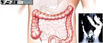
The main method of physical examination of S. is palpation, with the help of a cut in the right iliac region, a mobile, elastic, painless formation, semi-oval or oblong, 6-7 cm wide, is normally determined. The leading instrumental methods are X-ray and endoscopic examination (see Irrigoscopy, Colonoscopy). With contrast radiograph. a study with the introduction of barium sulfate using an enema or through the mouth determines the shape, location and displacement of the blood vessel, the presence of deformations, narrowings, filling defects, evacuation function and the condition of the ileocecal valve. Endoscopic examination allows us to clarify the nature of the patol. process, detect a tumor, fistulas, perform a biopsy, remove foreign bodies, polyps, etc.
Ileocecal angle
The ileocecal angle includes 4 elements.
The junction of the ileum and cecum is called the ileocecal angle. This term arose due to the specific type of such transition.
Indeed, depending on the anatomy of the body, the ileum can “flow” into the medial wall of the cecum at a right, acute or obtuse angle.
That is, technically, the above-mentioned term is usually understood as a whole “set” of organs. Thus, the ileocecal angle includes:
- terminal ileum;
- directly, the cecum;
- its vermiform appendage is the appendix (we will talk about it in more detail below);
- zones of connection of the listed organs.
Due to its specific structure, the ileocecal angle is capable of performing the function of a valve. It reliably isolates the small intestine from the large intestine, preventing the reverse flow of their contents. In addition, precisely because of the peculiarities of the attachment of the cecum in the abdominal cavity, this organ, if necessary, can be easily operated on without damaging neighboring tissues.
Watch the video for everything about the digestive system of the human body:
https://www.youtube.com/watch?v=RiU3Srab9kE
Active and inactive neuroendocrine neoplasms
Hormonally active and inactive neuroendocrine tumors are found in all parts of the gastrointestinal tract.
- Insulinomas release insulin into the blood and lower blood sugar levels. 95% of all insulinomas are benign.
- Gastrinomas increase gastrin production. They are found primarily in the duodenum and pancreas.
- Glucagonomas are very rare neoplasms that arise from the pancreas. They produce glucagon, an insulin “antagonist.”
Hormonally inactive neuroendocrine tumors occur throughout the gastrointestinal tract and are most often found in the stomach, appendix, and rectum. Inactive tumors are often discovered late because they grow slowly and in many cases do not cause discomfort.
Functionality
The main function of the cecum is to participate in the digestive process.
The main function of the cecum is its direct participation in the digestive process.
It is this organ that is responsible for the normal absorption of chyme (or rather, its liquid part). However, the work of the cecum cannot be called “irreplaceable”.
If its function is disrupted, the rest of the intestine will cope with the digestive process quite calmly.
A separate line should describe the functions of the appendix of the cecum. This organ does not take part in the digestive process. However, it plays a very important role in the formation of human immunity. This is where most of the lymphoid follicles are located. And the cells they produce, in turn, are responsible for protecting the body from any foreign agents.
Causes
A benign tumor of the cecum is localized in the first area and is often asymptomatic throughout life. The causes of the occurrence cannot always be determined. Epidemiological studies suggest that environmental causes are responsible for differences in incidence in geographically distinct populations. Differences in dietary fiber and antioxidant intake have been suggested as determinants of colon polyp development. Rare genetic causes:
- familial adenomatous polyposis;
- hereditary nonpolyposis colon cancer.
There is indirect evidence that consumption of red meat, fat and alcohol increases the risk of developing benign tumors. Conversely, calcium and folate supplementation may have a modest protective effect, especially in patients with colon polyps.
Typhlitis
Bloating can be a symptom of pelvic pathology.
This term refers to inflammation of the cecum in its typical manifestations, which is very similar to ordinary appendicitis, which makes diagnosing the disease very difficult.
The only difference between these two ailments is the nature of the pain that worries the patient. So, with typhlitis, unpleasant sensations begin to bother the patient some time after eating.
In this case, the source of discomfort is usually located directly in the iliac region.
It is also difficult to differentiate typhlitis from many disorders in the genitourinary area. To make sure that the patient’s problems are not related to renal colic, gynecological diseases or pelvic pathologies, the doctor pays attention to the following symptoms:
- density and soreness of the cecum (detected by palpation);
- bloating (especially on the right side);
- “splashing” in the abdomen (detected when “listening” to the patient with a stethoscope).
To clarify the diagnosis, doctors use modern research methods: x-rays and irrigoscopy.
Anatomical changes in the structure of the cecum, visible on photographs, usually help to confirm the suspicion of typhlitis. Thus, the organ seems to shorten, and the folds on its mucous membrane are smoothed out.
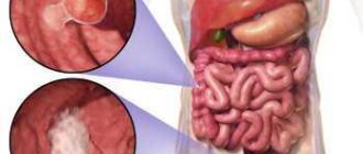
Diagnostics
The preliminary diagnosis of “cecal cancer” is established by specialists at the oncology clinic during the collection of complaints and anamnesis, and clinical examination. A large tumor can be detected during palpation of the abdomen. For the purpose of differential diagnosis, to clarify the location, shape and size of the tumor, and to identify metastases, additional research methods are carried out:
- Colonoscopy;
- Irrigoscopy;
- Computed tomography;
- Ultrasound screening;
- Diagnostic laparoscopy.
During an endoscopic or laparoscopic examination, doctors necessarily select material for histological analysis, which allows them to come to unambiguous diagnostic conclusions. The most informative method for diagnosing sigmoid colon cancer is rectoscopy. With sigmoidoscopy, up to 25 cm of the distal colon is examined.
The use of a flexible sigmoidoscope and colonoscope allows for more accurate preoperative diagnosis of cecal cancer. The X-ray method using a double contrast enema has great sensitivity. It allows you to detect small tumors. A malignant neoplasm manifests itself in the form of a characteristic narrowing or compaction, which is located in the contrast zone. In doubtful cases, doctors at the Yusupov Hospital repeat the examination or perform a colonoscopy.
Scanning computed tomography with air contrast is becoming increasingly widespread. This method is used when making a final decision about the need for surgical intervention. At the Yusupov Hospital, spiral computed tomography with a small slice thickness, the so-called “virtual colonoscopy,” is widely used to detect cancer of the cecum.
Colon carcinoma cells produce carcinoembryonic antigen (CEA), a tumor marker for cancer. However, it is not specific enough to serve as a reliable indicator of the existence of a tumor. Carcinoembryonic antigen is also found in pancreatitis, inflammatory bowel processes, in smokers and in people who abuse alcohol. The CEA test is used in patients with initially high levels of this tumor marker after surgery. Its level decreases after successful surgery, and an increase in CEA concentration in the postoperative period may be the first sign of tumor relapse.
Doctors at the Yusupov Hospital carry out differential diagnosis of cecal cancer with the following diseases:
- Diverticulosis of the colon;
- Ulcerative and ischemic colitis;
- Irritable bowel syndrome.
Other diseases manifested by rectal bleeding (hemorrhoids, polyposis) make diagnosis difficult. Pain in the right half of the abdomen may indicate the development of acute appendicitis. If the patient has positive symptoms of an “acute abdomen,” he undergoes urgent surgery, during which the true cause of the pain syndrome is determined.
Tubular adenoma of the cecum is a benign neoplasm. It may present with symptoms that resemble those of a cancerous tumor.
Make an appointment
Adenocarcinoma
Weakness and fatigue can be a symptom of some kind of disorder.
This oncological tumor disease is considered quite common among other similar pathologies. The main symptoms of this disorder are:
- anemia;
- weakness and fatigue;
- blood in stool;
- weight loss;
- flatulence;
- stool disorders;
- characteristic abdominal pain.
In the early stages, adenocarcinoma can be quite successfully treated with chemotherapy and radiation therapy. Thus, in 70% of patients who underwent these procedures, the disease did not recur for 5 or even more years.
Unfortunately, the later adenocarcinoma is diagnosed, the less chance the patient has for a successful and final recovery.
Classification
Classification by tumor origin:
- epithelial;
- connective tissue;
- neuroendocrine.
Polyps are benign growths that arise from the epithelial cells lining the colon. They are traditionally divided into 3 groups: hyperplastic, adenomatous and polypous. Connective tissue tumors (fibromas, lipomas, fibroids, angiomas, etc.) are extremely rare in the gastrointestinal tract. The most common tumors are fatty and smooth muscle tissues, as well as lymphatic vessels. Neuroendocrine tumors are divided into hormonally active and inactive. About half of them release hormones into the bloodstream. Inactive neuroendocrine tumors do not produce any hormones and often go undetected for a long time.