Differential diagnosis of adenomas with advanced colon tumors does not cause difficulties. Early invasive cancer may resemble a benign tumor, but has a number of characteristic features. Because Cancer invasion into the submucosal layer occurs through the stalk of the polyp, then with an increase in the volume of invasion it gradually thickens and shortens (eventually, the formation on the stalk loses it and is located on a broad base). The simultaneous disintegration of the tumor in the head region leads to its deformation and erosion (erosion is a characteristic sign of invasive cancer). Another characteristic feature is pronounced contact trauma. A retraction or “platform” on the surface of the lesion may also indicate invasive carcinoma [14].
Juvenile and Peutz-Jeghers polyps
Juvenile polyps occur primarily in children and adolescents. More often they are solitary, on a stalk and with a spherical head, have a bright red color and sometimes an eroded apex. Juvenile polyps are not lobulated. There are contradictions among morphologists regarding the nature of such polyps; a number of authors consider them as hamartromic, others point to their closeness to adenomatous polyps. Morphologically, irregularly shaped glands with cystic expansions containing mucin, exuded by cuboidal epithelium, are revealed. The stroma of such polyps is swollen, contains dilated vessels and inflammatory cells. It is believed that juvenile polyps do not become malignant.
Peutz-Jeghers polyps can either have a stalk or be located on a broad base, often lobulated and have a whitish color. Their distinctive morphological feature is the presence of branching smooth muscle bundles emanating from the muscular plate of the mucosa.
Diagnostic measures
If you experience discomfort in the anus, you should urgently contact a gastroenterologist.
If alarming symptoms appear, you should contact a gastroenterologist or proctologist. Diagnostics takes place in several stages:
- Examination and conversation with the patient. The doctor will be interested in the patient's complaints, past medical history and medical history of the next of kin. Then an examination and manual palpation of the rectum is performed. This will give the doctor information about the presence or absence of tumors in the patient.
- Colonoscopy - an endoscope is inserted into the patient's anus and the condition of the intestinal mucosa is examined. In addition, the device is equipped with forceps for collecting tissue samples. If during the procedure the doctor discovers a tumor, a biopsy will be performed immediately. The tissues will be immediately sent for analysis. This will allow you to determine or exclude the malignant nature of the polyp. Benign growths are removed during the procedure.
- Sigmoidoscopy - in essence, this manipulation is similar to colonoscopy. The device is also equipped with forceps for collecting samples, but the length of the tube is short. Therefore, it is impossible to examine the intestines in full.
- Sigmoidoscopy – the length of the endoscope tube is 60 cm. This allows a thorough examination of the sigmoid colon. The equipment is also capable of recognizing tumors larger than 10 mm. This procedure is less unpleasant than a colonoscopy.
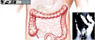
- Irrigoscopy - X-ray with contrast - barium salts. When examined, polyps look like defects in the filling of the intestine.
- Additionally, fecal examination is prescribed for the presence of occult blood. This is an indirect diagnostic method, but it confirms the presence of tumors in the intestines.
Inflammatory polyps
They represent a focus of inflammation. Often occur with nonspecific ulcerative colitis.
Their size and appearance can vary widely. We observed the formation of an inflammatory polyp in the area of utero-intestinal and enterovesical fistulas. They can form in the area of intestinal anastomosis. A large number of them in inflammatory bowel diseases can create difficulties in differential diagnosis with diffuse familial polyposis. Morphologically, the formation is represented by a different combination of granulation tissue and regenerative epithelium.
Preventive actions
Healthy eating will become an effective preventive measure.
This disease does not select the patient’s gender or age. But by following the rules of prevention, you can avoid this pathology. Doctors recommend:
- Adjust your diet. You should not get carried away with diets, it is enough to adhere to the principles of healthy eating.
- Quitting smoking, eating spicy foods, and drinking alcohol in large quantities. This will have a beneficial effect on all body systems.
- Maintaining a drinking diet.
- Watch your weight.
- When the first signs of pathology appear - blood or mucus in the stool - do not delay, but consult a doctor. This disease is treated by proctologists, coloproctologists, and gastroenterologists.
- According to indications, carry out the necessary operational measures.
A polyp is a benign neoplasm, but under certain conditions it can degenerate into malignant tumors. Don't delay your visit to the doctor. It’s easy to lose health, but it’s not always possible to restore it.
Additional methods for differential diagnosis of colon polyps
As mentioned above, the differential diagnosis of neoplastic and neoplastic polyps, as well as early cases of carcinoma, is quite difficult, especially for small tumors. To improve such diagnostics, S. Kudo et al. proposed a method for endoscopic diagnosis of the possible morphological structure of a tumor based on the pattern of glandular pits [11]. There are 5 types of patterns of glandular pits (Fig. 8), with a clear correlation between them and the morphological structure of the formation [11].
The first type, characterized by round, regularly spaced pits of almost the same size, is a sign of an unchanged mucous membrane. The second type (regularly located pits of large size and “star-shaped” or “bulbous” shape) occurs in 69.4% of cases with hyperplastic polyps, and in 30.5% with adenomatous polyps. The third type (S) is compactly located small rounded pits in 86.3% are adenomas, and 12.3% are carcinomas. The third type (L) - large, elongated pits - adenomas in 92.7% and carcinomas in 4.2%. The fourth type - pits of the type of branches or convolutions - in 74.9% of cases these are adenomas (usually glandular-villous), and in the rest - carcinomas. The fifth type - with partial or complete disruption of the structure of the fossae - carcinoma in 93.3% of cases and adenoma in 6.7%.
This pattern can be observed during a standard colonoscopy, but magnified colonoscopy provides a more accurate degree of diagnosis. Konishi K. et al compared the diagnostic yield of conventional colonoscopy and magnified colonoscopy based on 630 colonoscopies (330 with and 330 without magnification) [10]. The assessment of the pattern of glandular pits was carried out after irrigating the formation with a 0.2% solution of indigo carmine, according to the above classification. The accuracy of endoscopic diagnosis was then verified by morphological examination. This work included only patients with lesions no larger than 10 mm. Successful differentiation of neoplastic from neoplastic lesions was possible in 92% of cases with magnifying colonoscopy and only 68% of cases with conventional colonoscopy.
Symptoms of the disease
Discomfort in the anus may be a sign of polyps.
Polyposis of the sigmoid colon in the initial stages is asymptomatic. The patient can learn about the presence of pathology during a routine examination of other organs.
Multiple growths can cause some discomfort, but the first specific symptoms occur when the tumor reaches a size of more than 3 cm. Symptoms of sigmoid colon polyposis:
- pain, attacks of itching when trying to sit down or change body position;
- blood in stool after bowel movement. Some types of polyps can cause heavy bleeding;
- presence of mucus in feces;
- attacks of diarrhea alternate with delayed bowel movements. There may be a false urge to defecate;
- various signs of dyspepsia - nausea, vomiting, increased gas formation;
- large polyps may fall out of the anus;
- in severe cases - obstruction, fever, general weakness.
Diagnostics
Instrumental methods for diagnosing a neoplasm include:
- Total colonoscopy or sigmoidoscopy with biopsy is an informative study that allows you to detect a tumor, identify its size and location, and also obtain biological material for histological analysis. This requires 3-5 grips with standard endoscopic forceps. The specificity and effectiveness of the method increases with the use of modern technologies (magnifying and narrow-spectrum endoscopy, fluorescent diagnostics).
- Ultrasound using a rectal probe makes it possible to assess the degree of invasion of the walls of the rectum by the tumor.
- MRI of the pelvis, which is preferably performed in the presence of large tumors (more than 3 cm) and if malignancy is suspected. The method helps to clarify the main characteristics of the tumor and assess the condition of regional lymph nodes.
If it is impossible to perform an endoscopic examination, irrigoscopy or CT colonography is recommended.
Advantages of treating villous polyps at the Yusupov Hospital
In the proctology department of the Yusupov Hospital, all conditions have been created for the quick and comfortable treatment of patients. The wards are equipped with air conditioning, patients are provided with individual personal hygiene products and dietary meals. Proctologists take an individual approach to choosing the optimal treatment method for each patient. Complex cases of villous polyps are discussed at a meeting of the Expert Council with the participation of professors, doctors of medical sciences, and doctors of the highest category.
In the presence of a villous polyp of the rectum, the prognosis depends on the timeliness of diagnosis and the radicality of treatment. Surgeons at the proctology department of the Yusupov Hospital are fluent in all modern techniques of surgical interventions on the rectum. If there are indications and there are no contraindications, endoscopic operations are performed without opening the tissues of the anterior abdominal wall. The medical staff is attentive to the wishes of patients, ensures the implementation of all procedures and professional care in the postoperative period.
Gastrointestinal adenomas (adenomatous polyps)
| Adenoma of the ileum. Enteroscopy (Bunova S.S. et al.) |
Adenoma
is
a benign tumor arising from the glandular epithelium. Depending on the organ where it is located (localization), adenomas are distinguished: stomach, intestines, appendix, liver, kidneys, adrenal glands, lungs, as well as glands: salivary, prostate, thyroid, pituitary gland, parathyroid, mammary, sebaceous.
Adenomas of the gastrointestinal tract (GIT) are often called adenomatous polyps
. Adenomas can be either hereditary or non-hereditary. Hereditary ones include: familial adenomatous polyposis of the colon, Gardner's syndrome, Turcotte's syndrome and others (Kaibysheva V.O. et al.).
According to their structure, adenomas of the gastrointestinal tract are classified as:
- tubular
- villous (papillary)
- tubular-villous (papillotubular)
Tubular adenoma
has a clearly limited shape, crimson color and is characterized by slow growth. Consists of a glandular branch, which is limited by connective tissue. Microscopically, tubular adenoma is represented by proliferating adenomatous epithelium, built from branching and significantly convoluted glandular tubes, longer than in ordinary mucosa. In tubular adenoma, the presence of no more than 25% villous tissue is allowed. Adenoma has a mucous membrane-covered base consisting of connective tissue, smooth muscle cells and blood vessels. This is the most common type of adenoma and it can degenerate into malignant formations, so treatment must be carried out promptly and quickly.
Among epithelial tumors of the stomach, adenomas account for at least 3–13%. During endoscopic examination, typical adenomas usually look like polyp-like formations rising above the surface of the mucosa with a villous or lobulated surface.
| Tubular villous adenoma of the stomach. Severe dysplasia (×200) (Zvenigorodskaya A.P.) |
Villous adenoma
consists of thin finger-like outgrowths of the connective tissue of the lamina propria of the mucous membrane, covered with epithelium. Almost all villous adenomas contain a small number of glands. The amount of villous component is more than 75%. Macroscopically, villous adenomas have a wide base and a “shaggy” surface.
Tubular-villous type
adenoma - intermediate between tubular and villous. In tubular-villous adenomas, as a result of proliferation of the epithelium, the number of villi increases, which can be detected both on the surface of the polyp and inside large glands. The structure of the glands also changes: they lengthen, acquire an irregular shape, and fit tightly to each other. At the same time, the degree of epithelial dysplasia increases. Tubular-villous adenoma is considered to be one that has in its structure from 25 to 75% villous tissue. Macroscopically, tubular-villous adenoma differs from tubular adenoma in its more pronounced lobulation and the presence of small areas with very small lobules or villi (Borsuk A.D., Malaeva E.G.).
Gastric adenoma
Gastric adenomas, according to the modern classification of polyp-like formations of the stomach, are divided into (Pirogov S.S., Sokolov V.V.):
- intestinal type adenomas
- foveolar type adenomas
- pyloric adenomas
- fundal type adenomas
- inflammatory fibrous polyps
| Tubular adenoma of the stomach (×200) (Zvenigorodskaya A.P.) |
Of the gastric adenomas, intestinal type adenoma is the most common.
Develops against the background of chronic atrophic gastritis with intestinal epithelial metaplasia. The incidence of the disease increases with age. Intestinal-type gastric adenoma is an obligate precancerous disease. All gastric adenomas of the intestinal type are subject to endoscopic removal for therapeutic and diagnostic purposes, which is due to the high risk of their malignancy. Removed tumors must be carefully examined morphologically with serial sections performed every 2(1) mm (Pirogov S.S., Sokolov V.V.). Gastric adenoma is a precancerous disease with a risk of malignancy of 8-59% (Khomyakov V.M.).
There is an opinion that gastric ulcer, chronic atrophic multifocal gastritis, chronic atrophic multifocal gastritis with the presence of adenomatous polyps and intestinal type gastric cancer are stages of impaired cellular renewal of the gastric mucosa associated with the persistence of Helicobacter pylori
(Kogan N.Yu.).
Colon adenoma
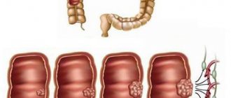
Adenomatous colorectal polyps are more common in men than in women.
Elderly patients have an increased risk of adenomas. During autopsies in people under 50 years of age, adenomas are detected in 17%, from 50 to 59 years old - in 35%, 60-69 years old - in 56%, 70 years and older - in 63% of cases. Data from screening colonoscopies showed similar results: at the age of 50-59 years, adenomas were diagnosed in 21-28%, 60-69 years - in 41-45%, 70 years and older - in 53-58% of patients. Colorectal adenomas can vary greatly in size, but small ones, less than 1 cm in diameter, are more common. As a result of 3371 colonoscopic adenomectomies, polyps of 0.5 cm or less were found in 38% of patients, 0.6-1.0 cm in 36%, and more than 1 cm in diameter in 26%. Among the examined patients, a single adenoma was detected in 60% of cases, and several adenomas were detected in 40% of cases. It is believed that solitary adenomas are facultative, and diffuse adenomatosis is an obligate precancerous disease. As patients age, the likelihood of detecting multiple adenomas increases. Adenomatous polyps, like colorectal cancer, are more common in the left colon. Clinical studies have shown that in more than 60% of cases, the removed adenomas are located distal to the splenic flexure of the colon. However, autopsy data from patients who died from other causes indicate that adenomas are more common in the proximal colon. Colorectal adenoma most often has the appearance of a polyp associated with the wall with a stalk or a wide base. The length of the stalk depends on the growth rate of the polyp and its location. Fast-growing polyps have a broad base, while slow-growing polyps tend to be pedunculated. The polyp stalk is formed as a result of peristalsis and traction of the polyp by a peristaltic wave. Some adenomas have a flat or indented appearance and do not rise above the surface of the intestinal mucosa.
Most colorectal adenomas are silent and are usually discovered incidentally. Sometimes adenomas can cause significant bleeding or lead to chronic anemia due to prolonged occult blood loss. Large rectal adenomas, in addition to bleeding, may be accompanied by tenesmus and mucus secretion. Producing large amounts of mucus can cause electrolyte disturbances. Distal adenomas of the rectum can partially prolapse through the anus (Borsuk A.D., Malaeva E.G.).
Observation of patients after ectomy of colon adenomas
The following observation intervals (control colonoscopy) are recommended for patients who, before the initial colonoscopy, did not have a burdened family history, hereditary polyposis syndromes, signs of colorectal cancer and hereditary diseases (Ivashkin V.T. et al.):
| After removal of colon adenomas, regular surveillance colonoscopy is required for early detection of colorectal cancer |
- very low risk (no colon polyps detected or only hyperplastic polyps detected) - after 10 years
- low risk (1-2 adenomas less than 1 cm each identified) - after 5 years, if there are no changes - every 10 years
- intermediate risk (3-4 adenomas smaller than 1 cm each or at least one adenoma measuring 1 cm or more were identified) - after 3 years, if there are no changes in two consecutive studies - every 10 years
- high risk (at least 5 adenomas smaller than 1 cm or at least 3 adenomas, at least one of which is at least 1 cm in size) - after 1 year, then every 3 years
- very high risk (a large polyp on a flat base of at least 2 cm or a malginized polyp with favorable prognostic characteristics was identified, complete removal was performed, no signs of damage to the edges were detected, no signs of low differentiation and involvement of blood and lymphatic vessels) - every 3 months, in the absence changes - every 3 years.
Adenomatous polyps of the esophagus
A long-term inflammatory process in the esophagus during reflux esophagitis leads to impaired repair and regeneration, as well as loss of control over epithelial proliferation. These processes underlie the development of metaplasia, dysplasia and polyps of the lower third of the esophagus and serve as the basis for neoplastic processes in the future. Adenomatous polyps were found in 8.3% of observed children and adolescents with erosive-ulcerative reflux esophagitis. In three patients, the surface of adenomatous polyps was lined with small intestinal epithelium. In two cases, we observed the development of adenoma of the abdominal part of the esophagus from foci of gastric metaplasia. During the treatment of reflux esophagitis, adenomatous polyps did not tend to regress (Semenyuk L.A., Sannikova N.E.).
Professional medical publications covering adenomatosis associated with gastrointestinal diseases
- Margaryan L.A. Comprehensive endoscopic diagnosis of gastric polyps. Abstract of dissertation. PhD, 14.00.27 - surgery. RMAPO, Moscow, 2009.
- Zvenigorodskaya L.A. Features of the clinical course and drug therapy of peptic ulcer in the elderly // Consilium medicum. – 2008. – T. 10. – No. 8. – P. 27–33.
- Kogan N.Yu. The role of markers of cellular renewal and apoptosis of gastric epithelial cells in the occurrence and progression of diseases associated with HP Abstract of thesis. PhD, 14.00.05 - internal diseases. VMI, Samara, 2008.
- Erdes S.I., Sergeeva T.N. Polyps of the cardio-esophageal junction in children // Pediatrics. – 2006. – No. 6. – p. 101–109.
- Firsova L.D., Masharova A.A., Bordin D.S., Yanova O.B. Diseases of the stomach and duodenum // – M: Planida. – 2011. – 52 S.
- Rudaya N.S. Modern approaches to diagnosis and choice of treatment tactics for chronic gastric erosions. Abstract of dissertation. Doctor of Medical Sciences, 01/14/17 - surgery. Siberian State Medical University, Tomsk, 2012.
- Burdina E.G. The role of persistent HP infection in the pathology of the upper gastrointestinal tract. Abstract of dissertation. Doctor of Medical Sciences, 14.00.05 - internal diseases. MC UDP RF, Moscow, 2007.
- Shatokhina I.V. Simultaneous operations in elderly and senile patients with prostate adenoma. Abstract of dissertation. PhD, 01/14/23 – urology. SSMU, Saratov, 2013.
On the website, in the Literature section, there is a subsection “Polyps”, containing professional publications on this topic.
Adenomatous polyps of the digestive system in ICD-10
Various adenomatous polyps of the digestive organs are classified under the following headings “Class II.
Neoplasms (C00-D48)”, group “D10-D36 Benign neoplasms” of the International Classification of Diseases ICD-10: In the three-character heading “D12 Benign neoplasms of the colon, rectum, anus [anus] and anal canal”: D12.0 Blind colon (including: ileocecal valve) D12.1 Appendix D12.2 Ascending colon D12.3 Transverse colon (including: hepatic and splenic flexures) D12.4 Descending colon D12.5 Sigmoid colon D12.6 Colon, unspecified part (including: adenomatosis of the colon, colon NOS, polyposis (congenital) of the colon) D12.7 Rectosigmoid junction D12.8 Rectum
In the three-character heading “D13 Benign neoplasm of other and unspecified digestive organs”: D13.0 Esophagus D13.1 Stomach D13.2 Duodenum D13.3 Other and unspecified parts of the small intestine D13.9 Ill-defined locations within the digestive system (including: digestive system NOS, intestines NOS, spleen) Back to section
Causes and risk factors for the development of hyperplastic polyps
Polyps most often develop in men between 40 and 60 years of age. In 80% of cases, doctors find single polyps when examining patients. Group and multiple neoplasms are less common. In 5-6% of patients, proctologists detect diffuse colon polyposis.
The development of hyperplastic polyps is provoked by the following factors:
- The nature of the diet with a predominance of refined foods in the diet, which contribute to constipation and prolonged retention of contents in the large intestine;
- Colon dysbiosis, which reflects a violation of local and decreased general immunity, contributes to changes in the differentiation and restoration of mucosal cells;
- Concomitant diseases of the biliary system and disorders of the production of bile acids, which have a mutagenic effect on the mucous membrane of the large intestine.
Active chronic inflammation and dysplasia of the mucous membrane play a certain role in the development of hyperplastic polyps.
Treatment of villous polyps of the rectum
Surgeons at the Department of Proctology use the most effective innovative method for treating villous polyps of the rectum: removal followed by histological examination. Proctologists will remove villous tumors using radio wave therapy. This is a painless, minimally invasive, absolutely safe method for removing villous polyps. Its principle is that electric current is converted into radio waves. The doctor directs them to the polyp and cuts it off using the heat that these tissues emit.
When using this method during surgery, there is no damage to the rectal tissue and no blood loss. In the postoperative period, the patient feels minimal pain. After the wound heals, scars do not form and the rectal lumen does not narrow. After the intervention, the doctor sends the element of the removed rectal polyp for histological examination.
Villous polyps of the rectum can be removed endoscopically using a rigid endoscope - a sigmoidoscope. During the operation, the surgeon, using special manipulators, cuts off the neoplasm to healthy tissue. After this, proctologists treat the wound of the rectal mucosa with high-frequency electric current. This prevents bleeding and prevents re-development of the tumor.
If the polyp is small, the patient goes home. After removal of large villous polyps of the rectum, the patient is hospitalized for 1-2 days in the proctology department. After such an intervention, the patient does not lose his ability to work. In order to undergo examination and treatment of a villous polyp of the rectum, make an appointment with a proctologist at the Yusupov Hospital online or call the contact center specialists any day of the week, regardless of the time of day.