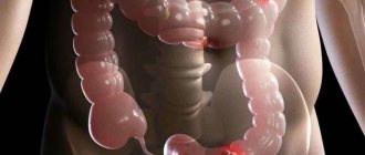
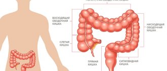
Intestinal obstruction is a pathological condition leading to disruption of the passage of a bolus of food through the intestines, circulatory disorders, intoxication of the body, and, if the outcome is unfavorable, to necrosis and perforation of sections of the intestinal wall and the death of a person. One of the causes of obstruction is intestinal volvulus - a situation when part of the large or small intestine twists around its own axis or around the axis of the mesentery. Places of torsion become a clamp not only for food mass, but also for nerve fibers, blood and lymphatic vessels.
Causes of volvulus and risk factors
For a turnaround to occur, atypical, force majeure circumstances must arise. These may be internal and external reasons:
- physical: abdominal injury, impact with water, sharp turn of the body, forced unusual position, compression, concussion, lifting heavy objects;
- food: overeating, especially after a long fast, eating unfamiliar exotic foods, large amounts of indigestible fiber in the diet, prolonged hunger;
- organic: adhesions, umbilical hernia, neoplasms, disruption of intestinal innervation due to damage to the central nervous system, pregnancy;
- chemical: lead or drug poisoning.
When analyzing patient data, factors were identified that increase the likelihood of intestinal torsion:
- age over 55 years (volvulus of the sigmoid colon often occurs in patients over 60 years of age, colon and cecum volvulus - after 50 years);
- regular constipation or diarrhea;
- sedentary lifestyle;
- hard physical labor;
- fanatical adherence to various fasting systems for weight loss, irregular nutrition;
- overeating, especially before bed.
Why do intestinal injuries occur?
Experts differentiate the most common causes of damage:
- Subjective characteristics of the body;
- Systematic lifting and carrying of heavy objects or professional loads;
- Malfunction of the intestines, constipation;
- Intense tension of the rectum during childbirth;
- Falling on protruding parts of trees, pipes, fences made of stakes;
- Damage from bone fragments during a pelvic fracture;
- Medical negligence during the use of an endoscope, enema, thermometer;
- Anal sex, use of intimate toys.
- Gunshot wound;
- Air injection;
Main symptoms
Intestinal volvulus is a health and life-threatening condition, so at the first sign of discomfort you should seek medical help.
Symptoms according to the degree of their appearance and increase:
- sharp, cramping pain in the abdomen;
- within 24 hours the pain becomes constant and difficult to bear, and can reach a state of shock;
- asymmetry of abdominal contours, bloating;
- nausea, repeated vomiting (may be with bile, and in severe cases with the smell or admixture of feces);
- weakness, pallor, low blood pressure, rapid pulse;
- headache, dizziness, possible confusion (and other signs of intoxication);
- dehydration (dry and cracked lips, sticky saliva, dull skin);
- no bowel movement.
The pathology develops quickly and the patient's condition rapidly deteriorates. A person will die within 2–3 days if emergency medical care is not provided.
The essence of the phenomenon
Most often, when people hear the diagnosis of “intestinal rupture,” they do not always understand what it is. “Intestinal rupture is a very general and broad concept. Intestinal rupture does not just happen; as a rule, it is an injury. If we talk about intestinal rupture, this is a very rare situation, for example in children. Most likely, this means ruptures that occur as a result of trauma: blunt trauma to the abdomen or penetrating injury to the abdominal cavity,” notes Alexey Stepanov. Read more
The specialist notes that blunt abdominal injuries can occur as a result of the following provoking factors:
- as a consequence of a traffic accident;
- when falling from a height;
- when falling on the handlebars of a bicycle and others.
At the same time, as Alexey Stepanov says, very rarely in these situations, intestinal rupture or separation of a section of intestine from the mesentery occurs. More likely, he says, such damage occurs with a penetrating wound to the abdominal cavity.
Intestinal infarction. Why does the attack occur? More details
How the disease develops
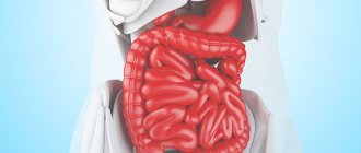
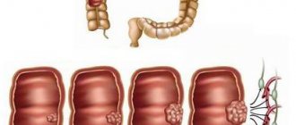
When the intestinal lumen is completely blocked, food masses and gases cannot escape naturally. The intestines contract, trying to move the contents, but cannot. Acute paroxysmal pain occurs. Gas and decay products accumulate, poisoning the nervous system. The compressed section of the intestine begins to swell from fermentation (hence the constant severe pain and visual enlargement of the abdomen). Blood flow and transmission of nerve impulses are disrupted. Peristalsis stops. Since there is no blood supply, the tissue does not receive oxygen and dies. Zones of necrosis are formed. The intestinal wall is corroded, ruptured, and the contents of the pathological focus are poured into the abdominal cavity, disturbing the internal balance in the peritoneum and seeding all organs with bacteria, causing inflammation (peritonitis).
Incomplete closure also brings suffering, but the course is not so rapid and the outcome is often favorable. Volvulus of the small intestine in adults is rare, but it is very severe.
Preventive actions
Of course, intestinal rupture is easier to prevent than to treat and correct. Therefore, notes Alexey Stepanov, the first option for prevention is the prevention of various abdominal injuries. “If we talk about the clinical picture, it is clear that in case of any blunt trauma to the abdomen, it is necessary first of all to contact a surgeon who can examine the patient and, in case of suspected damage to any organ, including the intestines, get his bearings in time, prescribe an appropriate examination and send the patient to the hospital,” emphasizes Alexey Stepanov.
What does the stomach live on? Mood and character are predetermined by intestinal microflora Read more
Diagnostics
A surgeon at the Yusupov Hospital conducts a visual, manual and instrumental examination, collects anamnesis and refers for further examination.
Intestinal obstruction due to intestinal volvulus can be confirmed or refuted using:
- X-ray contrast study with barium;
- ultrasound diagnostics;
- endoscopy;
- laparoscopy;
- laboratory diagnostics.
There are no specialized laboratory tests, but a general analysis and blood biochemistry can show the depth of intoxication and the inflammatory process.
Perforated ulcer of the stomach and duodenum
Scope of diagnostic and treatment assistance at the prehospital stage:
1. The most important task of a doctor who suspects a perforation of a stomach or duodenal ulcer is to organize the promptest possible hospitalization of the patient in the surgical department.
2. Basis for the diagnosis of a perforated ulcer with a typical clinical picture:
a) acute onset; b) “dagger pain” in the stomach; c) pronounced signs of irritation of the peritoneum in the initial period due to exposure to aggressive chemical factors; d) disappearance of hepatic dullness.
3. If the patient’s condition is serious and there are signs of shock, infusion therapy is carried out, vasopressors are administered, and oxygen is inhaled.
4. It is not recommended to administer narcotic analgesics, which can obscure the clinical manifestations of the disease and disorient the hospital surgeon.
Diagnostic protocol in a surgical hospital:
1. In the emergency department, a patient with a suspected perforated ulcer should be examined by a doctor first.
2. Body thermometry is performed, the number of leukocytes in the blood and the necessary laboratory tests are determined (blood type, Rh factor, blood glucose, etc.).
3. In all cases, an ECG is recorded to exclude the abdominal form of myocardial infarction.
4. A plain radiography of the abdominal cavity is performed to detect free gas. If the patient's condition allows, the study is carried out in a vertical position; if not, in a later position.
5. In addition to patients with a confirmed diagnosis of a perforated gastroduodenal ulcer, patients with questionable clinical symptoms are subject to hospitalization in the surgical department.
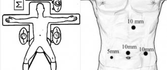
6. In the surgical department, the diagnosis must be completed and the diagnosis of a perforated ulcer confirmed or rejected. Laparoscopy can be used for this. If it is impossible to perform it for one reason or another, it is necessary to resort to diagnostic mid-median laparotomy.
In the surgical department, the patient should be explained the seriousness of the disease, the need for immediate surgical intervention, encouraged, reassured, and obtained consent for the operation. In this case, it is often necessary to tactfully and at the same time persistently convince the patient that there is no other way out of the current situation.
Indications for surgical intervention. The diagnosis of a perforated gastroduodenal ulcer is an absolute indication for emergency surgery. This also applies to covered perforation.
Conservative treatment has to be carried out in those extremely rare cases when the patient categorically refuses surgery. Therapy according to the Taylor method is as follows. Under local anesthesia
A thick probe is inserted into the stomach with a 1% dicaine solution, through which it is emptied of its contents. After removing the thick probe, a thin gastric probe is passed transnasally and connected to a device for continuous aspiration, which is carried out over several days. The patient is placed in the Fowler position. Place an ice pack on your stomach. Correction of water and electrolyte balance, complete parenteral nutrition, detoxification therapy are carried out and massive doses of antibiotics are prescribed for 7-10 days. Before removing the probe, water-soluble contrast is injected through it and X-rays are used to ensure that it does not flow beyond the contours of the stomach or duodenum. Meanwhile, even if the perforation zone of a gastroduodenal ulcer is delimited, the likelihood of the formation of local abscesses in the abdominal cavity is very high. Therefore, this method can be recommended in the most extreme cases, since if it is ineffective, time favorable for surgical intervention will be lost, and the patient will be doomed, despite his belated consent to the operation.
Preoperative preparation. Before surgery, the patient must have a tube inserted into the stomach and its contents aspirated. The bladder is catheterized. The surgical field is hygienically prepared. In the case of a serious condition of the patient caused by diffuse purulent peritonitis, intensive therapy is prescribed and carried out together with the anesthesiologist for 1-2 hours (for more details, see Chapter III).
Anesthesia. The operation is performed under combined endotracheal anesthesia. It is possible to use epidural anesthesia after correction of hypovolemia. In exceptional cases, suturing of the perforation is carried out under local anesthesia.
Access. An upper midline laparotomy is used. In the case of a covered perforated ulcer, with an erroneous incision in the right iliac region, a large tampon is inserted into this wound to drain the abdominal cavity for the entire period of the operation and a superomedial laparotomy is performed. The median wound of the anterior abdominal wall is sutured first at the final stage of the intervention.
Features of surgical intervention. How can perforation of the stomach or duodenum be detected during intraoperative exploration of the abdominal cavity? Quite often, immediately after incision of the peritoneum, a small amount of air comes out of the wound with a characteristic hissing sound. The fluid in the abdominal cavity is usually yellow-green, cloudy, mixed with mucus, and may contain pieces of food. Exudate is evacuated by suction, crumbly food masses are removed with tampons. If the perforation is not immediately detected, the stomach should be pulled to the left, after which the pylorus and duodenum become visible to a sufficient extent. In this case, on the hyperemic anterior wall of the stomach or duodenum, it is possible to identify a whitish, infiltrated area with a diameter of 1 to 3 cm, with a round or oval hole in the middle, with clear, as if stamped, edges, most often about 5 mm in diameter.
It is much more difficult to detect perforation if the ulcer is located low, on the duodenum, or, conversely, high, on the lesser curvature or on the posterior wall of the stomach. It is not easy to navigate when the surgeon encounters pronounced perigastritis, periduodenitis and extensive adhesions. In such cases, identifying the location of the perforation is facilitated by the methodical nature and sequence of the examination.
First, you need to carefully palpate areas that are difficult to examine, moving along the lesser curvature from the cardia to the descending branch of the duodenum. You should palpate not only the lesser curvature of the stomach, but also both of its walls, trying to enclose them between the thumb and index finger. The ulcer area is defined as a dense, rigid infiltration of the gastrointestinal wall.
Secondly, after the surgeon has found the infiltrate, but has not seen the perforation, you should grab this area with your fingers and try to carefully squeeze out the contents of the stomach or duodenum. In this case, only one drop of content may be released. Having detected inflammatory changes and crepitus in the retroperitoneal space, it is necessary to mobilize the duodenum according to Kocher in order to examine its posterior wall.
Third, when searching for the site of perforation, one should take into account the direction from which the effusion is coming. So, if it comes from the omental (Winslov) foramen, the perforation should be looked for on the posterior wall of the stomach, access to which opens after a wide dissection of the gastrocolic ligament. Every operating surgeon should not forget that
You may encounter cases where two ulcers perforate at the same time: on the anterior and posterior walls of the stomach. In the later stages after perforation, massive deposits of fibrin and accumulations of purulent exudate are found in a variety of places. In such cases, all parts of the abdominal cavity should be systematically examined and sanitized. To do this, the exudate is evacuated by suction, fibrin deposits are removed if possible (with tweezers and a wet swab), and its various parts are repeatedly washed with antiseptic solutions. Without fail, these manipulations must be performed in the subhepatic, right and left subphrenic spaces, lateral canals, and pelvic cavity. After evacuation of pus and initial washing of these areas, it is advisable to insert tampons into them for the period of intervention aimed at eliminating the main pathological process. After its completion, it is necessary to complete the sanitation of the abdominal cavity. The tampons introduced at the first stage of the operation are removed and all affected parts are re-treated. Leaving pus and fibrin films can lead to the formation of abscesses or the persistence and progression of peritonitis. If the surgeon, due to the “neglectedness” of the process, cannot fully sanitize the abdominal cavity during the primary surgical intervention, he should plan to perform a repeat sanitization operation (programmed relaparotomy after 24-48 hours).
After detecting a perforation, the surgeon must decide: whether to perform a gastric resection, suturing the perforation, or excising the ulcer followed by pyloroplasty and vagotomy?
Choosing a method of operation. The type and amount of benefits are determined strictly individually depending on the type of ulcer, the time elapsed since perforation, the severity of peritonitis, the patient’s age, the nature and severity of concomitant pathology, and the technical capabilities of the operating team. There are palliative operations (suturing of a perforated ulcer) and radical (resection of the stomach, excision of an ulcer with vagotomy, etc.). When choosing a method of surgical intervention, it should be borne in mind that the main goal of the operation is to save the patient’s life. Therefore, most patients are indicated for suturing of a perforated ulcer. This operation can be performed by any surgeon; in extreme cases, it can be performed under local anesthesia.
Suturing a perforated ulcer is indicated in the presence of diffuse peritonitis (usually when the perforation is more than 6 hours old), a high degree of surgical risk (severe concomitant diseases, old age), in young patients with a “fresh” ulcer without visual signs of a chronic process and an ulcer history, in the case of perforation of stress symptomatic ulcers.
“Youthful” ulcers after their suturing and antiulcer drug treatment tend to heal and have a disease-free course in 90% of cases. When determining the scope of surgery for perforated gastric ulcers, it should be borne in mind that they, especially in elderly patients, may be malignant. Therefore, whenever possible, gastric resection is advisable. If this is not possible, a biopsy must be taken.
The perforation in the stomach wall is “closed” with two rows of interrupted seromuscular sutures. Each of them is applied in a longitudinal direction to the axis of the stomach (intestines). In this case, a number of sutures are located in the transverse direction, which avoids narrowing of the lumen of the organ.
Perforated ulcers of the pyloroduodenal zone are preferably sutured with a single-row synthetic suture without capturing the mucosa, in the transverse direction, so as not to cause a narrowing of the lumen. If the walls of the ulcer in the circumference of the perforated hole are motionless, loose and applied sutures begin to cut through when tied, they can be reinforced by suturing a strand of the omentum or gastrocolic ligament on the leg.
Sometimes, when cutting sutures, it is necessary to use Polikarpov’s method, who suggested not tightening the edges of the ulcer with sutures, but freely plugging the perforated hole with a strand of omentum on the stem. Using a long thread, this strand is passed into the lumen of the stomach through a perforated hole, and then fixed with the same thread passed through the wall of the stomach back to the serous surface. When tying the ends of the thread, the gland seals the hole tightly. After this, around the ulcer and slightly away from it, the omentum is additionally fixed from the outside with separate sutures.
Retroperitoneal perforations are identified by the presence of air and bile impregnation in the paraduodenal tissue. To suture such an ulcer, preliminary mobilization of the duodenum according to Kocher is necessary. After suturing the perforated ulcer, the tissue is drained from the lumbotomy approach.
If, when an ulcer is perforated, a weakened patient also has pyloric stenosis, suturing the perforated hole must be supplemented with posterior gastroenteroanastomosis. As the experience of surgeons has shown, in this case it is also necessary to perform a vagotomy (from this it is clear that such an intervention cannot be considered optimal; in such situations it is better to perform excision of the ulcer with pyloroplasty (see below).
The final stage of surgery for a perforated ulcer of the stomach or duodenum should be a thorough toilet of the abdominal cavity. The more thoroughly the remnants of gastroduodenal contents and exudate were removed, the easier the postoperative period proceeds and the less opportunity there is for the formation of abscesses in the abdominal cavity.
If at the time of the operation there was a large amount of contents in the abdominal cavity, then, despite careful toileting, it is advisable to drain the abdominal cavity.
Endovideosurgical intervention. With appropriate equipment and qualifications of doctors, laparoscopic suturing of a perforated ulcer is possible. Detection of diffuse peritonitis, inflammatory infiltrate or signs of intra-abdominal abscess is an indication for switching to laparotomy.
The duodenal stump is sutured with a purse-string suture. An anastomosis is performed between the gastric stump and a loop of jejunum, passed behind the transverse colon through a “window” into the mesocolon.
Gastric resection is indicated in cases of chronic, callous gastric ulcers (especially if their malignancy is suspected), as well as in cases of decompensated pyloroduodenal stenosis. This operation is possible under the following conditions: 1) the absence of diffuse fibrinous-purulent peritonitis, which develops 6-12 hours after perforation; 2) the patient’s age is less than 60-65 years and the absence of severe concomitant diseases; 3) sufficient qualifications of the surgeon and the availability of conditions to perform this technically complex operation.
Resection is carried out, as a rule, according to the Billroth II method, as modified by Hoffmeister-Finsterer, and in particularly favorable conditions - according to the Billroth I method. For low duodenal ulcers, technical difficulties in processing the duodenal stump, it is advisable to perform an anastomosis according to Roux. Unimpeded evacuation of the contents of the duodenum avoids the insolvency of its stump. The technique of gastric resection is described in detail in special manuals and monographs. Here I just want to mention that it is preferable to apply a gastroenteroanastomosis with a single-row serous-muscular intranodular suture (Fig. 9.5), for good comparison and tissue regeneration. This avoids the development of anastomositis.
Excision of a perforated ulcer with pyloroplasty and vagotomy. Indicated for perforated ulcer of the anterior wall of the duodenal bulb without significant inflammatory infiltrate. The operation is performed under the same conditions as gastrectomy.
The operation is as follows. Two holders are placed on the edges of the duodenal ulcer so that they can stretch the intestine in the transverse direction. The ulcer is excised within healthy tissues along with the pylorus, in the form of a diamond, the length of which is directed along the axis of the stomach and duodenum. By pulling the holders, the defect in the duodenum is sutured in the transverse direction with a one- or two-layer suture, thus performing pyloroplasty according to Heineke-Mikulich. When perforation is combined with stenosis of the gastric outlet, the most adequate drainage will be provided by Finney pyloroplasty.
After sanitation of the abdominal cavity, vagotomy is performed. In conditions of emergency surgery, preference should be given to the most technically simple method - truncal vagotomy.
When perforation and bleeding are combined, a more reliable remedy is excision of the bleeding ulcer (or gastrectomy).
Pyloroantrumectomy with truncal vagotomy. Indicated for patients with duodenostasis (sharply dilated and atonic duodenum) or in the case of a combined form of peptic ulcer, when perforation of the duodenal ulcer and chronic gastric ulcer are detected.
Selective proximal vagotomy with suturing of a perforated ulcer is performed in young and middle-aged patients in the absence of peritonitis and gross cicatricial deformation of the pylorus and duodenum. This operation has limited use in emergency surgery.
Completing the operation. Carry out thorough sanitation and, if necessary, drainage of the abdominal cavity. In some situations, it is rational to install two tubes: one for nutrition (it is inserted into the jejunum), the other into the stomach for decompression.
Postoperative period. The experience of many surgeons convincingly shows the benefits of active management of patients after surgery. It includes rapid activation of the patient, breathing and therapeutic exercises and early, nutritious nutrition, which prevents the development of complications and accelerates regeneration processes.
Of the postoperative complications, bronchopneumonia is in first place in terms of frequency of occurrence, purulent complications are in second place, and disturbances in the evacuation of food from the stomach are in third place.
Subdiaphragmatic, subhepatic, interintestinal and Douglas pouch abscesses are complications often associated with insufficiently thorough toileting of the abdominal cavity during surgery. The clinical picture and diagnosis of these abscesses have been described in detail previously. We only emphasize that it is necessary to pay attention to the appearance of abdominal pain, persistent paresis of the gastrointestinal tract and control the nature of the temperature curve, pulse rate, and changes in the leukocyte formula.
Peritonitis that occurs in the postoperative period is usually associated with failure of the sutures after suturing the perforation or resection of the stomach and requires urgent reoperation. It should be noted that although the failure of the sutures is accompanied by repeated release of gas into the free abdominal cavity, its detection during X-ray examination at this stage loses its significance, since after laparotomy air is detected in the abdominal cavity for more than 10 days.
A more valuable diagnostic technique is to give the patient 1-2 sips of water-soluble contrast. If it extends beyond the gastrointestinal tract, it indicates failure of the sutures at the site of suturing a gastroduodenal ulcer or gastroenteroanastomosis.
It is impossible to ascertain the failure of the sutures of the duodenal stump in this way, since during resection using the Hoffmeister-Finsterer method, the contrast agent from the stomach does not enter the duodenal stump. In such cases, the presence of incompetence of the sutures of the duodenal stump will be indicated by a sharp pain syndrome, peritonitis and an increase in the amount of free gas when re-examined after 40-60 minutes.
Impaired gastric emptying in the postoperative period is manifested by regurgitation and vomiting. It may be due to the functional state of the gastrointestinal tract or be of a mechanical nature. For diagnostic and therapeutic purposes, in these cases, the introduction of a thin probe into the stomach and evacuation of its contents is indicated. At the same time, an active fight against postoperative intestinal paresis should be waged. The patient should be on parenteral nutrition and receive sufficient fluids, proteins and electrolytes.
If, after 5-7 days of conservative treatment, despite the elimination of intestinal paresis, the symptoms of stagnation in the stomach do not decrease, it is necessary to perform a gastroscopy to exclude a mechanical obstruction and decide on a repeat operation.
Mechanical reasons may be the following: 1) when suturing an ulcer: a) narrowing of the pyloric area - as a defect in the surgical technique, b) pronounced perigastritis and periduodenitis; 2) during resection of the stomach: a) narrowing of the anastomosis due to swelling of the walls and subsequent scarring, b) narrowing of the anastomosis due to inflammation and scarring of the mesentery of the transverse colon, c) flow of contents into the afferent loop with compression of the efferent loop, d) improper fixation of the gastric stump in the window mesentery of the transverse colon.
Outcomes. The main causes of mortality in perforated gastroduodenal ulcers are peritonitis, postoperative pneumonia and severe concomitant diseases. An unfavorable outcome is most often a consequence of the patient’s late seeking of medical help and untimely diagnosis. In recent years, in most medical institutions, mortality during surgical treatment of perforated ulcers of the stomach and duodenum has decreased and amounts to 5-7%. Long-term results depend not only on the type of operation, but also on the correctness of the chosen surgical tactics.
Treatment
Since the patient's condition with intestinal obstruction quickly deteriorates, only surgical treatment of intestinal volvulus is used
. The operation is abdominal, the surgeon manually straightens the formed intestinal loop; depending on the stage of development of the process, excises the affected area or the entire swollen area; returns the functional state.
For successful treatment and recovery after surgery, doctors at the Yusupov Hospital use a nasointestinal tube.
It is administered during surgery, after all major surgical procedures. The probe is passed through the esophagus and stomach into the operated section of the intestine and remains there until innervation, motility and blood circulation are restored, maintaining the correct position and free lumen of the intestine. Under medical supervision, it is removed after 3–7 days.
Drug treatment is symptomatic
: pain relief, relief of spasms and urge to vomit, restoration of water-salt balance.
Siphon enemas are also used for therapeutic purposes.
Not to be confused with perforation
It also happens that people equate concepts such as intestinal rupture and perforation. “No, intestinal perforation is a different situation, and it cannot be called an intestinal rupture. Intestinal perforation occurs with a variety of inflammatory lesions of the intestine, it happens with intestinal necrosis, as a result of the pathology that we will talk about later. For example, as a result of intestinal infarction. But these conditions can hardly be called intestinal rupture. These are local intestinal defects that occur against the background of an altered intestinal wall, as a result of any disease, including intestinal infarction, for example, with mesenteric thrombosis,” notes the surgeon.
How does an anal fissure appear?
The main symptom is pain, so it is difficult to ignore an anal fissure. Painful sensations can be of varying intensity, most often intensify during defecation, and gradually subside after it. The pain can be so severe that it makes you afraid to go to the toilet. Sometimes people with an anal fissure stop eating food because of this fear, hoping that this will delay the moment of defecation. This tactic often backfires.
In some situations, if you do not contact a coloproctologist for treatment for a long time, the intensity of the pain becomes so severe that it becomes painful to sit and walk. Most often this happens if an infection occurs.
There may be a feeling of “stiffness” in the anus, a foreign body in the anus.
Another equally frightening symptom is bleeding. Blood from an anal fissure is often bright red in color, may come out in drops or simply leave red marks on toilet paper. Sometimes traces of such blood can even be seen on underwear.
The appearance of blood, pain and any other unpleasant sensations in the anus should alert you. If you have the complaints noted above, you should immediately contact a specialist, since the sooner treatment is started, the faster it will be possible to get rid of the anal fissure completely.
What is required for differential diagnosis of this disease?
A fissure can occur with inflammatory diseases of the colon, such as Crohn's disease or ulcerative colitis. Infectious diseases (syphilis, HIV, herpetic infection, etc.) can also contribute to the development of anal fissure. Tumor diseases such as cancer, melanoma, leukemia can manifest themselves in the same way as an anal fissure. The patient will experience pain in the anus and bleeding from the anus.
Rice. 8. Cancer of the anal canal with transition to the perianal skin
Iatrogenesis
Ruptures may occur due to intraluminal endoscopic intervention in the colon. The complication most often occurs due to incorrect and abrupt insertion of the colonoscope. The risk increases if the patient suffers from serious bowel disease. If all safety rules are followed during an endoscopic examination, the risk of injury is minimal.
What tests do you need to undergo to start treatment?
Patients with an anal fissure are advised to undergo a colonoscopy and laboratory tests to clarify the cause of the disease. After all, if the crack is not an independent disease, then treatment of the crack may be ineffective.
There are cases when it is difficult to differentiate a disease without histological examination (taking a section of tissue to determine whether tissue cells are benign or malignant).
At KKMH (Clinic of Coloproctology and Minimally Invasive Surgery) you can undergo a full range of examinations that will help establish an accurate diagnosis.
A colonoscopy can be performed the next day after visiting a coloproctologist.
- Why not right away? - you ask.
The answer is simple:
— Because the study requires preparation (intestinal cleansing).
We will tell you how and how best to prepare for this study.
Laboratory tests are possible on the day of treatment (not always, because for some indicators you need to be on an empty stomach).
If you have diseases that require referral to related specialists, they will help you choose a doctor and make an appointment with him.