Gastrointestinal stromal tumors (GIST, GIST) appeared a little more than 20 years ago, when the electron microscope was improved, they were isolated from the company of various leiomyomas. GIST is a Russian abbreviation from English. These are tumors of mature age; up to the age of 40 they are very rare; men predominate in the mid-seventh decade of life. It is assumed that the incidence is not increasing, just diagnosis has become better.
Causes and risk factors of GIST
One of the main reasons for the development of the disease is hereditary predisposition. Some scientists point to a connection between the type of genetic mutation and the level of localization of neoplasia.
Gastrointestinal tumors of the intestine are formed when smooth muscle cells are disrupted. This occurs due to gene mutation and leads to the malignant degeneration of one’s own healthy cells and their abnormal growth. The following risk factors can provoke gene mutations:
- chronic stress, nervous tension;
- decreased immunity;
- smoking, alcohol abuse;
- poor nutrition;
- exposure to radiation;
- the presence of chronic diseases of the digestive system and a number of others.
Pathogenesis of the disease
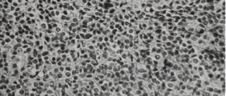
Interstitial cells of Cajal or corresponding progenitor cells are considered as the source of GISTs. 60–70% of GISTs are spindle cell tumors, 20–30% are epithelial or mixed tumors. The main immunohistochemical feature of GIST is the expression of KIT [CD117] and DOC-1, confirmed in 95% of tumors.
Mutations of the KIT gene are detected in 80–85% of all GISTs. In about 10–15% of cases, mutations in PDGF alpha receptors are detected. In the remaining 10%, mutations are not confirmed.
Hereditary predisposition is observed in very rare cases.
Risk factors
To date, no direct risk factors for the development of GIST have been identified.
Symptoms of GIST
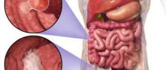
In 20% of cases, abdominal GIST is discovered by chance, during a routine examination or diagnosis of other pathologies. For a long time, the disease is asymptomatic and does not manifest itself in any way even when the tumor is already quite large. 50% of GIST cases are diagnosed at late stages of development due to the absence of specific symptoms and the difficulty of histological verification of the tumor Source: Yakubov Yu.K. Gastrointestinal stromal tumor. Clinical observations / Yu.K. Yakubov [et al.] // Norwegian Journal of Development of the International Science. - 2021. - No. 69. - P. 25-34..
The main reason for the absence of symptoms is the submucosal growth of the tumor, when the tumor spreads out in the wall of the internal organ and bulges into the abdominal cavity. By the time a tumor is detected, approximately every third patient already has metastases in the liver, peritoneum, and sometimes in the lungs. There are no specific symptoms. The clinical picture is similar to that of other gastrointestinal diseases.
The most common clinical manifestations include abdominal pain without clear localization (20–50%), acute bleeding from various parts of the gastrointestinal tract (50%) and intestinal obstruction (10–30%). The spectrum of symptoms also includes an increase in abdominal volume, weakness, anemia, a feeling of rapid satiety, weight loss, nausea, vomiting, palpable tumor masses Source: Kornilova A.G. Gastrointestinal stromal tumors: what's new in therapy? / A.G. Kornilova [and others] // Siberian Journal of Oncology. - 2015. - No. 2. - P. 81-87..
From the appearance of the first unpleasant symptoms to the detection of a tumor, it usually takes about six months.
Not everyone has pain. They are not strong, aching, there is no clinical picture of an “acute abdomen”. GIST does not grow into neighboring organs, like other types of cancer, but simply pushes them away from their anatomical location. In later stages, approximately 50% of patients develop symptoms of gastric bleeding (black loose stools, anemia, general weakness).
Lymphoma
The incidence of lymphoma reaches 20% among all neoplasms of the small intestine. The most common location is the distal jejunum due to the large amount of lymphoid tissue.
Risk factors include celiac disease, Crohn's disease, systemic lupus erythematosus, a weakened immune system, chemotherapy, or a history of other lymphoma.
The typical picture of small bowel lymphoma is wall thickening due to an infiltrating mass formation with aneurysmal dilatation of the lumen without obstruction. Aneurysmal expansion is caused by destruction of the intestinal wall and Auerbach plexus.
Lymphoma of the terminal jejunum
The images show a typical picture. Uneven thickening of the walls of the terminal ileum with aneurysmal dilatation of the lumen.
Less typical manifestations such as an intraluminal polyp or large eccentric mass with extension into surrounding tissue, sometimes with ulceration and fistula formation, occur.
As mentioned earlier, large adenocarcinomas and lymphomas can present a similar pattern. Marked enlargement of mesenteric or retroperitoneal lymph nodes and splenomegaly are signs that allow the diagnosis of lymphoma. Infiltration of mesenteric tissue contributes to the diagnosis of adenocarcinoma.
Lymphoma of the proximal jejunum
Here is a typical presentation of lymphoma with significant thickening of the proximal jejunal wall with accumulation of fluorodesoglycol (FDG). Expansion of the lumen in the tumor area and prestenotic expansion of the duodenum (red arrow).
First study the image, paying special attention to the first drawing.
Then continue reading.
Data received:
Decreased folding in the jejunum with increased folding in the ileum (jejunoileal reversed fold pattern) indicates celiac disease. Ileal intussusception (yellow arrow) in a patient with multifocal small bowel lymphoma (not all changes are shown here). Mesenteric lymphadenopathy (red arrows).
Enteropathic type T-cell lymphoma (EATL) A tumor of the jejunum with unclear contours and dilation of the lumen of the jejunum. The mesentery is infiltrated. Pathomorphologically, T-cell lymphoma of the enteropathic type was detected against the background of celiac disease.
This is enteropathic type associated T-cell lymphoma or EATL. This type of lymphoma affects the small intestine in patients with celiac disease.
This image is the following example of T-cell lymphoma in the setting of celiac disease.
Classification of gastrointestinal stromal tumors of the gastrointestinal tract
According to the International Classification of Gastrointestinal Tumors, 3rd revision, the following are distinguished:
- benign GIST;
- GIST with unspecified malignant potential;
- malignant GIST.
According to the international histological classification, the following types of GIST are distinguished:
- Spindle cell tumors (70%): sclerosing;
- sarcomatous;
- hypercellular;
- palisade-vacuolated.
- sclerosing, with syncytial structure;
Rare types of GIST include pleomorphic, signet ring cell, mesothelioma-like and oncocytic. Source: Kornilova A.G. Gastrointestinal stromal tumors: what's new in therapy? / A.G. Kornilova [and others] // Siberian Journal of Oncology. - 2015. - No. 2. - P. 81-87..
Depending on molecular genetic factors, they are distinguished Source: Yakubov Yu.K. Gastrointestinal stromal tumor. Clinical observations / Yu.K. Yakubov [et al.] // Norwegian Journal of Development of the International Science. - 2022. - No. 69. - P. 25-34.:
- GIST with c-KIT gene mutations. It has various localizations, occurs in most cases (85%), and is solitary in nature.
- GIST with mutations of the PDGFRa gene. It usually affects the stomach, reaches a gigantic size, has a favorable course (5-8%), and is solitary in nature.
- The familial form of GIST is a manifestation of hereditary mutations of the c-KIT or PDGFRa gene. Primary multiple tumors occur.
- GIST in hereditary syndromes in pediatrics, such as multiple GIST associated with neurofibromatosis type 1, in which there is an NF gene mutation not associated with mutations in the c-KIT or PDGFRa genes, and GIST in children with Carney's triad: gastric GIST, paraganglioma, chondroma lung not associated with mutations of the c-KIT or PDGFRa genes.
Lipoma
Well-circumscribed intraluminal tumor consisting of adipose tissue. Liposarcoma of the small intestine is extremely rare.
CT scan shows the formation of fatty density in the duodenojejunal flexure area. Low MR signal intensity from a mass on T2 fatsat (lower right image). Endoscopic picture of a lipoma (top right image).
Stages
By identifying the stage of the disease, specialists assess the extent of the spread of the tumor process, which is identified as a result of diagnostic studies. It is extremely important to correctly determine the stage of GIST, since the choice of treatment tactics, health and life of the patient depend on this.
For staging GISTs, the TNM system developed by the International Union of Cancer is used. The letters in the name of the classification mean different criteria by which the growth of a tumor is assessed. Staging is necessary for a clear assessment of the prognosis and selection of the optimal method of therapy.
The most important factors for prognosis in GIST are the location, size of the neoplasm, and the mitotic index.
Symbol T (assess the size of the tumor):
- T1 - in its greatest dimension, the neoplasm is no more than two centimeters in size;
- T2 - neoplasm more than two centimeters, but not more than five centimeters in the greatest dimension;
- T3 - the tumor is more than five centimeters in size, but not more than ten centimeters in its greatest dimension;
- T4 - the neoplasm in its greatest dimension is more than ten centimeters in size.
Symbol N (assess the absence or presence of metastases in the lymph nodes):
- N0 - no metastases;
- N1 - the patient has metastases in regional lymph nodes.
Symbol M (assess the presence of distant metastases to other organs):
- M0 - no metastases;
- M1 - there are distant metastases.
In addition to the TNM system, when determining the stage of GIST, the localization of the neoplasm and the mitotic index (low or high) are taken into account. When the tumor is localized in the stomach, the prognosis is more favorable, therefore all GISTs are further divided into two groups - extragastric and gastric. The distribution by stage for each of these groups is carried out separately.
Intestinal polyposis
Intestinal polyposis falls into the following main categories: familial adenomatous polyposis (eg, Gardner's syndrome), hamartomatous polyposis (eg, Peutz-Jeghers syndrome), and other/other rarer polyposis syndromes. Patients with these syndromes often have multiple small intestinal polyps. Large polyps can, becoming malignant, simulate a primary tumor of the small intestine.
A patient with Peutz-Jeghers syndrome with an ileal polyp causing intestinal obstruction.
The images show a patient with Peutz-Jeghers syndrome with multiple jejunal polyps.
A large polyp in this patient was removed endoscopically.
Diagnosis of GIST
The diagnosis is made based on the results of the anamnesis, examination of the patient, clinical picture, laboratory and instrumental studies.
Basic methods for diagnosing GIST:
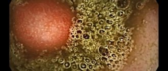
- Esophagogastroduodenoscopy (EGDS)
. An endoscope is inserted into the patient through the mouth, with the help of which the esophagus, duodenum, and stomach are examined. If a neoplasm is detected, then simultaneously with the diagnosis, a small piece of the tumor is taken (biopsy) for histological examination. - Computed tomography (CT)
using a contrast agent taken orally or injected intravenously. Using this study, the localization of GIST on CT, the size of the tumor, its prevalence, and the presence of metastases are determined. - Endosonography
. This is an ultrasound examination performed through an endoscope. With its help, you can accurately determine the location of the tumor and identify its nature. This data is needed to determine the tactics of the operation. - MRI
. Typically, this study is prescribed if the tumor is located low.
If the presence of metastases is suspected, the patient may be prescribed radiography, CT of the chest, radiography of the spine, scintigraphy of skeletal bones, and other studies.
If possible, use PET-CT
. Using this diagnostic method, you can accurately determine the extent of GIST prevalence, as well as small metastases that are not visible during other studies.
The final diagnosis is made based on immunochemical and histological examination of tissue samples
tumors taken during endoscopy. In addition, differential diagnosis with other neoplasms of the digestive system is carried out.
Crohn's disease
Thickening of the walls of the small intestine due to inflammation or infection must be differentiated from malignancy. The hallmarks of the inflammatory process (Crohn's disease), with the exception of proliferation with coarse trabeculations and excess visceral fat around the small and large intestine (creeping fat sign), are ulceration and hypervascularity of the vascular arcades of the adjacent mesentery (comb sign).
There is a clear connection between Crohn's disease and adenocarcinoma of the small intestine.
Preoperative differential diagnosis of these diseases is challenging due to the lack of characteristic imaging features.
An indicator of malignancy in small bowel obstruction is the lack of response to drug therapy.
History of resection of the cecum and jejunum. Local thickening of the jejunal wall with contrast enhancement in Crohn's disease.
Multifocal lesion in Crohn's disease (marked with arrows).
Crohn's disease in the active stage. An extended segment of jejunum with thickened walls, a comb sign, and transmural contrast enhancement.
Treatment methods for gastrointestinal stromal tumor
The main treatment for this type of tumor is surgery
. The extent of surgical intervention depends on the location and extent of spread of the tumor. In most cases, the tumor is radically removed along with several centimeters of surrounding healthy tissue.
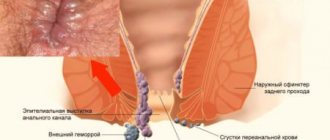
The removed tumor is urgently sent for histology. If cancer cells are detected, the affected area is excised along the incision line. With this disease, metastases to the lymph nodes are extremely rare, so lymph nodes are not removed (except for rectal GIST, which metastasizes to the lymph nodes in 30% of cases).
In the presence of single metastases in the liver, their surgical removal or radiofrequency thermal ablation is indicated.
If the tumor is inoperable, the patient is prescribed chemotherapy
, after which a re-examination is carried out. If the tumor has shrunk to the desired size, then surgery is performed. In all other cases, the course of chemotherapy is continued.
Hemangioma
Most interstitial hemangiomas are localized in the jejunum.
They can be pedunculated or broad-based, and are characterized by lacunar contrast enhancement in the arterial phase, homogeneous in the delayed phase.
Pathologically confirmed hemangioma: coronal post-contrast T1 FS and coronal T2 images show a well-enhancing, circumscribed endophytic lesion.
Prognosis for GIST
The prognosis for gastric GIST and intestinal tumors depends on the size, location and extent of the tumor. The average five-year survival rate for GIS is 48%. Half of the patients survive up to five years from the date of surgery. If the tumor is larger than 10 centimeters, the survival rate drops to 20%.
GIST has a high probability of recurrence. Approximately 80% of patients experience relapses of the disease within two years after surgery. For inoperable tumors, the average life expectancy is 10-21 months.
Desmoid
Desmoid (aggressive fibromatosis) is a rare, benign connective tissue tumor. It is a primary tumor of the mesentery and can simulate malignant tumors of the intestine and mesentery. Desmoid may occur sporadically but may be a component of Gardner's syndrome. Often there is a history of abdominal surgery. Desmoid is a tumor that does not metastasize, but is prone to recurrence. Non-radical surgical treatment contributes to a high relapse rate.
Desmoid in the mesentery usually shows minimal contrast enhancement. The vessels of the small intestine and mesentery move apart or become overgrown with desmoid. Because this tumor is very dense, percutaneous biopsy may be difficult.
Prevention of GIST
The main objective of surveillance is early diagnosis of the disease, which significantly facilitates subsequent treatment and improves the prognosis. After surgery to remove a tumor, you must visit a doctor for a routine examination. In the first two years, this should be done every 3-6 months. After 3-5 years, you need to visit a specialist every 6-12 months. After 5 years from the date of surgery, it is enough to visit the doctor once a year or when health problems arise.
A routine examination usually includes the following steps:
- history taking, physical examination;
- Abdominal ultrasound - performed every 3-6 months;
- colonoscopy or FGDS - performed every 3-6 months;
- X-ray of the chest organs (once a year);
- CT scan of the pelvic and abdominal organs using a contrast agent is performed every 6-12 months.
The exact timing is determined by the attending physician depending on the risk of disease progression.
Sources:
- Bredikhina E.V. Diagnosis and treatment of gastrointestinal stromal tumors (literature review) / E.V. Bredikhina, E.M. Bredikhin // Medical news. - 2022. - No. 2. - P. 4-7.
- Ivanov Yu.V. Features of the clinical course, diagnosis and treatment of gastrointestinal stromal tumors / Yu.V. Ivanov // Clinical practice. - 2012. - No. 1. - P. 59-64.
- Klimentov M.N. Gastrointestinal stromal tumor (clinical observation) / M.N. Klimentov [and others] // Academy. — 2016.
- Kopp M.V. Multidisciplinary approach in the diagnosis and treatment of gastrointestinal stromal tumors / M.V. Kopp, I.A. Koroleva // Malignant tumors. — 2013.
- Kornilova A.G. Gastrointestinal stromal tumors: what's new in therapy? / A.G. Kornilova [and others] // Siberian Journal of Oncology. - 2015. - No. 2. - P. 81-87.
- Kotlyarov P.M. Radiation diagnostics of gastrointestinal stromal tumors / P.M. Kotlyarov [and others] // Bulletin of the Russian Scientific Center of X-ray Radiology of the Ministry of Health of Russia. — 2012.
- Mazurin V.S. Gastrointestinal stromal tumors. Standards of diagnosis and treatment / V.S. Mazurin [etc.] // Almanac of Clinical Medicine. — 2007.
- Snigur P.V. Gastrointestinal autonomic neurogenic tumor of the stomach / P.V. Snigur [and others] // Bulletin of the Russian Cancer Research Center named after. N. N. Blokhin RAMS. — 2005.
- Yakubov Yu.K. Gastrointestinal stromal tumor. Clinical observations / Yu.K. Yakubov [et al.] // Norwegian Journal of Development of the International Science. - 2022. - No. 69. - P. 25-34.
Information verified by an expert
Mikhailov Alexey Gennadievich operating oncologist, doctor of the highest qualification category, Ph.D. experience: 21 years
The information in this article is provided for reference purposes and does not replace advice from a qualified professional. Don't self-medicate! At the first signs of illness, you should consult a doctor.
Typhlitis
Target symptom in a patient with neutropenic enterocolitis and sepsis.
Neutropenic enterocolitis is a potentially life-threatening condition characterized by an inflammatory process that can progress to necrosis, is more common in patients with leukemia and is manifested by damage to the mucous membrane during cytostatic therapy (8). "Typhlitis" (from the Greek "typhlon," cecum) is a neutropenic enterocolitis in the ileocecal region; we prefer the broader term, "neutropenic enterocolitis", since other parts of the small/large intestine are often also involved (8).