Colon polyp - symptoms and treatment
The main methods for diagnosing colon polyps are stool occult blood testing and colonoscopy.
Determination of occult blood in feces. The study is carried out as part of colorectal cancer screening and medical examination of the population as a whole. This is the safest and simplest laboratory method for diagnosing colon polyps, which is based on the determination of hemoglobin in stool. Even minimal hemoglobin concentrations may indicate hidden, clinically not manifested bleeding from the gastrointestinal tract.
Using this method, approximately every third polyp less than 1.0 cm and every second one more than 1.0 cm are diagnosed. In 80% of cases, a negative result indicates that there really is no hidden bleeding [13].
Because the test can produce false-positive and false-negative results, it may not be sufficient to make a diagnosis. To confirm or refute the diagnosis, you need to perform a colonoscopy.
Colonoscopy. This is the most informative method for diagnosing colon polyps. It is recommended for everyone to do it at the age of 45, and if there are risk factors (polyposis, colorectal cancer in relatives) - earlier. This is an instrumental method in which the doctor examines the mucous membrane of the colon in real time using a flexible endoscope. During a colonoscopy, you can examine the pathological formation in detail, take material for histological examination, or completely remove the tumor.
The study is performed after thorough cleaning of the intestine from the contents. You should follow a low-fiber diet 3-5 days before the test. This means that you need to exclude products of plant origin: fruits, berries, vegetables, herbs, cereals, bran. You should also use laxatives. It is advisable to use large or small volume preparations (4 or 2 liters of solution) based on polyethylene glycol. The regimen can be two-stage or one-stage:
- With a two-stage regimen, the dose of the drug is divided into two parts, and the patient takes them in turn: the evening before the test and the morning of the day, for example, 2 liters of a large volume solution or 1 liter of a small volume.
- The morning one-stage regimen consists of sequentially taking the entire dose of the drug solution on the day of the study.
In 95% of cases, one of these regimens is enough to cleanse the intestinal mucosa. You just have to take into account that the two-stage regimen is easier to tolerate by patients, since you need to take a smaller volume of the drug at a time.
Complications of colonoscopy include bleeding and perforation, i.e. disruption of the integrity of the intestine. However, they occur extremely rarely: in asymptomatic patients undergoing screening colonoscopy, complications are observed in 0.007-0.008% of cases [18].
Modern technologies used in endoscopy make it possible to examine the mucous membrane in various lighting spectra, with digital image processing and optical magnification. Due to this, during the study it is possible to determine the histological structure of the polyp with an accuracy of 85.5% [19].
Sigmoidoscopy is a study in which the doctor examines not the entire intestine, as with colonoscopy, but only its initial sections: the rectum and sigmoid colon. Has the same accuracy as a colonoscopy. This method is rarely used due to the widespread use of colonoscopy.
If it is not possible to perform a colonoscopy, then other diagnostic methods can be used:
- CT colonography is an X-ray method for examining the colon, which allows you to identify space-occupying formations. To prepare for the study, you need to clear the intestines of contents with enemas or laxatives to clean water. With CT colonography, the intestinal lumen is filled with air or carbon dioxide, computed tomography is performed, followed by processing of the resulting images [13].
- Irrigoscopy is an X-ray examination of the colon, which is performed after cleansing the intestine, followed by rectal administration of a contrast agent and air (double contrast). This method makes it possible to detect 60–80% of polyps less than 1.0 cm and 75–90% of polyps more than 1.0 cm. Disadvantages of radiological methods: low detection of formations less than 1 cm, radiation exposure and the impossibility of taking a biopsy [15].
- Capsule endoscopy of the colon is a method of diagnosing pathology of the small and large intestine using a capsule-shaped device that the patient takes orally. Passing through all parts of the gastrointestinal tract, the device conducts continuous video recording. The endoscopist subsequently reviews and interprets this recording [16].
Causes of the disease
Tubular adenoma of the rectum
Tubular adenoma is believed to form as a result of an abnormal process of cell proliferation and apoptosis. The process of cell growth is not limited to the intestinal walls, so the tumor grows towards the intestinal lumen and takes on the appearance of a polyp.
Many studies have shown a significant risk of an adenoma developing into rectal carcinoma as it grows. Thus, it is believed that such polyps can have malignant degeneration within 4 years after their formation. Also, an increase in the number of polyps significantly increases the risk of cancer.
Possible reasons for formation:
- Genetic factors. Relatives of patients with polyps have an increased risk of developing carcinoma, so regular screening is important in this case.
- Lifestyle and diet. Products and auxiliary substances that prevent the growth of adenoma include dietary fiber, plant components, carbohydrates and folate. Excess fat and alcoholic beverages in the diet increases the risk of developing adenoma. There is also a link between smoking and the incidence of polyps in patients under 60 years of age.
- Acromegaly. Patients with this disease have an increased risk of developing adenomas and rectal cancer.
- Streptococcus bovis infection and bacteremia. These conditions are also associated with a high risk of tubular adenoma and carcinoma in patients.
- Atherosclerosis and high cholesterol. Data from many studies indicate a high risk of developing polyps in atherosclerotic pathology of intestinal vessels.
- Inflammatory bowel diseases. Patients suffering from irritable bowel syndrome and other chronic inflammations are at significant risk of developing adenomatous rectal polyps. The risk of malignant degeneration of such polyps also increases.
- History of breast cancer.
- Consequences of cholecystectomy.
- Obesity and type 2 diabetes.
Molecular studies of rectal adenoma indicate high activity of certain oncogenes. The progressive accumulation of multiple genetic mutations leads to the degeneration of the normal intestinal mucosa into an adenoma.
Recent research has helped identify a number of genes and their mutations responsible for the occurrence of the disease.
Complications
The most dangerous complication of polyps is malignant degeneration of the polyp cells. The likelihood of colon cancer depends on:
- size (the larger the polyp, the greater the risk);
- type of neoplasm (adenomatous and serrated polyps are more likely to degenerate);
- time of detection (the earlier polyps are detected, the less the threat).
Fortunately, polyps grow slowly. In most cases, colon cancer begins to develop 10 years after the formation of a small polyp. The exception is hereditary diseases, in which malignancy occurs much faster.
Treatment
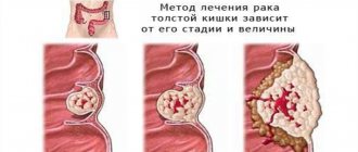
The main treatment method is removal of the tubular adenoma. Sometimes it is also necessary to remove part of the affected intestine.
Possible surgical options:
- Removal of individual intestinal polyps (polypectomy). The indication for such an operation is a polyp size of more than 1 cm. The advantage of such an operation is rapid rehabilitation.
- Laparoscopic removal of polyps is a minimally invasive technique that allows you to quickly and safely remove rectal adenoma.
- Removal of part of the colon and rectum. Such treatment is required in rare cases with severe disease.
Before choosing a surgical technique, a thorough endoscopic and histological examination is performed. If malignant degeneration of the polyp is confirmed, more extensive surgery may be required to completely remove neoplastic cells.
This video will show you how to remove a tubular adenoma of the rectum:
What are intestinal polyps?
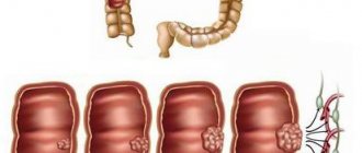
Intestinal polyps are small benign neoplasms that grow asymptomatically on the inner (mucous) lining of the intestine.
Polyps of the large intestine are the most common. This is a fairly common disease, affecting 15-20% of people. The size of polyps is usually less than 1 cm, but can reach several centimeters. They grow alone or in groups. Some look like small bumps, others have a thick or thin stalk with a seal in the shape of a mushroom or a bunch of grapes. The polyps themselves are benign formations that rarely worsen a person’s well-being. But they can transform into malignant tumors that are difficult to treat. Therefore, when polyps are identified, they are recommended to be removed.
The diagnosis of intestinal polyps can be made to people of any age, gender, or race. It is found somewhat more often in men, and the most typical age of patients is 50 years and older. People of the Negroid race are more prone to the formation of polyps and their malignant degeneration than Caucasians.
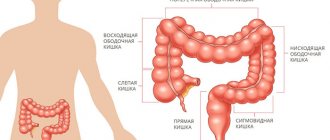
Figure 1. Polyps in the descending colon. Photo: freepik.com
Symptoms
In the early stages there are practically no symptoms
An asymptomatic course is typical in the early stages of adenoma growth. Often, polyps are discovered accidentally during an intestinal colonoscopy.
Possible symptoms and signs:
- Rectal bleeding. This is a nonspecific symptom that may also indicate rectal cancer, hemorrhoids, and traumatic injury to the intestinal mucosa.
- Change in stool color. The presence of blood in the stool can cause black or crimson-colored stools.
- Intestinal dysfunction. As polyps grow, symptoms such as constipation, diarrhea and flatulence may develop. Diarrhea may appear suddenly and not go away within a week.
- Pain in the rectal area. A large polyp can also cause intestinal obstruction, which increases pain.
- Iron-deficiency anemia. This is an important sign of rectal adenoma, developing against the background of chronic bleeding. Constant bleeding causes iron deficiency in the body, which ultimately makes it difficult for red blood cells to carry oxygen. Anemia causes symptoms such as weakness, fatigue and dizziness.
If you experience bleeding, abdominal pain and black stools, you should immediately consult a doctor. Rectal adenoma rarely causes dangerous bleeding, but there is a possibility of other consequences.
Types of polyps
- adenomatous - the most common, approximately 2/3 of all neoplasms belong to this group. In some cases, these polyps degenerate into cancerous tumors or become malignant, as doctors say. Not all of them are capable of malignancy, but if colon cancer comes from a polyp, then an adenomatous polyp is to blame in 2 out of three cases;
- serrated - depending on the size and location, they have a different likelihood of malignancy. Small polyps located in the lower part of the colon (hyperplastic polyps) rarely develop into cancer. But large, flat (sessile) ones, located in the upper part of the intestine, are most often transformed;
- inflammatory ones occur after inflammatory bowel diseases (ulcerative colitis, Crohn's disease). Prone to malignant degeneration.


