- home
- Coloproctology
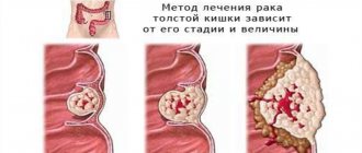
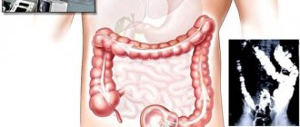
- Colon cancer
- Technique of laparoscopic right hemicolectomy
Fig.1. Layout of the operating team, trocar insertion points.
Fig.2. Intersection of the ileum and transverse colon with the ENDO-GIA-30 device.
Fig.3. Intracorporeal technique for anastomosis formation. Stages of the operation.
Rice. 4. Formation of an end-to-end anastomosis.
Rice. 5. Immersion of the anastomosis into the abdominal cavity.
Technique of laparoscopic right hemicolectomy for colon cancer.
The world's first laparoscopic right hemicolectomy was performed by Jacobs in 1990. The technique of laparoscopic colon resections includes standard steps: intracorporeal mobilization and devascularization, followed by resection and anastomosis.
Most researchers prefer the extracorporeal method of applying an interintestinal anastomosis, which is explained by the simplicity of the technique, convenience and speed of execution. In this case, the mobilized section of the intestine is removed through a minilaparotomy incision 5-6 cm long onto the abdominal wall and anastomosis is performed in the usual way.
A number of authors give preference to the completely intracorporeal technique of intestinal resection and anastomosis formation and believe that only in this case “the basic principles of laparoscopic colon surgery remain intact.” We adhere to the “golden” mean - when, without compromising the quality of the operation, it is possible to perform a complete intracorporeal resection of the right flank, we perform it. In the case of a large tumor, short mesentery, we perform the operation using a combined approach - mobilization of the intestine (mesorectumectomy) laparoscopically, and anastomosis extracorporeally.
Indications for laparoscopic hemicolectomy on the right side are neoplasms of the right flank of the colon, Crohn's disease, and in rare cases, diverticulosis, polyposis, and other diseases.
Watch a video of operations performed by Professor K.V. Puchkov. You can visit the website “Video of operations of the best surgeons in the world.”
Doctor's comment
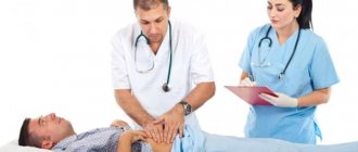
If you are undergoing a hemicolectomy and are experiencing understandable anxiety associated with surgery, you should know that the operation is performed under anesthesia. Our clinic uses only modern techniques and drugs for anesthesia, so patients do not experience pain either during the procedure or after it. In addition, in 90% of cases we manage to do without an intestinal stoma; during the procedure, a colorectal anastomosis is formed, so the function of natural movement of intestinal contents and emptying is preserved. If the optimal solution is the formation of a temporary colostomy, reconstructive surgery is prescribed after 2-3 months. For diseases of the large intestine, both the choice of treatment method and the result depend on how thoroughly the diagnosis is carried out. At our clinic you can undergo a comprehensive examination - in a short time and without tiring queues. Do not delay your visit to the doctor, make an appointment; a timely operation significantly increases the chances of minimally traumatic treatment and a quick recovery.
Head of the surgical service at SwissClinic Konstantin Viktorovich Puchkov
Operation technique
Intracorporeal technique
For benign diseases, the operation begins with mobilization of the cecum and ascending colon along the right lateral canal, pulling the right flank of the colon medially and upward.
Continuing further isolation of the distal part of the ascending and hepatic flexure of the colon, the hepatocolic ligament is isolated and divided using a blunt and sharp method. Then the duodenum and right ureter are retracted. Simultaneously with the omentum, the transverse colon is isolated in the desired area.
The retroperitoneal space is gradually exposed. At this stage, you should be very careful not to damage nearby organs (right ureter, kidney, etc.).
The mesentery of the right flank of the colon is mobilized. The ileocolic, right and descending branches of the middle colic artery are divided in one of the described ways.
The levels of intersection of the ileum and transverse colon are determined, which are identified within the viable areas. The level of anastomosis is determined by several signs, the most important of which is the visually traceable pulsation of the marginal vessels. At this point, the intestine is intersected with the ENDO-GIA-30-60 apparatus (Fig. 2).
Rice. 6. Formation of end-to-side anastomosis.
Fig.7. Formation of ileo-transverse anastomosis using the GIA-55 device.
Fig.8. Stitching with the TA-60 device followed by resection of the specimen.
Rice. 9. Formation of an end-to-side anastomosis using a circular stapler.
Fig. 10. Suturing the stump of the transverse colon with the TA-60 device.
Next, ileo- and colotomies are performed, the jaws of the ENDO-GIA-30(60) device are inserted into the holes, and stitching is performed. The holes are sutured with a hardware or manual continuous suture on an atraumatic needle (Fig. 3.).
The abdominal wall is dissected in the right hypochondrium, and the resected specimen is removed, previously placed in a special plastic container. All ligated and coagulated vessels are carefully examined. After this, the wound and trocar punctures are sutured layer by layer.
Extracorporeal technique
After mobilizing the right flank of the colon, the abdominal wall is incised in the right hypochondrium or along the midline. The mobilized sections of the colon and the distal part of the ileum along with the tumor are removed into the wound. Resection is performed with manual or hardware formation of anastomosis.
Options for extracorporeal anastomosis formation
Hand stitch (end to end)
A 2-row or single-row anastomosis is formed. The inner row (with a 2-row suture) is formed with a continuous suture with an absorbable thread on an atraumatic needle, the outer row with an interrupted gray-serous suture. The discrepancy between the diameters of the lumens of the ileum and transverse colon is eliminated by incising the antimesenteric wall of the ileum in an oblique direction. This type of manual anastomosis requires the least amount of time (Fig. 4). The formed anastomosis is immersed in the abdominal cavity (Fig. 5).
Hand stitch (end to side)
The stump of the transverse colon is immersed with a 2-row suture. A colotomy is made along the tenia, approximately 2 cm long, and the terminal ileum is sutured (Fig. 6).
Hand stitch (side to side)
The stump of the ileum and transverse colon is sutured with a manual blanket suture and additionally immersed with 2 half-purse-string sutures (it is possible to stitch the stump with a linear stapler). The anastomosis is performed at a distance of 4-8 cm from the edge of the stump. It is more advisable to dissect the transverse colon to form an anastomosis along the taenia libera, since the intestinal wall in this place is the most durable. Preference is usually given to a 2-row anastomosis. The first (inner) row is formed with a continuous suture using an absorbable atraumatic thread, the second (outer) row is formed with an interrupted gray-serous suture. After this, the stump of the ileum and transverse colon is fixed to the opposite colon with several gray-serous sutures to prevent intussusception into the anastomotic area with the development of obstruction. At the final stage of the operation, the “window” of the mesentery is carefully sutured.
HARDWARE SEAM (side to side, using linear twisting machines).
The most widely used method is the two-device method. The terminal sections of the ileum and transverse colon are juxtaposed side to side antiperistaltically. The jaws of the GIA-60 device are inserted into their lumen, and suturing is performed while simultaneously crossing the walls (Fig. 7). In the transverse direction, at the intended level of resection, the TA-60(90) device is applied, and simultaneous suturing of the ileum and transverse colon is performed (Fig. 8). The part of the intestine to be removed is cut off along the branch of the device.
Hardware seam (end to side; using linear and circular staplers).
The main part of the circular stapler CEEA-21(25) is inserted into the lumen of the transverse colon; the sharp part perforates the side wall of the transverse colon in the area of the taenia libera at a distance of 8-10 cm from the edge. A purse-string suture is placed on the terminal ileum and tightened after insertion of the head of the CEEA-21 apparatus into the lumen. The head is adapted to the main part of the circular stapler, and stitching is performed (Fig. 9).
After this, the terminal part of the transverse colon is sutured with a TA-60 device (Fig. 7.10).
Recovery
After the operation, the patient is transferred to the hospital for constant monitoring of the progress of recovery.
For the first two days, the patient will be fed through a tube. Then liquid food is allowed. If there are no complications, solid food can be included in the diet 7-8 days after surgery.
Discharge usually occurs 2 weeks after the intervention, but strict dietary restrictions remain. Typically, full adaptation requires at least six months. Often, hemicolectomy is the patient’s only chance for recovery. This intervention falls into the category of complex procedures that require increased attention, experience and skill of the surgeon. Trust your health to professionals - come for an appointment at a medical office.
Features of right hemicolectomy for cancer
The principles of oncological radicalism require maximum removal of the mesentery of the colon, omentum, and retroperitoneal tissue en bloc with the tumor. Paracolic, intermediate, basal and main lymph nodes must be eliminated. Removal of a single block includes the concept of mesocolonectomy - removal of the intestine with fiber and lymph nodes in a single fascial case. All manipulations of instruments with the tumor should be minimized. Prevention of trocar metastases should also be carried out: washing wounds with solutions of antiseptics and cytostatics; performing a minilaparotomy adequate to the size of the tumor; removing the drug in a special plastic bag, etc.
The operation begins not with mobilization of the right flank of the colon along the right lateral canal, but with high ligation of the great vessels. First, the ileocolic artery and vein are isolated and clipped, then the right colic vessels. Then, the parietal peritoneum is dissected in a single block from bottom to top directly behind the dome of the cecum. The volume of lymph node dissection corresponds to the location of the tumor: in case of cancer of the ascending, right flexure and initial part of the transverse colon, para-aortic lymph node dissection is performed, and in case of damage to the cecum, we additionally remove ileal tissue on the right. The peritoneum is dissected along the right lateral canal, and the mesentery of the colon is isolated along the prerenal layer of fascia retroperitonealis, completely removing the prefascial tissue. If tissue dissection is performed in the desired layer, then this stage is almost bloodless. The hepatocolic and gastrocolic ligaments are divided, and the greater omentum is removed. During intraperitoneal resection, the drug is removed in a special plastic container. If an extracorporeal anastomosis method is used, then it is necessary to reliably isolate the wound from contact with the tumor (for this we usually use a sterile plastic bag). In case of a locally advanced tumor, after the end of the operation, the abdominal cavity is washed with an antiseptic or cytostatic solution (5-FU; mitocin, etc.).
K. V. Puchkov: “Oncological disease is not a death sentence”
From our own experience, we were convinced that adequate lymph node dissection is easier to perform when mobilizing the right parts of the colon along the lateral canal from the bottom up, with isolation and skeletonization of the right ureter, aorta and inferior vena cava.
With this method of mobilizing the right flank of the colon, the principles of casing and zonality are fully preserved (mesocolonectomy). Ligation of the great vessels is carried out as high as possible. The intersection of the afferent and efferent colons is also advisable to carry out extracorporeally using stitching devices. Considering the fact that in order to extract the removed drug it is necessary to make an incision adequate to the size of the tumor, performing the final stages is technically simpler and more convenient to perform using extra-abdominal access.
K. V. Puchkov, D. A. Khubezov. Minimally invasive surgery of the colon K. V. Puchkov, D. S. Rodichenko. Hand suture in endoscopic surgery
Why is it better to have a hemicolectomy at the Swiss University Hospital?
- The clinic’s surgeons were among the first to begin performing minimally invasive operations on the intestines; each specialist has performed more than 600 successfully performed surgical interventions.
- During the operation, we use the most modern techniques and equipment, for example, to reduce blood loss, the LigaSure device (USA), ultrasonic scissors, absorbable suture material, modern stitching devices that minimize the risk of complications in the postoperative period, etc.
- Our Center was one of the first to begin performing simultaneous (one-step) operations: in the presence of concomitant pathologies, one anesthesia can get rid of several diseases at once.
- We have the opportunity to undergo a high-quality and comprehensive examination, the clinic is equipped with the most innovative expert-class equipment, and the reception is conducted by experienced specialists of the highest or first category. Every year we consult more than 5,000 people, and more than 5 thousand types of research are available to patients.
List of published works on the topic of laparoscopic right hemilectomy
- Puchkov K., Filimonov V., Titov G., Chubezov D. Laparoscopic technology in coloproctology // Proktologia. - 2001. - Suppl. No. 1. - P.97.
- Puchkov K.V., Khubezov D.A., Khubezov A.T., Titov G.M. The use of laparoscopic access in colorectal cancer surgery // Pacific Med. magazine - 2002. - No. 2 (special issue). — P.64 — 65.
- Puchkov K.V., Khubezov D.A., Khubezov A.T., Titov G.M. On the prospects for the introduction of laparoscopic operations in coloproctology // Specialized medical care. Issue 3. - Ryazan: OKB, 2002. - P. 114 - 115.
- Puchkov K.V., Khubezov D.A., Khubezov A.T. Laparoscopic radical operations for colorectal cancer // Problems of coloproctology. Issue 18. - M., 2002. - P.406 - 409.
- Puchkov K.V., Khubezov D.A., Khubezov A.T., Titov G.M. Laparoscopic symptomatic operations for colorectal cancer // Problems of coloproctology. Issue 18. - M., 2002. - P.414 - 416.
- Puchkov K.V., Khubezov D.A., Khubezov A.T., Yudina E.A. Laparoscopic access in colon cancer surgery. Technique of lymph node dissection // Current issues in coloproctology: abstract. report 1st Congress of Coloproctologists of Russia with international. participation / edited by G.I. Vorobyova, G.P. Kotelnikova, B.N. Zhukova. - Samara: GP: SamSMU, 2003. - P.394 - 395.
- Puchkov K.V., Khubezov D.A., Yudina E.A., Yudin I.V. Boundaries of laparoscopic resection for colon cancer // Current problems of coloproctology: abstract. report scientific conf. with international participation, dedication 40th anniversary of the State Research Center of Coloproctology, Moscow, February 2 - 4. 2005 / ed. G.I. Vorobyova, I.L. Khalifa. - M., 2005. - P.279 - 280.
- Puchkov K.V., Khubezov D.A., Yudina E.A., Yudin I.V. Laparoscopic subtotal colectomy in the treatment of diffuse familial adenomatosis // Current problems of coloproctology: abstract. report scientific conf. with international participation, dedication 40th anniversary of the State Research Center of Coloproctology, Moscow, February 2 - 4. 2005 / ed. G.I. Vorobyova, I.L. Khalifa. - M., 2005. - P. 280 - 281.
- Puchkov K.V., Khubezov D.A.. Minimally invasive colon surgery: a guide for doctors. - M.: Medicine, 2005. - 280 p.
- Puchkov K.V., Khubezov D.A., Yudin I.V. Technical aspects of performing laparoscopic lymph node dissection // Journal. obstetrics and women's diseases. - 2005. - T.54 (special issue). — P.87 — 88.
- Puchkov KV, Khubezov DA, Yudin I. Boundaries of laparoscopic resection at cancer of colon // Proktologia. — Supplement No. 1. - 2006. - P.100.
- Puchkov K.V., Khubezov D.A. Limits of laparoscopic resection for colon cancer // First International. conf. thoraco-abdominal Surgery: Sat. abstract – Moscow, 2008. – P.64.
- Khubezov DA, Puchkov KV, Ogoreltsev AY Laparoscopic right hemicolectomy for colon cancer // Abstracts book of the 17th EAES Congress 2009, 17 - 20 June, 2009, Prague, Czech Republic. – P.155.
- Puchkov K.V., Ogoreltsev A.Yu., Lukanin R.V. Features of laparoscopic lymph node dissection for cancer of the right colon // Russian School of Colorectal Surgery. Abstracts of scientific works. Moscow, March 15, 2010 - P.59.
- Puchkov K.V., Khubezov D.A., Ogoreltsev A.Yu., Lukanin R.V., Andreeva Yu.E. Single-port laparoscopic (SILS) colproctectomy with ileoanal reservoir anastomosis // Scientific-practical. conf. with international participation in “Technology of single laparoscopic access in abdominal surgery.” - M.: Publishing House "MEDPRACTIKA-M", 2011 - pp. 26–28.
- Puchkov K.V., Khubezov D.A., Ogoreltsev A.Yu., Lukanin R.V., Semionkin E.I. Single-port laparoscopic (SILS) colproctectomy with ileoanal reservoir anastomosis // Coloproctology - 2011. - No. 2 (36). - P. 9-12.
- Puchkov K.V., Khubezov D.A., Ogoreltsev A.Yu., Lukanin R.V. Options for laparoscopic lymph node dissection depending on the location of cancer of the right colon // Fifth International Conference. Russian school of colorectal surgery. "Multidisciplinary approach in the treatment of rectal cancer." Conference materials. - Moscow, June 23-24, 2011 - P. 48.
- Puchkov K.V., Khubezov D.A., Ogoreltsev A.Yu., Lukanin R.V., Andreeva Yu.E. Single-port laparoscopic (SILS) colproctectomy with ileoanal pouch anastomosis // Fifth International Conference. Russian school of colorectal surgery. "Multidisciplinary approach in the treatment of rectal cancer." Conference materials. - Moscow, June 23-24, 2011 - P. 48.
- Puchkov K.V., Khubezov D.A., Ogoreltsev A.Yu., Lukanin R.V. Experience of laparoscopic SILS colproctectomy with ileoanal reservoir anastomosis and preventive ileostomy // Materials of the 3rd All-Russian Congress of Coloproctologists, October 12-14. 2011, Belgorod, Coloproctology - 2011. - No. 3 (37). - P. 115.
- Puchkov K.V., Khubezov D.A., Ogoreltsev A.Yu., Lukanin R.V., Puchkov D.K. Selection of the volume of laparoscopic lymph node dissection depending on the location of cancer of the right colon // Almanac of the Institute of Surgery named after. A.V. Vishnevsky. T.7, No. 1 - 2012. “Materials of the XV Congress of the Society of Endoscopic Surgeons of Russia.” – Moscow, 2012. – P. 315-316.
- Puchkov K.V., Khubezov D.A., Puchkov D.K. On the issue of the technique of performing right-sided hemicolectomy for cancer of the right colon // Materials of the IV Congress of Surgeons of Kazakhstan “New Technologies in Surgery”. – Almaty, 2013. – P. 67.
- Puchkov K.V., Khubezov D.A., Puchkov D.K. Results of using the NOSE technique in laparoscopic colon surgery // Endoscopic surgery. – 2014. – T20. - No. 1. – P. 328 – 330.
- Puchkov K.V., Khubezov D.A., Puchkov D.K. Possibilities of using the NOSE technique in laparoscopic colon surgery // Materials of the International Scientific and Practical Conference "Endovideosurgery in a Multidisciplinary Hospital", St. Petersburg: Publishing House "Man" and his health”, 2014 – pp. – 93 – 94. ISBN 978-5-9905495-4-8.
- Puchkov K.V., Khubezov D.A., Puchkov D.K. Results of using the NOSE technique in laparoscopic colon surgery // Almanac of the Institute of Surgery named after. A.V. Vishnevsky. T.10, No. 1 - 2015. “Materials of the XVIII Congress of the Society of Endoscopic Surgeons of Russia.” – Moscow, 2015. – P. 341-342.
- Pat. 2013117186 RF MPK8 A61 B17/02. A method of temporary fixation of abdominal and pelvic organs during laparoscopic operations and a device for its implementation / K.V. Puchkov, V.V. Korennaya, D.K. Puchkov. - No. 2013117186/14; application 04/15/2013; publ. 10/27/2014, Bulletin 30.
Professor K.V. Puchkov performs laparoscopic resection of the rectum with aorto-iliac lymphadenectomy (Moscow, 1st Clinical Hospital of the Administration of the Mayor and Government of Moscow, September 29, 2007) Laparoscopic resection of the rectum (Professor K.V. Puchkov September 18-20, 2013 year in Kiev (Ukraine)