Polyps in the large intestine are benign neoplasms formed from glandular epithelial cells. Normally, tissues from this type of cells line the inside of the intestine, but under the influence of certain factors they begin to grow uncontrollably, forming single or multiple mushroom-shaped, spherical or branched outgrowths directed into the intestinal lumen. In the international classification of diseases ICD-10, colon polyps are designated by code K63.5. (colon polyps).
Most often, the pathology is diagnosed in men and women of the older age category. The greatest surge in incidence is observed after overcoming the 60-year mark. In childhood, polyps are found more often in boys aged 3 to 6 years (in 80% of cases), and most often neoplasms are localized in the distal intestine.
The main danger of polyps in the large intestine is the high risk of malignancy (degeneration of a benign neoplasm into a malignant one). That is why, regardless of what polyps (type, shape and size) are detected, they are considered precancerous neoplasms with varying degrees of malignancy.
Types and sizes of colon polyps
In medicine, a branched classification of polyps of the large intestine has been formulated depending on the cellular structure, appearance, nature and number of tumors. The size of the polyps also plays a role in diagnosis, since this parameter is directly related to the risk of intestinal cancer.
Polyps are divided into 4 types (forms) depending on their structure:
- Adenomatous is the most common and dangerous form of polyps, which can differ in appearance. There are tubular (tubular), tubular-villous and villous. The neoplasms are penetrated by many tiny vessels and often bleed. Regardless of appearance, they have a high level of malignancy (malignancy). It is diagnosed mainly in older people.
- Hyperplastic - a small glandular polyp, slightly rising above the surface of the intestinal mucosa, has a color natural to the surrounding epithelium. It is quite rare to transform into a malignant tumor. Often diagnosed in patients over 40 years of age.
- Hamartomatous - white-pink neoplasms that consist of several types of cells that develop disproportionately. Rarely reaches large sizes, as they grow evenly and very slowly. Such a neoplasm often bleeds and has a low risk of malignancy.
- Inflammatory - a glandular polyp that forms against the background of an acute inflammatory process in the intestine.
Based on the number of neoplasms in the intestine, single, multiple and diffuse (hundreds of rapidly growing and spreading neoplasms) polyps of the large intestine are distinguished. Diffuse and multiple polyps have the greatest tendency to transform into cancerous tumors.
Good to know! Affects the rate of malignancy and the size of polyps. Thus, small formations with a diameter of up to 1 cm have a degree of malignancy from 3 to 9%, while large polyps with a diameter of more than 3 cm transform into a cancerous tumor in 30% of cases.
Causes of rectal polyps
The causes of polyps are ambiguous; provoking factors are:
- psycho-emotional disorders;
- chronic inflammation of the mucous membrane of the intestinal wall;
- hereditary factor;
- bad ecology;
- physical inactivity (low physical activity);
- poor diet, low fiber content in food.
There are also theories that polyps may appear as a result of chronic inflammatory diseases in the colon. The risk of polyp formation increases with diseases such as:
- ulcerative colitis;
- enteritis;
- dysentery;
- haemorrhoids;
- frequent constipation and digestive system disorders;
- hereditary predisposition;
- viral diseases;
- excessive consumption of animal fats;
- smoking and alcohol abuse;
- age.
1 Diagnosis of polyposis in MedicCity
2 Diagnosis of polyposis in MedicCity
3 Diagnosis of polyposis in MedicCity
Symptoms of rectal polyps
There are no characteristic symptoms of rectal polyps (as well as colon and colon polyps). The disease can be asymptomatic for a long time. Symptoms appear if the tumor is injured or an inflammatory process appears at the site of its formation. The following symptoms of rectal polyps are distinguished:
- anal itching;
- frequent urge to go to the toilet, pain during bowel movements, presence of blood and mucus in the stool;
- bleeding when a polyp is injured;
- chills, fever when an inflammatory process occurs;
- prolapse of a pedunculated polyp during defecation (the person feels pain as the polyp is pinched by the sphincter), etc.
There is a direct dependence on the size of the polyps and the severity of the disease. With polyps exceeding 2-3 or more centimeters (for example, with villous adenomas), the patient may experience the following manifestations:
- blood in the stool;
- pathological impurities in the form of mucus;
- stomach ache;
- constipation and a feeling of a foreign body in the anus with the appearance of large polyps in the intestine that interfere with the passage of feces;
- weight loss and exhaustion of the body with pronounced water and electrolyte disturbances (with giant polyps).
If, with such symptoms, the patient does not seek help from a proctologist, then serious complications of polyps may arise. A person becomes weak and his hemoglobin decreases. Infection of polyps leads to the formation of anal fissures and paraproctitis. Rectal polyp disease is a precancerous condition of the intestine and can develop into a malignant tumor.
If you believe strict medical statistics, then every fifth polyp can degenerate into a cancerous tumor. Therefore, mandatory observation and, if necessary, removal of polyps in the rectum are necessary!
1 Anoscope
2 Anoscope
3 Gastroscopy in MedicCity
Causes of colon polyps
Doctors cannot name the exact reasons for the formation of benign tumors in the large intestine. Previously, it was believed that their formation occurs under the influence of bad habits - alcoholism, smoking. However, a considerable number of patients diagnosed with colon polyposis follow the principles of a healthy lifestyle, which raised some doubts about the validity of such judgments.
Recent studies have found that bad habits as such are not the main risk factor. They play the role of a trigger, but in most cases the disease occurs in those whose close relatives suffered or suffer from polyps in various parts of the gastrointestinal tract. In addition, colon polyps threaten people with acute and chronic diseases of the digestive system, excess weight, and chronic stool disorders due to non-compliance with norms and diet.
According to statistics, approximately 50% of men and women over 60 years old have polyps in the large intestine. This indicates that age-related changes are the most common cause of the disease.
Important! Certain types of polyps are formed due to inflammation of the intestinal mucosa.
Risk factors
Risk factors and mechanisms of colon polyps are being actively studied. Among the environmental factors, the nature of nutrition is significant, with a predominance of refined foods in the diet, which contribute to constipation and prolonged stasis of intestinal contents. Dysbacteriosis of the colon has some influence on the occurrence of polyps, reflecting a violation of local and decreased general immunity, contributing to changes in the differentiation and regeneration of mucosal cells. The role of concomitant diseases of the biliary system and impaired production of bile acids, which have a mutagenic effect on the mucous membrane, is also noted as a risk factor. Active chronic inflammation and mucosal dysplasia play a certain role in the occurrence of polyps.
Symptoms and signs of colon polyps
At the initial stages of development, polyps do not appear in any way, and therefore they are discovered by chance when examining the intestines for other diseases. Clinical manifestations of polyposis appear if there are several benign formations or they are more than 1 cm in diameter. In this case, the symptoms can be very diverse and nonspecific. Most often, patients complain of the following phenomena:
- unstable stool with frequent alternation of diarrhea and constipation, sometimes within one day;
- irritation of the mucous membrane of the anus and the skin of the perianal area, which occurs against the background of abundant mucus secretion from the rectum (a symptom characteristic of polyps located in the lower part of the colon);
- scant rectal bleeding that is not accompanied by acute pain in the anus (a symptom characteristic of villous polyps located in the lower part of the large intestine);
If you have these problems, you should urgently contact a gastroenterologist or coloproctologist, since they are the ones who diagnose and treat the disease.
Tubular adenoma of the colon with dysplasia
Each of the identified adenomas, regardless of type and structure, has signs of dysplasia. There are three degrees of dysplasia - mild, moderate and severe (or high). The degree of dysplasia is determined by histological examination of the removed adenoma based on a number of characteristics. Predominantly adenomas with a mild degree of dysplasia are diagnosed. The larger the size of the adenoma and the older the patient, the greater the risk of developing an adenoma with signs of severe dysplasia. The degree of dysplasia is one of the most important factors in the malignancy of an adenoma, along with its size, location, duration of the disease and replacement of the tubular structure with a villous one. An extreme degree of dysplasia is usually equated to cancer in situ. At the same time, epithelial dysplasia itself is not cancer, but since it is essentially a violation of the normal tissue structure of a part of an organ, in this case the colon, it can, with further progression and an increase in its severity, lead to the appearance of tumor cells.
Diagnostics
Very often, the symptomatic manifestations of colon polyps are similar to other diseases of the gastrointestinal tract. When making a diagnosis, differential diagnosis is used, during which the doctor excludes other pathologies.
First of all, the doctor collects anamnesis and conducts a survey of the patient, during which he establishes the characteristics of the patient’s symptoms, diet and gastronomic habits.
Important! To make a correct diagnosis, a specialist will need information about previous diseases and diseases of the colon that immediate relatives have suffered.
Next, the coloproctologist conducts a digital examination of the rectum, during which it is possible to determine the presence of polyps in the area of the rectum remote from the anus, their size, method of attachment to the mucous membrane (pedunculated or wide base), shape, consistency and degree of displacement.
In addition to the listed studies, patients with suspected colon polyposis will have to undergo a number of laboratory tests:
- general blood test (in some situations, low hemoglobin levels indicate polyposis);
- stool occult blood test (often replaced by the Colon-View occult blood screening test).
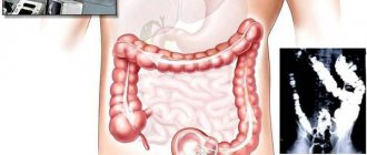
To establish the exact location, size, number and shape of polyps in the colon, instrumental diagnostic methods are used:
- sigmoidoscopy - a visual examination using endoscopic equipment equipped with a fiber optic system, a light source and forceps for taking a biopsy;
- colonoscopy is a method of visual examination of the intestines using a flexible probe (colonoscope) equipped with a camera, a lighting device, an air injection tube and forceps for taking a biopsy;
- irrigoscopy - x-ray examination of the colon using a contrast agent;
- magnetic resonance or computed tomography.
After colonoscopy and sigmoidoscopy, a study of biological materials (parts of the mucous membrane of the colon or the polyp itself) is carried out to determine their cellular structure and the risk of malignancy.
Differential diagnosis
A rectal polyp must be distinguished from other pathologies of the pelvic organs, such as:
- lipoma (can be located in the submucosal layer of the right half of the colon or, if it grows to a large size, throughout the intestine);
- a tumor of non-epetial size (they do not have a stalk);
- large fibroids (growth in the muscle layer, creates difficulties in intestinal patency, is rare);
- angioma, vascular tumors;
- actinomycosis of the colon;
- Crohn's disease (located in the upper parts of the large intestine, can manifest as pseudopolyposis).
To correctly diagnose this disease, it is necessary to conduct a histological analysis.
Treatment methods for colon polyps
Conservative methods of therapy for colon polyps are not used, as they are not effective. Doctors call surgical removal of tumors the only way to eliminate them. Depending on which polyps are detected during diagnosis, the following methods are used:
Surgical colonoscopy is used to remove single and multiple polyps. It is performed using a colonoscope equipped with a loop electrode. During the procedure, a loop is placed on the stalk of the polyp and tightened until it fits as tightly as possible. Then high-frequency currents are passed through the loop, as a result of which the head is cut off and the leg is charred. The resulting wound is cauterized.
If the polyp is located on the mucous membrane of the colon on a broad basis, an endoscopic operation is performed, the so-called endoscopic resection of the mucosa, with coagulation of the base of the wound. The operation is performed using a special electric knife, which is used to resect a section of the mucous membrane along with the polyp. Surgeries for resection of colon polyps can be quite lengthy, the duration depends on the size of the polyp, but they are minimally traumatic and do not require long-term hospitalization. Typically, the hospitalization period is from 1 to 3 days.
Radio wave and laser coagulation. Used to remove one or more polyps. The tumor is exposed to a narrow beam, causing its tissue to die. To prevent bleeding, coagulation of blood vessels is performed. The procedure, like the previous one, is low-traumatic, does not cause side effects and does not require long-term rehabilitation and recovery.
Abdominal surgery. For diffuse familial polyposis, a subtotal or total colectomy is performed, i.e. resection of the colon with the affected area, subsequently an anastomosis is formed.
Immediately after surgery, a gentle diet with limited salt, sour, and spicy foods is recommended. Do not eat too hot or cold foods, mushrooms, fatty meat and fish, fried and smoked foods, alcohol and strong coffee. You will have to stick to the diet for at least 4 weeks.
After removal of types of polyps prone to malignancy (villous, multiple and large), a year after treatment, the patient is shown an endoscopic examination of the intestine to monitor the condition of the mucous membrane. Medical supervision is recommended for two years.
If there are no symptoms of polyposis, colonoscopy is subsequently performed every three years.
How does an intestinal polyp appear?
Cancer in the intestine begins, as a rule, not out of nowhere, but against the background of a benign tumor, popularly called a “polyp.” All benign tumors are divided according to their structure into:
- adenomatous polyps
- and adenomas: tubular, villous and tubular-villous.
Adenomatous polyps are the most common, accounting for almost a third of all tumors.
The risk of cancer due to a polyp is about 15%, maybe more, so all polyps are removed. A quarter of all existing villous adenomas can become cancer, so they are considered the most “harmful”. Of course, not all adenomas become malignant, but the chance of cancer cells appearing in one or another part of the polyp increases with the polyp, its size and age.
A polyp is a tumor that is benign for the time being. They should be distinguished from hyperplastic polyps, which are not polyps at all, but the result of inflammation of the mucous membrane. This name has been preserved since the time when in medicine everything was determined by eye without histological examination, so they were mistaken for true tumors.
Real polyps consist of connective tissue and irregular and deformed glands of the mucous membrane; they are not just a growth, like a hypertrophic polyp, but a complex tumor. And not just a benign tumor, but a precancer that can become a real cancer.
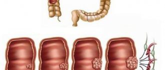
It is not known why polyps occur in the colon; it is assumed that diet, life and genetic factors are to blame. The intestinal mucosa consists of villi and depressions - crypts; for each villi there are several crypts. At the bottom of each crypt, intensive division of stem cells occurs, replenishing the lost cells of the villi and the crypts themselves. A polyp on the villus appears when the balance between proliferation—replenishment of cells—and their differentiation—specialization of newborn cells by function—is disturbed.
The size and shape of polyps vary even in one intestine; they can look like warty protrusions, or they can be a ball on a thin stalk or a fungus sitting on a fold. They can cluster in one place, forming a cluster, or they can be located a meter from each other. It is believed that adenomatous polyps accumulate additional mutations in genes that should normally suppress emerging cancer cells, or in genes responsible for regulating proliferation, and the polyp becomes malignant - becomes malignant, and this is early colon cancer.
Complications of the disease
In patients who ignore the signs of the disease, colon polyps can provoke various complications:
- intestinal bleeding;
- intestinal obstruction;
- severe forms of anemia.
The most dangerous complication of colon polyposis is colorectal cancer. This disease poses a serious threat to the patient’s life and requires complex combined treatment - surgery, chemotherapy, and radiation therapy.
About the risks of colorectal cancer and the importance of timely diagnosis of colon polyps, watch the video:
The only complication that threatens patients who have undergone the procedure for removing polyps in the large intestine is intestinal perforation, when a hole forms in the intestinal wall at the site of polyp removal. As a result of the effusion of intestinal contents into the abdominal cavity, peritonitis may develop. Fortunately, such complications occur extremely rarely.
Prevention of the development of tubular adenomas
In many cases, colon adenoma is a sign of a hereditary disease, so patients with a family history of colon diseases, including cancer, form a risk group that should be under close medical supervision and regularly undergo screening tests for the early detection of adenomas.
Since chronic diseases of the large intestine, such as colitis, can contribute to the development of tubular adenomas, treatment or compensation of these diseases will also prevent the formation of adenomas.
Since nutritional factors such as high fat content, especially refined fat, and low dietary fiber content in the daily diet are directly related to the appearance of intestinal adenomas, diet correction will help not only prevent the appearance of tubular adenomas, but also have a positive effect on the growth dynamics of existing ones adenoma.
A connection has also been identified between smoking and the development of tubular adenomas, and the number of adenomas is directly proportional to the duration of smoking, so giving up this bad habit will have a positive effect not only on the lungs, but also on the colon.
Finally, since the development of colon adenomas is promoted by a sedentary lifestyle and excess body weight, exercise and weight management may also be a reasonable recommendation for the prevention of the development of tubular adenomas.
In the case of already identified tubular adenomas, dynamic monitoring of patients using endoscopic methods is indicated. The frequency of examinations is determined individually, based on the specific clinical situation, but the following regimen is considered optimal:
- after removal of large adenomas on legs and narrowed bases: in the first year - every 6 months, subsequently - once a year;
- after removal of large wide-based adenomas and tubular adenomas with dysplasia (regardless of their macroscopic properties): in the first year - once every 3 months, in the second year - once every 6 months, thereafter - once a year.
Tubular adenoma of the colon is a benign neoplasm that can be the source of a malignant neoplasm - colon cancer. Therefore, in no case should one be dismissive of this pathology. Euroonko has all the capabilities to conduct comprehensive diagnostics, professional removal of adenomas and follow-up with highly qualified specialists.
Book a consultation 24 hours a day
+7+7+78