Postcholecystectomy syndrome (PCES) is a functional disorder that develops after removal of the gallbladder (cholecystectomy) and is largely associated with dysfunction of the sphincter of Oddi (SO). PCES also includes other functional disorders (duodenal dysfunction - duodenostasis, chronic biliary insufficiency, bacterial overgrowth syndrome (SIBO), functional intestinal disorders, reflux of bile into the stomach - duodenogastric reflux).
Postcholecystectomy syndrome develops in almost half of the operated patients.
Forms of PCES
The following variants of PCES are distinguished:
- spastic variant
- variant with CO deficiency, chronic biliary insufficiency and fat digestion disorders
- variant with SIBO, duodenal hypertension, sphincter of Oddi dysfunction (SOD), stagnation of bile in the ducts
- variant with SO dysfunction, with chronic biliary insufficiency, fat digestion disorder, intestinal hypertension, SO spasm
- the same with duodenogastric reflux, reflux gastritis.
Factors influencing the formation of PCES can be divided into two main groups:
- Contributing (sphincter of Oddi dysfunction - DSO).
- Allowing:
- bacterial overgrowth syndrome
- duodenal hypertension (increased pressure in the duodenum)
- chronic biliary insufficiency
- intestinal and abdominal hypertension (increased pressure in the intestines and abdominal cavity)
- duodenogastric reflux.
Functions
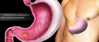
The gallbladder is an elongated sac that is located under the liver. It is necessary for the accumulation of bile and the regulation of its flow into the ducts. Its capacity reaches 50 ml. Increased bile formation is observed after eating fatty foods.
Given the direct connection with the liver, the inflammatory process from the bladder (cholecystitis) quickly spreads to the gland, causing hepatitis. The gallbladder has the following main functions:
- accumulative, due to which the bile becomes concentrated;
- excretory in response to the entry of food into the digestive tract.
In turn, bile:
- has antibacterial properties;
- helps in the breakdown of fats;
- neutralizes the acidity of the food bolus that comes from the stomach;
- stimulates peristalsis;
- increases the production of hormones (cholecystokinin, secretin) that activate digestive enzymes;
- removes toxins, bilirubin, and cholesterol.
Bile, produced in liver cells, enters the ducts and accumulates in the bladder, where it gets rid of water and becomes more concentrated. It contains acids, salts, pigments, cholesterol, protein structures and phospholipids. After food enters the digestive tract, the bile duct contracts, pushing the contents into the common bile duct (common duct) and duodenum.
Diagnostics of PCES
The diagnostic range of studies should include:
- Ultrasound of the abdominal organs
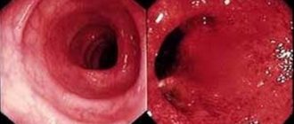
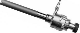
- esogastroduodenoscopy with targeted examination of the nipple of Vater
- ultrasound examination with assessment of the functional state of the sphincter of Oddi (nutritional and pharmacological tests)
- endoscopic ultrasonography
- dynamic echo-choledochography
- dynamic cholescintigraphy with T 99
- hydrogen breath test
- X-ray examination.
Further rehabilitation after gallbladder removal
To quickly return to your normal lifestyle and prevent the development of complications, you need to do the following within a month:
- follow the prescribed diet;
- avoid overheating, hypothermia, increased physical activity, lifting weights over 3 kg;
- refuse to visit baths, saunas, solariums, swimming and relaxing on the beach;
- exclude sexual contacts;
- refrain from drinking alcohol;
- perform light exercises every day without straining the abdominal muscles;
- use a belt-bandage made of elastic fabric.
After 7 days, you can go for half-hour leisurely walks.
The action of a unique technique used in our clinic by Dr. Borisov, called Selective Chronophototherapy (SPT), which is based on photodynamic therapy, is capable of activating a cascade of biochemical and cellular reactions to regulate the level of immune status indicators, demonstrates excellent results both when used independently and in combination with other methods. This significantly reduces the risk of relapse of cancer and the severity of side effects from cytostatic agents, while enhancing their effect on the affected cells.
Dr. Borisov’s gentle technique can be used repeatedly; a course of treatment is recommended. The number of procedures is regulated by the treatment regimen and achievement of the desired result.
We recommend taking VIALIFE capsules or VIALIFE solution, because they contain the highest possible concentration of chlorophyll, which:
- strengthens the immune system;
- enhances cell regeneration;
- saturates tissues with oxygen;
- has antioxidant, anti-inflammatory, detoxifying effects.
Treatment of PCES
After the diagnosis has been made and the mechanisms of disease development have been deciphered, treatment is prescribed, the general principles of which can be reduced to the following stages.
I. Dietary therapy is considered an important component of treatment. There are several stages according to the timing:
- Diet of the early postoperative period: split 6 meals a day with the last meal before bedtime, limiting fat, adding foods containing fiber to the diet, vegetables and fruits must be thermally processed.
- Diet of the period of functional adaptation: fractional nutrition is maintained with the restoration of existing restrictions.
- Diet for the period of uncompensated biliary insufficiency and impaired digestion of fats. It is necessary to restore fats in the diet in combination with replenishment of bile acids and enzymes.
II. Pharmacotherapy depends on the form and severity of PCES and includes various groups of drugs that relieve spasms, motor regulators, anti-inflammatory and antibacterial agents, bile acid preparations, and enzyme agents.
Therapy should be continuous (duration and intensity are selected individually).
III. In cases of ineffectiveness of complex treatment, the issue of surgical interventions is decided - papillosphincterotomy.
Syndrome after cholecystectomy in the practice of a general practitioner and gastroenterologist
The problem of recurrent pain that develops or recurs after cholecystectomy has appeared recently in historical terms, just as the first cholecystectomy was recently performed - in 1882 by the German surgeon C. Langebuch. At the same time, he formed the concept that made it possible to carry out this operation: “The gallbladder should be removed not because it contains stones, but because it produces them.” The attractiveness of this concept is that it creates a false impression of the therapeutic effect of removing the gallbladder, and not of the costs that the disease leads to and which force the removal of a very important functional organ. Already 40 years after the start of surgical removal of the gallbladder, it became clear that not all patients experience improvement (in some patients, pain persists, in others new sensations appear that worsen the quality of life). In 1926 [1], the French surgeon Mally-Guy proposed the term “postcholecystectomy syndrome” (PCES), which included purely surgical aspects (surgical injury to the ductal system of the liver; leaving a long stump of the cystic duct, operations in the “acute period” of the disease, etc. .). The very experience of that time (6000 cholecystectomies), when 497 reoperations were required (which amounted to 8.1%), led to the understanding that even in a purely surgical concept, PCES is heterogeneous. In addition to surgical heterogeneity, functional disorders began to be recorded, based not on surgical errors, but on the operation itself [1]. It became clear that some of the reasons were associated with insufficient preoperative examination of patients and after surgery the clinical manifestations returned again, some of the manifestations worsened, and new ones appeared. Recent statistics (Zvyagintseva T.D., Shargorod I..I., 2011 [7]) show that only 46.7% of operated patients experience improvement, in 25% the operation does not bring relief, and in 28% a return is recorded attacks [7]. PCES began to increasingly take on general medical significance, and not only surgeons, but also gastroenterologists and therapists began to be involved in its study [2–4]. From today’s point of view, “postcholecystectomy syndrome” is a collective concept that unites a variety of pathological conditions that arise directly or indirectly after cholecystectomy and sometimes have no causal connection with the absence of a gallbladder in the patient, but are accompanied by chronic recurring pain without a clear source [5, 6] . At the same time, the Rome Consensus II (1999) proposes to consider postcholecystectomy syndrome as a purely functional disorder, characterized by dysfunction of the sphincter of Oddi (SDO), caused by disturbances in its contractile function, complicating the normal outflow of bile into the duodenum in the absence of organic obstacles.
From this time on, a new (therapeutic) period begins in the study of PCES and successes in its treatment begin to largely depend on pharmacotherapeutic effects.
What makes up and what explains the physiology of bile movement:
If you have a gallbladder
- hepatic secretion;
- rhythmic activity of the sphincters of the terminal part of the common bile duct;
- normal functioning of the gallbladder sphincter;
- normal operation of the cystic duct valve;
- absorption function of the mucous membrane of the gallbladder and ducts.
All these anatomical and functional formations participate in the formation (form) of the pressure gradient, which is the engine of bile. The movement of bile through the duct system also depends on the phase of digestion:
- outside the digestion phase, bile enters the gallbladder at the moment of closure of the sphincter of Oddi (SO), while in small portions it also enters the duodenum, so the SO is not permanently closed;
- during the digestion phase, the CO opens, the gallbladder contracts and bile enters the duodenum, participating in the emulsification of fats and the activation of digestive enzymes, triggering intraluminal digestion [8, 9].
With the removal of the gallbladder from the system that forms the pressure gradient, a very important link in this important (to a large extent) self-regulating system is excluded and the entire system begins to function according to the laws of pathophysiology.
The main pathogenetic links are represented by the following provisions:
- A necessary condition for ensuring local self-regulation is the presence of CO and the gallbladder.
- In the absence of the gallbladder (cholecystectomy), the process of regulation and self-regulation is disrupted, the main self-regulating formation becomes the SO, which, in the absence of the gallbladder, shows constant readiness for dysfunction.
- The work of the CO begins to largely depend on the pressure gradient (liver, ducts, duodenum).
- With low CO tone, bile is constantly discharged into the duodenum and in a short time chronic biliary insufficiency develops with indigestion of fats.
- In some patients, RS tends to spasm, which forms pain symptoms (paroxysmal, constant).
- At different periods of time, SO dysfunction can be of a different nature and form different clinical symptoms.
- In some patients, at different times after cholecystectomy, bacterial overgrowth syndrome (BOS) develops, leading to duodenal hypertension, changing the pressure gradient and passage of bile (with stagnation in the ducts).
- In some patients, duodenal hypertension develops due to chronic biliary insufficiency and intestinal digestion disorder, leading to intestinal hypertonicity.
- In some patients, impaired intestinal tone and abdominal hypertension lead to duodenogastric reflux and reflux gastritis.
Based on the above and the recommendations of the Rome Consensus II and III, which recommended considering PCES as a consequence of dysfunction of the sphincter of Oddi, the following options for PCES can be distinguished:
- spastic variant;
- variant with CO deficiency, chronic biliary insufficiency and fat digestion disorders;
- option with SIBO, duodenal hypertension, DSO, stagnation of bile in the ducts;
- option with CO dysfunction, with chronic biliary insufficiency, fat digestion disorders, intestinal hypertension, CO spasm;
- the same with duodenogastric reflux, reflux gastritis [9].
Based on the above and relying on the recommendations of the Rome Consensus II and III, the recommendations of the Society of Gastroenterologists, we proposed our own definition of PCES in 2012.
PCES should be interpreted as a functional disorder that develops after cholecystectomy and is largely associated with dysfunction of the sphincter of Oddi, not only of the biliary type, but also of the pancreatic type. PCES also includes other functional disorders (duodenostasis, chronic biliary insufficiency, bacterial overgrowth syndrome, functional intestinal disorders, duodenogastric reflux).
From our point of view, this definition has greater practical significance, since it clearly defines the nature of treatment and allows all factors influencing the formation of PCES to be divided into two main groups:
- Contributing (sphincter of Oddi dysfunction).
- Allowing:
— bacterial overgrowth syndrome; - duodenal hypertension; — chronic biliary insufficiency; - intestinal and abdominal hypertension; - duodenogastric reflux.
Identification of these factors allows you to most adequately create an examination program, formulate a diagnosis and prescribe treatment.
Statistics
- Every year, about 2.5 million planned and emergency operations are performed on the biliary tract around the world.
- There are 600,000 cholecystectomies performed annually in the United States.
- In Russia, until 2012, 340,000 cholecystectomies were performed. In the last 2 years, 500,000 cholecystectomies have been performed.
- Postcholecystectomy syndrome develops in 5–40% of operated patients (both are correct and depend on the time of group formation and the nature of the operations) [10].
The statistics presented are very important, because they, unfortunately, indicate an increase in operations for cholelithiasis, which, in turn, leads to an increase in the number of patients suffering from PCES.
Since the main factor contributing to the formation of PCES is SO dysfunction, we would like to present in this report the recommendations of the Rome Consensus II and III on this disorder. The Rome Consensus II (2002) proposes to distinguish two clinical types of DSO:
- biliary;
- pancreatic.
The biliary type has three subtypes.
The first is characterized by an attack of pain in the right hypochondrium (with or without irradiation) in combination with the following three signs:
a) an increase in AST and (or) alkaline phosphatase by 2 or more times with double measurements (during an attack); b) delayed release of contrast agent during endoscopic retrograde pancreatocholecystography (ERCP) (more than 45 minutes); c) expansion of the common bile duct more than 12 mm.
The second is an attack of pain with one or two signs.
The third is characterized only by attacks of pain.
The pancreatic type of DSO can be represented by classic pancreatitis with epigastric pain and increased serum amylase and lipase. In less severe forms of pain there is, but no increase in enzymes.
In cases where ERCP excludes the absence of stricture pathology, manometry of the biliary and pancreatic sphincters is indicated.
The Rome Consensus III (2006) retained the main types of dysfunction, but recommended excluding instrumental studies as more dangerous than the pathology itself (ERCP, manometry).
In this regard, the diagnostic range of studies should include:
- transabdominal ultrasonography;
- esogastroduodenoscopy with targeted examination of the FS;
- ultrasound examination (ultrasound) with assessment of the functional state of the mucus (nutritional and pharmacological tests);
- endoscopic ultrasonography;
- dynamic echography;
- dynamic cholecystography with T 99;
- hydrogen test;
- X-ray examination (if gastroduodenostasis syndrome is suspected, the clinical manifestations of which are heaviness and pain in the epigastric region with possible irradiation to both the right and left hypochondrium, a feeling of nausea, rapid satiety, vomiting, which brings relief).
X-rays show an increase in the volume of the stomach and duodenal bulb, and impaired evacuation from the stomach, mainly due to low muscle tone. The lumen of the duodenal bulb is expanded, the progress of contrast is slow, the backflow of contents from the duodenum into the stomach is recorded, peristalsis is sluggish, propulsive ability is sharply reduced [11].
Establishment of biliary insufficiency. Biliary insufficiency (BI) is a symptom complex of digestive disorders that develops either as a result of a decrease in the production of bile and bile acids, or as a result of irreplaceable losses of bile acids. BN is recorded by measuring the amount of bile and the decrease in bile acids entering the intestine 1 hour after the introduction of the irritant.
For patients after cholecystectomy, we proposed dynamic ultrasonography with food load. The technique for doing it is as follows.
- The diameter of the common bile duct (CBD) is searched and determined on an empty stomach.
- A food load is given: 20 grams of butter, cheese, sweet tea - 6.5 g/sugar, white bread.
- 30 minutes after the food load, the CBD diameter is searched and determined.
- 1 hour after the food load, search and determine the CBD diameter again.
- Clinical symptoms, their appearance, increase, duration or absence are also recorded. All these data are entered into the study protocol [8].
Interpretation of the research results:
30 minutes after the food load.
1. Dilation of the common bile duct after a food load may indicate either a spasm of the CO or organic stenosis. 2. A reduction in the diameter of the common bile duct indicates the normal functioning of the CO (an increase in bile synthesis after a food load changes the pressure gradient, and the CO opens - a physiological reaction). 3. The absence of fluctuations in the diameter of the common bile duct after a food load may indicate either CO hypotension or CO due to the adhesive process.
60 minutes after the food load:
4. The dilation of the duct increases, pain appears and intensifies. This is most likely evidence of SO stenosis. 5. The original diameter of the common bile duct is preserved, or the existing expansion is preserved without pairs (confirms point 2 - normally functioning CO). 6. Preserved original diameter of the common bile duct.
This study fully meets the requirements of the Rome Consensus III (so that the study itself is no more dangerous than the pathology that is being diagnosed) and provides information sufficient to make a treatment decision.
After establishing (suspicion) the presence of chronic biliary insufficiency, its degree is determined. The characteristics are presented in table. 1.
The importance of this definition is related to the physiological role of bile in digestion.
- Neutralization of acidic food gruel coming from the stomach into the duodenum.
- Activation of intestinal and pancreatic enzymes.
- Emulsification of fats, due to which the action of lipase is carried out over a larger surface.
- Activation of lipase, which promotes hydrolysis and absorption of fat digestion components.
- Facilitates the dissolution in water and absorption of fat-soluble vitamins A, D, E, K.
- Bilirubin, cholesterol, metabolic products of sex hormones, the thyroid gland and adrenal glands are removed from the blood with bile.
- Activation of intestinal motility.
- Excretes salts of heavy metals, poisons, medicinal and other substances.
Since duodenal intubation after cholecystectomy is associated with technical difficulties, this interpretation can be based on the clinical equivalents presented in Table. 2.
Our experience in diagnosing and treating patients with PCES
We studied 140 patients in whom pain in the right hypochondrium, epigastric region or left hypochondrium persisted, appeared, intensified, or changed at different times after cholecystectomy (from 6 months to 19 years, mostly women 110/30, age from 19 to 74 years). The nature of the survey is presented above.
As a result of the study, it was established:
- 60 patients had either SO spasm or its insufficiency (according to the prevailing clinical and instrumental symptom complex);
- 50 patients had a predominant failure of the mucous membrane with chronic biliary insufficiency;
- in 20 patients, in addition to DSO, gastroduodenostasis and SIBO were recorded;
- 40 patients - DSO, biliary insufficiency, indigestion (mainly fats), intestinal dysfunction with increased intra-abdominal pressure, gastroduodenostasis, reflux gastritis.
The research results and analysis of clinical manifestations allowed us to make the following diagnoses (within the framework of one nosology - PCES).
- Postcholecystectomy syndrome: spastic dysfunction of the sphincter of Oddi.
- Postcholecystectomy syndrome: hypokinesia of the sphincter of Oddi, chronic biliary insufficiency I, II, III degrees.
- Postcholecystectomy syndrome: dysfunction of the sphincter of Oddi (biliary 1, 2, 3; duodenogastrostasis, bacterial overgrowth syndrome 1st, 2nd, 3rd stage).
- Postcholecystectomy syndrome: DSO (biliary type), chronic biliary insufficiency, digestive disorder (mainly fats), intestinal functional disorder with a predominance of hypertension, duodenostasis.
- Postcholecystectomy syndrome: dysfunction of the sphincter of Oddi with a predominance of hyperkinesia, duodenogastrostasis, reflux gastritis.
After the diagnosis has been made and its pathogenesis has been deciphered, treatment is prescribed, the general principles of which can be reduced to the following provisions.
- Dietary therapy is considered an important component of treatment. In terms of timing - diet of the early postoperative period: split 6 meals a day with the last meal before bed, limiting fat, adding foods containing fiber to the diet, vegetables and fruits must be thermally processed. The diet of the period of functional adaptation preserves the fragmentation of nutrition with the restoration of existing restrictions. Diet for the period of uncompensated biliary insufficiency and impaired digestion of fats. It is necessary to restore fats in the diet in combination with replenishment of bile acids and enzymes.
- Pharmacotherapy depends on the form and severity of PCES (both main and associated symptoms are taken into account):
- anticholinergics;
- nitrates;
- calcium channel blockers;
- myotropic antispasmodics;
- “motor regulators”;
- anti-inflammatory (mainly antibiotics, antibacterial drugs are non-absorbable when SIBO is established);
- ursodeoxycholic acid preparations (if chronic biliary insufficiency (CBF) is established, the dose of which depends on the degree of CBI);
- enzyme preparations (for indigestion, mainly fats).
- Therapy should be permanent (duration and intensity are developed individually).
Own data
The first 2 variants of PCES - 60 patients (30 men, 30 women), age from 20 to 77 years. The duration of cholecystectomy ranges from 6 months to 19 years. All patients had pain in the right hypochondrium or epigastric region associated with eating. Pain occurred at different times after cholecystectomy, in 3 patients it was progressive in nature (in some patients the pain was paroxysmal, in some it was constant, aching, worsening after eating).
The patients underwent an ultrasound of the abdominal organs (with food load), and it was found that in 23 patients the common bile duct was 1.1–1.2 cm; in 37 patients the common bile duct was 0.5–0.6 cm.
Results of the study of the CBD after a food load:
| A) after 30 minutes 1 - 25 patients 2 - 21 patients 3 - 14 patients | B) after 60 minutes 4 - 4 patients 5 - 34 patients 6 - 22 patients |
Thus, spasm of the sphincter of Oddi or its organic stenosis was detected in 38 patients, which amounted to 60.38%; in the remaining 22 patients, insufficiency of the sphincter of Oddi was detected. This group of patients had no signs of gastroduodenal stasis and SIBO. It should be noted that the tendency of the CO to spasm is typical for patients in the early stages after cholecystectomy (the early period after surgery is the 1st year and the initial period of adaptation is 1–3 years; in subsequent years, CO deficiency was more often recorded).
Treatment of patients with the 1st variant of PCES - spastic
Choice of myotropic antispasmodic
The characteristics of drugs that have an antispasmodic effect are presented in Table. 3. It also shows the zone of distribution of the effect, which is important for treatment, since the drug should be characterized by the most selective effect.
As can be seen from the table presented, hymecromone (Odeston) has this effect.
Odeston (hymecromone) is a synthetic analogue of umbealopherin found in anise and fennel fruits, which are used as antispasmodics (papaverine-like effect). The second effect is related to its relationship with cholecystokinin (it affects the gallbladder and ducts as an antagonist, and has a synergistic effect on the sphincter of Oddi).
The initial dose of Odeston was 200 mg (1 tablet) × 3 times with a possible dose increase to 1200 mg/day, the duration of treatment was 3 weeks (as monotherapy). In 34 patients, pain decreased significantly during the 1st week of treatment; 11 patients subsequently required an increase in the dose of the drug to 800–1000 mg/day. In 4 patients, despite treatment, pain persisted. The lack of effect was regarded as a consequence of organic stenosis. They conducted a contrast study of the biliary ducts, established stenosis of the common bile duct (or the terminal part of the common bile duct), and performed papillosphincterotomy with effect.
These data allowed us to conclude that:
- Ultrasound with food load in patients after cholecystectomy makes it possible to establish the type of DSO and select an adequate antispasmodic.
- Odeston is a selective antispasmodic in relation to SO and is most effective for the treatment of type 1 PCES (spastic).
- The lack of effect of treatment with Odeston is the basis for a contrast study of the biliary ducts in order to establish organic stenosis and its degree and subsequent surgical treatment.
Treatment of patients with the 2nd variant of PCES (hypokinetic dysfunction of CO and chronic biliary insufficiency)
This group included 22 patients who, in addition to hypokinesia SO and CBN, had no signs of indigestion, SIBO and gastroduodenostasis. The treatment used was the motility regulator Itomed (Czech Republic), the mechanism of action of which is the blockade of D2 receptors and blockade of cholinesterase (thereby potentiating the effect of acetylcholine). Acting throughout the gastrointestinal tract, it affects the pressure gradient, restoring the passage of bile and increasing the tone of CO. Ursosan, a preparation of ursodeoxycholic acid (URDC) (Czech Republic, Pro-Med), was used to replenish bile acids. The daily dose of Itomed was 50 mg × 3 times, the dose of Ursosan depended on the degree of biliary insufficiency (7 patients had a mild degree - they received 500 mg/day, 15 patients had a moderate degree of biliary insufficiency - the dose of Ursosan was 750 mg/day, which they received in one reception at night). The duration of treatment is 3 weeks.
As a result of the treatment, pain (discomfort in the epigastric region and right hypochondrium) was relieved by the 10th day of treatment. Patients receiving 750 mg of Ursosan had an effect within the same time frame, but it was characterized by greater stability.
Discussion . Considering the impossibility of changing the anatomical and functional situation and the failure of the SO, therapy with “motor regulators” should be resorted to when pain appears. The duration of their use should be determined by the stability of pain relief. Biliary insufficiency requires constant replenishment with bile acid preparations. The present period of time is characterized by the development of doses and duration of administration. We are studying similar options (10 days of each month, a month of each quarter, constant use of small doses of 250–500 mg/day). Each of these regimens may be sufficient for a particular patient. The criterion for the effectiveness of treatment is the absence of progression of biliary insufficiency and the absence of signs of impaired digestion of fats.
Treatment of patients with the 3rd variant of PCES
CO dysfunction (according to the predominant component), duodenogastrostasis, SIBO. The observation group consisted of 40 patients (mostly women 30/10), age from 40 to 70 years. Alpha Normix was used as a basic therapy in a daily dose of 200–400 mg (every 8–12 hours), the dose of the drug and duration of treatment depended on the severity of SIBO (1st, 2nd, 3rd degree), duration of treatment 7 -10 days. The effectiveness of treatment was monitored using a hydrogen test. The convenience of the test is the speed of obtaining an answer. Since SO dysfunction was multidirectional, Trimedat (trimebutine), which belongs to the group of opiate peptides and is an agonist of peripheral μ-, δ- and κ-opiate receptors, was used as a motility regulator. Since these receptors form multidirectional effects, the overall effect is regulatory in nature. In this version of PCES, the objects of influence are DSO, duodenogastrostasis, formed and maintained by SIBO. In addition, the drug changes visceral sensitivity, which is important in the treatment of functional disorders. The daily dose of Trimedat was 600 mg (3 tablets). The total duration of treatment is 3 weeks.
Efficacy: pain, discomfort, rapid satiety, bloating of the upper abdomen and bowel disorders were relieved within 10 days. The speed of onset of the effect depended on the relief of SIBO, which, in all likelihood, maintained gastroduodenostasis and provoked DSO.
Thus, the basic drug in the treatment of the 3rd option is a non-absorbing antibiotic (Alpha Normix), which stops microbial contamination in the stomach and duodenum and helps relieve gastroduodenostasis. “Motility regulators” can be used as a means of restoring motility, and the drug of choice is Trimedat, which restores the tone of the CO and upper gastrointestinal tract.
The hydrogen test was carried out by department employees V. A. Loginov and M. A. Kruchinina.
Treatment of patients with the 4th variant of PCES
DSO, chronic biliary insufficiency, digestive disorder (mainly fats), duodenogastrostasis, intestinal functional disorder with a predominance of hyperkinesia.
The observed group consisted of 20 patients. The complex of drugs used: Duspatalin 600 mg/day (mebeverine hydrochloride, the advantage of which is a predominant effect on intestinal tone, without a significant effect on peristaltic activity); Ursosan - 500–750 mg/day; Pangrol is an enzyme preparation containing 10,000 and 25,000 IU of lipase (we used 25,000 × 3 times per meal). Normalization of intestinal microflora, sufficient enzyme and “bile acid replacement” led to the restoration of pressure in the stomach and duodenum and the restoration of CO tone. Subsequently, maintenance treatment with UDCA and enzyme preparations should be permanent. This eliminates risk factors for DSO and prevents recurrence of pain in the epigastrium and right hypochondrium. A sufficient maintenance dose of Pangrol is 10,000 units, and Ursosan is 500 mg/day with the development of the rhythm of administration.
Treatment of patients with the 5th variant of PCES
DSO, duodenogastrostasis, reflux gastritis.
There were 20 patients under our supervision. This option, in addition to the accepted examinations, requires studying the acid-producing function of the stomach (endoscopic pH-metry is sufficient) and, if hypersecretion is established, introducing it into a treatment complex that includes motility regulators Trimedat, Itomed, Ursosan, and also gastric secretion blockers. The basic drug in this situation is Ursosan, which, on the one hand, compensates for the lack of bile acids, and on the other, converts the synthesis of bile acids to increase the production of non-toxic ones, since the main damaging factors are bile acids, lysolecithin and pancreatic enzymes thrown into the stomach. The effect of treatment (elimination of pain and gastric dyspepsia is recorded by the end of the 1st week of treatment). UDCA drugs are used as maintenance therapy.
General conclusion
We must agree with the conclusion of the Rome Consensus II and III that the development of PCES is based on dysfunction of the sphincter of Oddi. The basis of this dysfunction is the removal of the gallbladder, an organ that is very important functionally. Surgical errors in the operation can hardly be the basis of PCES, since they are not directly related to dysfunction of the CO, which maintains the pressure gradient and normal passage of bile through the ductal system. Also, diseases that developed before surgery are not directly related to this.
Our materials show that there are several variants of PCES (we have identified 5), and they are formed as a result of various functional disorders that directly or indirectly lead to disruption of bile passage (gastroduodenostasis, indigestion, dyskinesia, intestinal dysfunction, SIBO, reflux gastritis). These disorders are risk factors for DSO and its maintenance. The therapeutic approach and the choice of pharmacotherapy that most adequately relieves the exacerbation of PCES depends on their presence.
Essentially, work on studying the therapeutic approaches of PCES and treatment has just begun, it needs to be continued and intensified with the hope of obtaining the desired effect.
Literature
- Malle-Guy P., Kestels P. J. Syndrome after cholecystectomy. M., 1973.
- Lazebnik L. B., Konanev M. I., Ezhova T. B. Comparative study of the quality of life in patients with cholelithiasis and postcholecystectomy syndrome. Materials of the 5th Slavic-Baltic Scientific Forum, St. Petersburg, Gastro-2003, p. 93.
- Kovalev A.I., Sokolov A.A., Akkuratova A.Yu. Postcholecystectomy syndrome: causes. Tactics of surgical treatment // News of surgery. 2011, vol. 19, no. 1, p. 20–21.
- Ilchenko A. A. Why does cholecystectomy not always improve the quality of life? // Pharmateka. 2012, 17, p. 23–29.
- Zimmerman Ya. S., Kuisman T. G. Postcholecystectomy syndrome: a modern view of the problem // Clinical medicine. 2006, no. 8, pp. 4–11.
- Sheptulin A. A. Roman criteria for functional disorders of the gallbladder and sphincter of Oddi: controversial and unresolved issues // Russian Journal of Gastroenterology, Hepatology, Coloproctology. 2005, No. 4, p. 70–74.
- Zvintseva T. D., Shargorod I. I. Postcholecystectomy syndrome: dysfunction of the sphincter of Oddi // Faces of Ukraine. 2011, no. 2, p. 100–106.
- Minushkin O. N., Maksimov V. A. Biliary-hepatic dysfunction. Toolkit. M., 2008.
- Minushkin O. N. Use of the drug “Odeston” in clinical practice, M., 2014.
- Vetshev P. S., Shkrob O. S., Beltsevich D. G. Cholelithiasis. M., 1998.
- Sokolov L.K., Minushkin O.N., Savrastov V.M., Ternovoy S.K. Clinical and instrumental diagnosis of diseases of the hepatopancreatoduodenal zone. M., 1987.
- Maksimov V. A. et al. Biliary insufficiency. M., 2008. P. 232.
O. N. Minushkin, Doctor of Medical Sciences, Professor
FSBI UMTS UDP RF, Moscow
Contact Information
FAQ
Is the development of PCES caused by surgical errors?
As we studied the mechanism of development of this process, it was found that errors in surgical intervention are in last place among the reasons for the formation of this condition.
Is there a cure for PCES?
The disease has a tendency to relapse, so treatment must be carried out in courses, sometimes for life.
How often does PCES develop after gallbladder removal?
On average, the disease develops in 30% of people who undergo cholecystectomy.
Is it possible to limit oneself to following a diet in the treatment of PCES?
Compliance with dietary recommendations is a mandatory, but not the only step in the treatment of this condition.
Are bile acid preparations used in the treatment of PCES?
Yes, if there are signs of a violation of the composition, formation and secretion of bile.
What is cholecystectomy
The operation is carried out after a thorough examination of the patient. Its indications include stone formation, malignant lesions, empyema or gangrene of the gallbladder. Cholecystectomy can be performed openly or using laparoscopic instruments. The latter technique is characterized by fewer complications, a shorter rehabilitation period and a good aesthetic effect.
During the patient’s conversation with the anesthesiologist, the doctor talks about the features of anesthesia and, if necessary, prescribes additional examination (coagulogram, blood glucose).
If the patient is taking anticoagulants (drugs that inhibit the coagulation system), they must be discontinued 3-7 days before surgery.
12 hours before cholecystectomy, the patient is prohibited from eating and drinking.
Contraindications include:
- pregnancy;
- acute infectious diseases;
- severe blood diseases;
- decompensated cardiovascular, respiratory or renal failure.
Case histories
Story No. 1
Patient Sh., 56 years old, came to the EXPERT Clinic with complaints of nagging pain in the right after eating, bitterness in the mouth. From the anamnesis it is known that 7 years ago the gallbladder was removed on a planned basis due to cholelithiasis. Subsequently, the patient followed a diet. She made no complaints. For the last 3 years she has not followed a diet and has gained 6 kg. Over the past year, I began to notice these complaints, which was the reason for contacting a doctor. An ultrasound of the abdominal organs at the place of residence revealed no serious abnormalities in the structure of the organs. The gastroenterologist at the EXPERT Clinic prescribed a comprehensive laboratory and instrumental examination, including an assessment of the function of the bile ducts and sphincter of Oddi (dynamic choledochography). Based on the results of the examination, a secondary functional disorder of the sphincten of Oddi (SPED) was established. Diet therapy and a course of treatment aimed at restoring the function of the biliary system in the absence of the gallbladder were prescribed. As a result of the therapy, her health has improved, the patient has no complaints, and continues treatment and observation by a physician-supervisor in the EXPERT Clinic.
Story No. 2
Patient P., 48 years old, came to the EXPERT Clinic with complaints of girdling pain after eating and nausea. From the anamnesis it is known that 3 years ago the gallbladder was removed on a planned basis due to cholelithiasis. I tried to follow the diet with minor errors. I did not abuse alcohol. When contacting a doctor at his place of residence, the condition was regarded as chronic pancreatitis and courses of pancreatic enzyme preparations were prescribed without a significant effect, and therefore the patient contacted the EXPERT Clinic. When conducting a comprehensive laboratory and instrumental examination prescribed by a doctor at the EXPERT Clinic, it was found that these complaints were not caused by an inflammatory process in the pancreas, but by the formation of a functional disorder of the sphincter of Oddi of the pancreatic type. For this reason, treatment was prescribed, which included not only enzyme preparations, but also antispasmodic agents and bile acid preparations. With this treatment, the complaints stopped. The patient continues a course of anti-relapse therapy.
Diet and diagnosis after surgery
To save yourself from complications, you should follow a simple diet and doctors’ recommendations after surgery in advance. What risks will be reduced? Life will become better, metabolism will accelerate, metabolism will be restored.
Diet rules after gallbladder removal surgery:
- Drink water on a regular basis. Before eating, drink a glass of water; this will improve the digestion process, making it easier for the organs of the digestive system affected by surgery. Water will help your liver function. Slags and toxins are eliminated with reduced acidity.
- Eat 5-6 times a day. Frequency and proper diet will help bile to be released evenly. The intestines will not suffer from increased acidity, and the risks of complications will decrease. Eating 5-6 times a day does not imply a large amount of food. Food products are divided into breakfast, lunch and dinner. Every 2-3 hours between main meal intervals you can eat fruits, nuts, and vegetables. Don't overload your body. Light snacks needed.
- All food does not exceed normal temperatures. It is impossible and not recommended to eat food that is very hot or very cold. Ice cream, hot tea, pizza from the oven - you will have to give up in favor of healthy, warm, optimally chilled food.
- Do not sit at a banquet table immediately after surgery. Portions during the week should be small, the size that fits in the palms of your hands. The body needs a fasting day to adapt to new digestive techniques.
- After the first week, focus on protein foods. Chicken, meat, legumes are products that dilute bile. Remember, the body is not used to the constant secretion of bile into the abdominal cavity. Improper nutrition with foods of high acidity leads to the operating table.
- Forget the word “fried.” Steam, stew, eat raw - the principle after the procedure. Avoid fatty, fried, smoked foods.
- Drinking coffee and strong tea is prohibited. Hibiscus, mint tea, green teas, mineral water are substitutes for usual drinks.
- Fast food, soda, carbonated drinks, alcoholic beverages, drugs with high iron content, activated carbon. Do not touch the substances for more than six months.
- Hot spices, salt, pepper, oregano, cloves, paprika, hot sauces, ketchup, mayonnaise, garlic, onions are now prohibited.
- Sausage and sausage products.
- Fat and blue cheeses.
- Meals should be simple and balanced, with more vegetables. Fruits are eaten in small quantities - frequent consumption sometimes leads to bloating. There is no need to disturb the intestines.
The right side hurts after surgery in 3 out of 20 cases. Removal surgery is performed frequently, so the risk of complications is low. If you notice fever, breathing problems, sharp and aching pain (right rib), attacks in the right side and under the shoulder blade (cramps) - immediately consult a doctor for help! Complications do not wait for time, the situation is getting worse every moment. Be careful.
Main pathologies of the biliary system
Why does my gallbladder hurt and have to be removed? Experts divide the most common gallbladder diseases into the following groups:
| № | Helpful information |
| 1 | dyskinesia (disturbances in normal functioning) |
| 2 | inflammation (cholangitis and cholecystitis) |
| 3 | pathologies caused by metabolic disorders (cholelithiasis, polyposis) and so on |
The course of all of these groups of pathologies is accompanied by pain, heartburn, loose stools and other symptoms that are extremely unpleasant for a person appear, causing him serious discomfort. In many cases, drug therapy does not give the desired result, and the bladder has to be removed.
However, even after the removal of this organ, in many cases the patient continues to be bothered by pain and digestive disorders (colitis, pancreatitis, and so on). This is due to the fact that surgery often eliminates the consequences and not the cause of the pathology (for example, with calculous cholecystitis, the root cause is a violation of normal metabolism, and surgery does not eliminate this problem).