Content:
- Causes of abdominal pain during a hangover.
- Gastritis in alcohol addicts.
- Gastric ulcer in alcoholics.
- Is it possible to recover at home?
- Treatment in a hospital setting.
- Conclusion.
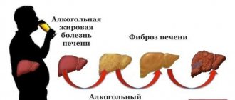
During a binge, all parts of the digestive system are the first to take the “blow” from the aggressive effects of alcohol.
Therefore, most people feel pain in the stomach after heavy drinking during the hangover period, when the withdrawal syndrome gains strength (it is also called withdrawal syndrome). Binge drinking is an exacerbation of chronic alcoholism. With this disease, not only the stomach suffers; systematic consumption of alcohol-containing drinks leads to a malfunction of all organs and systems (gastrointestinal tract, brain, nervous system, etc.). In the gastrointestinal tract, the functioning of the esophagus, stomach, liver, pancreas, gall bladder and intestines is disrupted.
Introduction
Chemical burns remain the most common acquired disease of the esophagus in childhood. Children, unlike adults, take the chemical in small quantities. However, accidental ingestion of even one sip of a concentrated toxic substance can lead to severe scarring of the esophagus with various complications, long-term treatment, repeated operations, and disability.
Early consequences of chemical burns of the esophagus (COB) include laryngeal edema, exotoxic shock, bleeding, necrosis of the esophageal or stomach wall, mediastinitis and the formation of cicatricial stenosis. Late complications of COP: gastroesophageal reflux (GER), hiatal hernia (HH), impaired motility, candidiasis, long-term malignancy [6, 14, 15, 21, 25, 29, 31].
Cicatricial esophageal stenosis (ESS) is the main problem faced by pediatric surgeons in chemical burns. The treatment outcome is essentially determined by the nature of the cauterizing agent [1, 2, 9, 10, 15-17, 23, 24, 29, 31]. Particularly severe lesions are observed in burns caused by concentrated alkalis [12, 15-17, 18, 23, 29, 31].
Currently, the most common chemicals accidentally drunk by children are vinegar essence and alkaline solutions used in everyday life to clean glass-ceramic surfaces or pipes (Mole, Schumanite, battery fluid, caustic soda, other substances). Very rarely, technical acids play a role in the etiology of burns of the upper digestive tract. In recent years, the number of children with damage to the esophagus as a result of swallowing disk batteries has been growing, which, in addition to burns of the esophagus, lead to the formation of tracheoesophageal fistulas and bleeding [22, 30].
There are “non-surgical” ways to prevent the formation of RSP: early lavage of the esophagus and stomach [4, 23], hormone therapy [4, 28], “local” exposure to drugs [4, 11], the use of ozone, laser therapy, upperbaric oxygenation, physiotherapeutic methods [ 4, 6]; treatment of GER [1, 6, 14, 25, 29, 31, etc.].
“Surgical” prevention of narrowing of the esophagus during burns in the early period (up to 4-6 weeks) includes stenting [7, 8, 26], early preventive bougienage [3, 9, 10, 19].
In order to judge the effectiveness of a particular burn treatment method, it is necessary to know exactly who will have esophageal stenosis. As is known, RSP occurs exclusively in third degree burns [1, 9, 10, 13, 15-17], so it is extremely important to identify this particular group of patients.
In the clinic of pediatric surgery of the Russian State Medical University on the basis of Children's Clinical Hospital No. 13 named after. N.F. Filatova has accumulated extensive experience in treating children with COP over a period of more than 60 years. The work of S.D. was devoted to the development and improvement of methods for diagnosing and treating OCP over the years. Ternovsky, O.V. Blagoveshchenskaya, S.I. Vozdvizhensky, N.I. Kondrashina, Yu.F. Isakova, E.A. Stepanova, V.I. Geraskina, S.S. Mostovoy, G.S. Vasilyeva, A.Yu. Razumovsky and others.
Over the course of many decades, our clinic has improved the endoscopic differential diagnosis of grade II and III COP, clarified the indications for preventive bougienage, introduced other methods of dilatation therapy, etc.
Material and methods
Every year in Children's Clinical Hospital No. 13 named after. N.F. Filatova treats about 200 children with suspected chemical burns of the digestive tract.
Over a 22-year period, 4252 children with suspected COP were admitted to the clinic (Table 1).
During primary esophagoscopy, a burn of the esophagus was excluded in 65.3%; degree II-III COP was detected in 20.8% of children. A third degree burn of the esophagus (cicatricial stenosis) was diagnosed in 5.9% of patients who came to the clinic with suspected chemical injury to the esophagus. An isolated third-degree gastric burn was detected in 0.19% of cases.
Patients aged 1 to 3 years accounted for 82%. Boys (69%) traditionally outnumber girls (31%). All patients took the chemical accidentally, due to adult oversight.
The cause of the third degree burn of the esophagus was vinegar essence (67%), alkali (28.6%), manganese crystals (2%), batteries (1%), unknown substance (1.4%). The etiology of isolated gastric burns included technical acids and concentrated alkalis.
It should be emphasized that over the last decade, according to our clinic, the number of patients with third-degree burns of the esophagus caused by vinegar essence has almost doubled, and the number of burns caused by alkali has increased by more than 3.5 times. It is noteworthy that in recent years there have been no severe burns caused by manganese crystals, which is due to the ban on the sale of this drug in Moscow pharmacies. However, in recent decades there has been a trend towards an increase in the number of children with esophageal lesions from disk batteries.
For the period 1985-1995. in Children's Clinical Hospital No. 13 named after. N.F. Filatov there were 530 children with COP II-III degrees. The patients underwent traditional treatment, adopted in our clinic after the introduction of fiber-fiber technology into practice [10, 13, 15-17, 19].
For the period from 1996 to the present, we have been using a new treatment tactic, in which we abandoned early preventive bougienage in all children with grade II-III COP after introducing the method of bougienage using a conductor string in our department. It should be emphasized that bougienage using a metal conductor string in our country was developed and introduced into surgical practice in adult patients in 1965 at the Russian Research Center for Surgery by E.N. Vantsyan and R.A. Toshchakov [5].
The following tactics were adopted. After relief of the general symptoms of the burn, the children are discharged. We hospitalize them again 4-6 weeks after the burn for a control FEGDS. If dysphagia occurs, patients are hospitalized earlier. When a third degree burn is confirmed, we begin bougienage. The first bougienage of the esophagus is carried out along a conductor string. We use hollow thermoplastic bougies from Neoplast (France), currently we use bougies. 2-3 days after bougienage using a guide string, we switch to “blind” bougienage according to the scheme adopted in our clinic for the treatment of grade III COP.
If patients in the acute period have persistent dysphagia and intoxication, which indicate deep damage to the esophagus, as well as the formation of extensive stenosis of the esophagus (alkali burn), then we perform gastrostomy according to Kader for subsequent bougienage by the thread.
All patients with RSP undergo a complex of examinations to identify GER. Patients with GER are prescribed drug antireflux therapy. In cases of developed hiatal hernia, laparoscopic Nissen gastrofundoplication was performed.
The results of the new tactics depended on the chemical agent that caused the third degree burn of the esophagus.
results
All children with grade III COP caused by vinegar essence, after bougienage of the esophagus using a guide wire under anesthesia, were transferred to direct bougienage. The effectiveness of dilation therapy was 100%. There was no perforation of the esophagus, and gastrostomy or reconstructive surgery was not required in any case. All children recovered.
In 43% of patients with a third degree burn of the esophagus caused by alkali, extensive stenosis of the esophagus developed. Dilation therapy was ineffective in these patients; they underwent coloesophagoplasty. Perforation of the esophagus during bougienage occurred in 5 cases, including in one case the esophageal rupture was sutured, in one case a cervical esophagostomy was performed, and in three cases the esophagus was extirpated. Subsequently, all patients underwent coloesophagoplasty. However, in 57% of patients with third-degree esophageal burns caused by alkali, dilation therapy was effective. The most aggressive alkalis were Mole, caustic soda, concentrated solution of Whiteness and Schumanite.
Three patients with isolated gastric lesions underwent Billroth-I resection. On the 3rd day, one child with total gastric necrosis (soldering acid burn) underwent gastric extirpation, placement of a hanging enterostomy, then Roux-en-Y esophagojejunostomy.
Discussion
In the 40s of the 20th century, severe burns from alkalis, such as caustic soda (caustic soda), office glue and ammonia, prevailed in children. Over the next 50-60 years, the nature of cauterizing substances changed towards less aggressive ones. The number of burns to the esophagus caused by alkali has decreased significantly due to the cessation of the use of caustic soda in everyday life. Chemical burns caused by vinegar essence began to predominate. The number of burns to the pharynx and esophagus caused by manganese crystals has increased.
The number of patients with chemical burns of the esophagus has increased in recent years. This is due to the chemical industry’s production of new alkali-containing products in beautiful bottles used for cleaning glass ceramics and pipes, as well as the continued production of concentrated acetic acid and careless storage of chemicals by adults in everyday life.
In recent decades, the number of children with esophageal lesions caused by disk batteries has increased, due to the increase in the number of electronic toys and devices. The incidence of battery ingestion into the esophagus is approximately 10 per million people per year, and 1 in every 1000 battery ingestions causes serious injury [22].
Button-type batteries contain heavy metals such as mercury, silver, lithium and concentrated alkali. The mechanism of damage when a battery enters the esophagus includes the corrosive effect of leaking electrolyte, toxic effects, external current, and necrosis due to pressure [30]. If a battery enters the esophagus as a result of the corrosive effect of the leaking electrolyte and pressure, a chemical burn and necrosis of the mucosa is possible with the subsequent formation of a cicatricial stricture, perforation, esophageal-tracheal anastomosis, and even an aortoesophageal fistula [22, 30, 31]. If a disk battery gets into the esophagus, tissue necrosis and wall perforation can occur within 4-6 hours. If a battery is detected, given the high risk of life-threatening complications, emergency removal is indicated.
For a long time, rigid esophagoscopy was considered contraindicated for chemical burns in children due to the risk of injury and bleeding. The feasibility of using diagnostic esophagoscopy in the acute period of COP in children was substantiated in the works of E.A. Stepanova. Thanks to rigid esophagoscopy, starting from the middle of the 20th century, it became possible to differentiate in the acute period large groups of children who do not have COP and patients with first-degree burns, and therefore, save them from prophylactic bougienage [20].
Since the 50s of the last century, our clinic has adopted the method of early preventive bougienage of the esophagus for all children with II-III degree burns [3, 9, 10, 19, 20]. According to publications of our department, as of 1978, the method was effective in 97% of observations [19].
However, due to the impossibility of differentiating grade II and III COP in the early period, prophylactic bougienage was started in all patients with fibrinous deposits. This led to the so-called “unnecessary” bougienage of the esophagus in patients with second-degree burns [10, 15-17, 19, 28]. In addition, early bougienage creates a risk of esophageal perforation, slows down reparative processes, and causes emotional trauma to the child [7, 8, 26].
The introduction of modern fiber endoscopy into pediatric practice (80s) made it possible to develop a new approach to the diagnosis and treatment of COP. In our clinic, esophagoscopic signs have been developed that allow us to differentiate between grades II and III COP in the acute period [10, 13, 15-17]. Endoscopic signs indicating the possibility of the formation of a narrowing of the esophagus (III degree) are circular, rough, gray fibrinous deposits, upon removal of which the underlying tissues bleed for a long time, and the removal itself is difficult; rigidity of the walls of the esophagus when inflated with air.
It was also found that healing of the esophageal wall in case of a second degree burn does not always fit into the “traditional” 3 weeks, but can be delayed up to 4-6 weeks or more without the formation of scars that narrow the lumen of the esophagus.
The developed endoscopic criteria for the differential diagnosis of COP made it possible to improve the diagnosis of degrees of esophageal burns in the early period and apply a differentiated approach to the treatment of children with COP II and III degrees in the acute period. In 50.5% of patients, the diagnosis of a second degree burn of the esophagus was accurately established and early bougienage was not performed [10, 15-17].
The idea of abandoning early bougienage for COP in children has existed for a long time. However, by refusing preventive bougienage, we “deliberately” get a group of patients with developing esophageal strictures.
In our clinic, it was accepted: if, in case of a II-III degree burn, prophylactic bougienage was not started on time (in the first 7-10 days after the burn) or was carried out incorrectly, then direct bougienage is dangerous and contraindicated. Such patients did not undergo dilation therapy within 4-6 weeks from the moment of the burn. Then, if the developing stenosis of the esophagus was confirmed, a gastrostomy tube was placed and bougienage by the thread began (from the 6th week after the burn).
The introduction of bougienage of the esophagus using a guide wire allowed us to abandon preventive bougienage. The method makes it possible to safely dilate the developing RSP and relieve a large group of children with second-degree burns from “unnecessary” bougienage (Table 2).
The undoubted advantage of bougienage using a guide string over bougienage using a thread is that there is no need for gastrostomy. Then we move on to supportive bougienage of the already restored lumen of the esophagus according to the scheme adopted in our clinic for the management of patients with grade III COP.
However, we believe that patients with extensive stenosis of the esophagus caused by alkali solutions are advised to apply a gastrostomy and bougienage with a thread. If this treatment is ineffective, esophagoplasty is necessary.
Isolated lesions of the pharynx, according to our clinic, in 98% of cases occurred after a burn with manganese crystals. In case of burns with other substances, stenosis of the pharynx is combined with stenosis of the underlying parts of the digestive tract. Children with cicatricial stenosis of the pharynx make up 0.5% of all patients with cicatricial narrowing of the esophagus [2]. The introduction of prolonged nasotracheal intubation (PNTI) into pediatric toxicology practice has increased the effectiveness of treatment for severe chemical burns of the hypopharynx. PNTI method, according to data from Children's Clinical Hospital No. 13 named after. N.F. Filatov, helped reduce the incidence of complications by 4.5 times and is an alternative to early tracheostomy for severe burns of the hypopharynx with breathing problems.
We consider the following features of bougienage in children with pharyngeal stenosis to be fundamental: carrying out bougienage by thread with large-sized bougiens, regardless of age (No. 44-60 on the Charrière scale); large bougie sizes correspond to the size of the child’s throat; Direct bougienage in case of pharyngeal stenosis with bougies of this size in children is impossible.
A method of endoscopic ultrasonography of the esophagus has been developed for the purpose of early diagnosis of the depth of damage to the esophagus [1].
Most specialists in the treatment of chemical burns in children and adults note a significant influence of GER on the results of treatment of RSP [1, 14, 15, 25, 29, 31]. With COP, cicatricial shortening of the esophagus develops, secondary cardiac hiatal hernia, cardia insufficiency and reflux esophagitis occur. Patients are prescribed complex antireflux therapy against the background of maintenance bougienage, including proton pump blockers. Persistent GER promotes further progression of scarring. In such situations, the bougienage effect is only temporary, since the effect of refluxed gastric juice promotes rapid re-scarring [25, 29, 31]. In this case, antireflux surgery is indicated.
It has been proposed to use bioabsorbable stents as a step-by-step treatment for esophageal stenosis in children [27].
In our clinic, the bougie sizes have been revised, and we currently use larger bougie sizes when treating RSP (Table 3).
Thus, the endoscopic criteria for a third degree burn of the esophagus, as opposed to a second degree burn, in the first weeks are the presence of circular fibrinous deposits; wall rigidity during air insufflation; rough, gray fibrinous deposits on the burn surface, upon removal of which the underlying tissues bleed for a long time, and removal itself is difficult.
For a second degree burn, specialized therapy is indicated, aimed at local treatment and relief of general symptoms of poisoning. For a third degree burn, in addition to the treatment mentioned above, bougienage of the esophagus is indicated.
Blind bougienage for cicatricial stenosis of the esophagus is contraindicated. Direct bougienage is possible as an early preventive measure in cases of II-III degree COP or as a supportive measure in cases where the lumen of the esophagus has already been restored using other dilatation methods.
The use of bougienage of the esophagus along a guide wire made it possible, in case of a third-degree burn, to carry out the procedure from the 4th to 6th week, when the scarring process in the wall of the esophagus already begins, and this tactic made it possible to replace early bougienage and dilatation by a thread with the preliminary application of a gastrostomy.
For third-degree burns caused by vinegar essence, the use of new tactics turned out to be effective in 100% of children. After string dilatation, all children were transferred to “blind” bougienage according to the treatment regimen for third-degree burns. No gastrostomy was required in any observation.
In half of the cases with grade III COP, there was a need for gastrostomy and thread bougienage. In case of extended stenosis, esophageal plasty is indicated.
In the presence of GER, drug antireflux therapy is indicated. When a hiatal hernia develops or when conservative treatment is ineffective, gastrofundoplication is indicated.
Causes of abdominal pain during a hangover
Anyone who has tried strong alcoholic drinks has felt a burning sensation in the mouth and esophagus. This is how the mucous membranes react to the irritating effect of ethyl alcohol. This is a small chemical burn that does not cause much damage if used infrequently.
Things are different for people with chemical dependency. With regular use, ethanol and its breakdown products are constantly in the stomach, corroding its mucous membrane and causing pain. After withdrawal from the binge period, alcohol addicts experience even more severe discomfort.
It is known that the mucus that is produced in the stomach has protective properties against various types of aggressive influences (hydrochloric acid of gastric juice, substances ingested with food and drinks). Regular consumption of alcohol disrupts the production of protective mucus, so hydrochloric acid, pepsin and ethanol itself “attack” the mucous membrane and destroy it. The presence of pain indicates serious damage to the gastric wall.
Causes and nature of symptoms
More than other organs, the human stomach experiences the consequences of alcohol consumption. The next day, the pain is accompanied by nausea, heartburn, a feeling of heaviness, an unpleasant taste in the mouth, and a headache. Reasons that may cause abdominal pain after drinking:
• increased production of gastric juice with slow gastric secretion; • rapid absorption of toxic substances into the blood caused by an increased amount of gastric juice; • decrease in the protective functions of the stomach walls; • gradual destruction of capillaries.
Already existing chronic diseases of the digestive system can also provoke attacks of the disease and cause pain and discomfort in the stomach under the negative influence of alcohol.
The following diseases may occur as a result of exposure to alcohol:
• gastritis; • burn of the stomach and esophagus; • acute poisoning; • pancreatitis.
Gastritis in alcohol addicts
An addict may experience pain in the stomach area both during and after binge drinking. Moreover, during heavy drinking, the anesthetic (pain-relieving) effect of ethyl alcohol simply drowns out the pain. After a binge, when the flow of ethanol into the body stops, pain begins to be felt.
With alcoholic gastritis, on endoscopic examination, doctors observe a picture of inflammation of the gastric mucosa, and the appearance of small bleeding erosions is possible.
People complain of stomach pain and nausea. Sometimes vomiting occurs, stool is disrupted (most often constipation appears). Painful episodes can be either acute or aching in nature. If gastritis is not treated adequately, ulcerations may occur. In some cases, tissue degeneration occurs and a tumor process occurs.
Burns of the esophagus. Review article
Ivanova Elena Yurievna
Otorhinolaryngologist
April 25, 2021
Chemical burns of the esophagus in children occupy a special place in the practice of an otolaryngologist.
Burns of the esophagus are usually accompanied by burns of the oral cavity and pharynx. They can be thermal or chemical.
Thermal burns are divided into burns caused by vapors and gases and burns caused by hot liquids. Chemical burns of the pharynx and esophagus are more severe both in their course and in their consequences.
The most common chemical burns of the oral mucosa, pharynx and esophagus occur between the ages of 1 and 2.5 years. And we must not forget about teenagers.
Why exactly this age?
At one year old, a child begins to walk and explore the world on his own. Children experience the world differently than adults. You want to pick up bright, beautiful packaging, twirl it, and if you manage to open an “unidentified object,” you want to try it “on your tongue.”
ATTENTION PARENTS!!! Protect your child from pain, fear and torment...
What do I need to do?
Make sure all strong cleaning products are kept out of reach of the child. “Mr. Muscle”, “Mole” should be placed in a place inaccessible to the child, because sometimes the consequences are hard to imagine. A drop of Mole liquid causes severe burns to the mouth, larynx, and esophagus; sometimes it is enough for a child to lick the lid of this product. Unfortunately, this leads to cicatricial narrowing of the esophagus, which requires constant bougienage, and in some cases, esophageal plastic surgery.
Symptoms: usually difficulty swallowing, increased salivation, and sometimes vomiting. Call an ambulance immediately. In the oral cavity, pharynx, and esophagus, the mucous membrane is hyperemic and has necrotic deposits that bleed easily. After a few days, these signs subside, and an imaginary improvement occurs (usually for 8-12 days). The child begins to eat not only liquid, but also solid food. However, after 2-4 months, a narrowing of the esophagus develops.
Burns have a seasonality: in the spring, as a rule, in March, they begin to prepare seedlings for the dacha. Sometimes, for better growth, various liquid fertilizers and potassium permanganate are used.
Potassium permanganate crystals cause severe burns. Children, out of curiosity, and first of all, out of oversight, pour beautiful crystals into their mouths. Potassium permanganate crystals, as a rule, fall on the soft palate, buccal mucosa, tonsils, and sometimes on the epiglottis. It is urgent to rinse your mouth with ascorbic acid; swelling of the mucous membrane increases within 2 hours.
In the fall, preparations are made. Be careful, vinegar essence!
Plants at home.
When buying house plants, if you have a small child at home, be sure to consult with flower growers. For example, Diefenbachia juice, which is contained in the leaves, causes burns to the oral cavity.
Dear parents, take care of your children and protect them from the dangers that surround them at home and in the country.
Gastric ulcer in alcoholics
There is a misconception that strong alcohol can cure the stomach. However, this is not true; alcohol, as an anesthetic, somewhat muffles pain impulses and creates the illusion of well-being. But during heavy drinking, the destruction of the stomach walls occurs so intensely that ulcerations of significant size and depth can form.
After binge drinking, alcohol addicts complain of sharp cutting pain in the epigastrium (upper abdomen). The pain is most severe on an empty stomach and 1-1.5 hours after eating . Episodes of hunger pain are one of the surest signs to suspect the presence of ulcers.
In addition to pain, the addict may experience nausea and vomiting of blood. General weakness is explained by both blood loss and impaired blood microcirculation in the gastric wall. The ulcerative process is accompanied by changes in stool (constipation, diarrhea). Under no circumstances should you ignore or leave your loved one without a medical examination if they have the symptoms described.
The most dangerous complication of an ulcer is bleeding . It can manifest itself both externally (vomiting blood, black feces) and internally. Internal bleeding may cause a sharp drop in blood pressure, a thready pulse, and even loss of consciousness. With such symptoms, you should immediately call an ambulance, as death is possible.
Publications in the media
Chemical burns of the esophagus are caused by concentrated alkalis and acids. Causes. Accidental or deliberate (with suicidal intent) ingestion of concentrated acids (acetic essence, battery electrolyte) or alkalis (ammonia, caustic soda).
Pathogenesis • Local action •• Acids cause coagulative necrosis with the formation of a dense scab, which prevents the substance from penetrating deeper and reduces its penetration into the blood •• Alkalies cause liquefaction necrosis, which promotes the transfer and spread of alkali to the underlying initially undamaged tissues • General toxic effect with the development of multiple organ failure ( primarily hepatic-renal).
Pathological anatomy • Alkali burns are characterized by deeper and more widespread damage to the wall of the esophagus • The most pronounced changes occur in places of physiological narrowing • Stages of the burn •• I - hyperemia and edema of the mucous membrane •• II - necrosis and ulceration of the mucous membrane •• III - formation of granulation tissue •• IV - scarring • By the end of the first week, necrotic areas are rejected, forming ulcerations. Superficial ulcers epithelialize within 1–2 months, deep ulcers within 2–6 months with the formation of connective tissue.
Clinical picture • Stage I (acute) - 5–10 days •• Pain in the mouth, pharynx, behind the sternum, in the epigastric region •• Hypersalivation •• Dysphagia •• Shock in the immediate hours after injury •• Burn toxemia, the symptoms of which begin prevail in a few hours • Stage II (imaginary well-being) - 7–30 days. As a result of the rejection of necrotic tissue, swallowing becomes somewhat freer. Complications: •• esophageal bleeding •• perforation of the esophageal wall •• sepsis • Stage III (stricture formation) - from 2 to 6 months, sometimes for years. On the wall of the esophagus, areas of varying length are formed with a sluggish healing process, the wound surfaces are covered with scabs and bleed easily. Dysphagia to the extent of complete obstruction of the esophagus; laryngospasm, cough, suffocation caused by food entering the respiratory tract.
TREATMENT Diet. Liquid, tube and parenteral nutrition are recommended. Conservative treatment • Narcotic analgesics - trimeperidine, omnopon • Washing the oral cavity and stomach with solutions of antidotes in the first hours after the chemical enters the esophagus • Drinking plenty of fluids (water, milk) followed by inducing vomiting as first aid at the scene • Mandatory early (in the first hours) gastric lavage (fluid volume from 1 to 5 liters depending on age) • Intensive antishock therapy • Antihistamines with a pronounced sedative effect (promethazine, chloropyramine) • Detoxification therapy • With the development of acute renal failure - extracorporeal blood purification • Infusion therapy with forced diuresis, antibiotic therapy • GK • Fish oil, vegetable oil orally • Early start (2 months after the burn) of bougienage of the esophagus for 1–1.5 months in combination with GK and lidase • At the stage of stricture formation, bougienage is the main method of treatment • • Blind •• Hollow radiopaque bougies over a metal guide •• Under the control of an esophagoscope •• Based on the principle of bougienage without end (bougienage with a sequential increase in the diameter of the bougies during one procedure using a gastrostomy - the bougies are passed through the oral cavity, esophagus and gastrostomy) •• Retrograde (through gastrostomy tube).
Surgical treatment • For segmental strictures - partial plasty of the esophagus • For extensive strictures - total plasty of the esophagus with presternal or intrasternal placement of a graft from the small or large intestine. Complications • Cicatricial narrowing of the esophagus • Intravascular hemolysis in burns with vinegar essence • Aspiration pneumonia • Psychosis • Sepsis.
ICD-10 • T28.6 Chemical burn of the esophagus
Is it possible to recover at home?
It is no secret that alcohol addicts not only do not admit that they have problems with alcohol, but are also very reluctant to turn to doctors for other reasons. Even with severe pain, they may categorically refuse professional therapy.
Relatives often do not understand the severity of the patient’s condition, so they try to help him on their own. After the end of the binge, when pain appears in the epigastric region, you can give an available pharmaceutical sorbent (Enterosgel, activated carbon, etc.). It is advisable to eat low-fat broths, drink plenty of fluids, and follow a gentle diet.
Many try to alleviate the patient's situation by giving him laxatives and inducing vomiting. This allows you to remove any remaining alcohol that has not yet been absorbed into the blood.
But self-medication is still undesirable, since only a qualified specialist can make the correct diagnosis and prescribe adequate therapy. Therefore, if you have pain in the epigastric region of an unknown nature, you should contact a gastroenterologist (this is a specialist who treats diseases of the gastrointestinal tract).
What to do if your stomach hurts after drinking alcohol?
The main principles of treating the consequences of drinking alcohol are the elimination of hangover syndrome and relieving a person of pain, as well as the rapid elimination of toxins and the prevention of dehydration.
During treatment, in order to calm the stomach mucous membranes, the patient must definitely stop drinking alcohol, follow a gentle diet, and take prescribed medications.
The patient must be given first aid so that he can get rid of the unpleasant consequences. If symptoms of poisoning persist for a long time, and blood is observed in the vomit, it is imperative to call an ambulance.
Medicines
Treatment involves taking medications that will help relieve attacks of pain and are aimed at restoring the normal state of the mucous membranes.
Drugs that can be used to treat the consequences of drinking alcohol: • drugs with antispasmodic effects (Papaverine); • sorbents for removing toxins (activated carbon, Smecta); • means for normalizing the functioning of the digestive system (Loperamide); • antiemetics (Motilium); • painkillers (No-spa).
To relieve a hangover and eliminate pain, many people take a small dose, supposedly to equalize the level of alcohol in the blood. Such methods of “treatment” not only do not bring any benefit, but can even worsen the situation.
Folk remedies
Traditional medicine also has a number of recipes that can relieve pain without drugs.
They are easy to prepare at home. The ingredients for the recipes can be found in any home and in the home medicine cabinet. Effective folk methods include: • tea brewed from currant leaves; • tincture made from chamomile flowers; • consumption of flax or sea buckthorn oil.
Treatment in a hospital setting
Since pain in the epigastric area (especially after heavy drinking) can be caused by a number of reasons, after heavy drinking you must first conduct a thorough examination .
The doctor examines and interviews the person (or his relatives). It is important to know how long the binge lasted, approximately how much alcohol the addict consumed, and whether there are any concomitant diseases.
In instrumental research, fibrogastroduodenoscopy (FGDS) plays an important role. The doctor can immediately determine the source of pain impulses, the presence of gastritis, ulcers or tumors.
Based on the examination data, survey, laboratory and instrumental studies, the gastroenterologist makes appointments. Complex therapy includes the placement of droppers (the composition is determined in each case individually), drugs that reduce stomach acidity, enzymes, and anticholinergics. According to indications, laxatives and diuretics are prescribed.
Medical Internet conferences
Relevance: According to WHO, the number of acute and chronic poisonings in children in economically developed countries is increasing from year to year, the cause of death of 20% of children under the age of 15 years is exogenous intoxication. In our country, over the past 5 years, the number of poisonings in children has doubled (an annual increase of 3-15%). Among nosological forms, more than 80% are poisonings with medicinal substances, food and household chemicals, with the maximum number of poisonings (from 77.2% to 85%) occurring between the ages of 1 and 3 years. Accidental use of various chemical substances by children is often accompanied by the development of burns of the upper digestive tract (UGT) of varying severity. In recent years, there has been a trend toward an increase in the number of chemical burns of the esophagus in children, which is explained by an increase in the number and types of aggressive substances, as well as their easy availability to the consumer.
Purpose: to study the outcomes of damage to the upper parts of the VOPT due to the accidental use of various chemical substances by children.
Materials and methods: According to the archives of the KB named after. S.R. Mirotvortsev SSMU conducted a retrospective analysis of 612 case histories of children aged 9 months to 15 years hospitalized with suspected oral poisoning by various chemicals and drugs (2009 – 2013). Signs of poisoning were diagnosed in the emergency room in 544 children and sent for treatment to the children's intensive care unit (DORIT). Due to a suspected chemical burn of the VAPT, 68 children were urgently hospitalized at the children's surgical clinic (DSC). In the conditions of the DHC, all children (100%) underwent a general clinical laboratory examination (CBC, OAM, ECG); instrumental examination was carried out in 43 (63.2%) patients: FGDS - 38 (55.8%) patients, fluoroscopy of the upper digestive tract (UGDT) with Susp. BaSO4 – 5 (7.3%) patients. The children were consulted by an ENT doctor and a pediatrician. The depth of tissue damage in a burn of the esophagus (endoscopic picture) was assessed according to the classification (S.D. Ternovsky, E.N. Vantsyan, 1971). Both conservative and surgical treatment methods were used (bougienage of the esophagus with COOK bougies, gastrostomy placement).
Results. All patients hospitalized at DHC ingested the chemical accidentally due to adult carelessness. Gender distribution: 45 boys (66.2%), 23 girls (33.8%); by age: up to 1 year – 1 (1.5%) child; from 1 year to 3 years - 54 (79.4%) people; from 3 to 6 years – 10 (14.7%) people; 6 to 9 years old – 3 (4.4%) people (diagram 1); average age of patients – 2.17±0.12 years.
A burn to the esophageal mucosa was detected when using acetic essence (acetic acid) in 36 (52.9%) children, alkali - in 10 (14.7%) patients, potassium permanganate crystals - in 17 (25%) patients, and other chemicals ( glue, ammonia-anise drops, ammonia, tincture of iodine, etc.) – in 5 (7.4%) children.
Chemical burns of the mucous membrane of the oral cavity with acid and alkali were clinically manifested by swelling and hyperemia of the lips, hyperemia and bleeding of the oral mucosa, hypersalivation; when consuming potassium permanganate crystals, the mucous membranes of the mouth and tongue turn black. In severe cases, fibrin deposits on the oral mucosa, dysphagia, and dysphonia were noted in 8 (11.7%) people. Complications in the form of acute renal and liver failure were not registered in these children.
Primary FGDS was performed in 47 (69.1%) patients, 4 patients underwent fluoroscopic examination of the VOPT with Susp. BaSO4 within 1 to 7 days. Parents of 12 children refused endoscopic examination (FGDS). The endoscopic picture of a 1st degree burn was diagnosed in 12 children (25.5%), a 2nd degree burn in 8 patients (17%), a 3rd degree burn in 11 patients (23.4%), a burn in 16 cases (34.1%). the esophagus was not endoscopically confirmed (diagram 2). As a result of FGDS, the localization of the burn surface was verified: a burn of the mucous membrane of the upper third of the esophagus was diagnosed in 12 patients, a burn of the mucous membrane of the middle third of the esophagus - in 4 children, a burn at the border of the upper and middle third of the esophagus - in 3 patients, a burn at the border of the middle and lower third of the esophagus - in 1 child, burn of the mucous membrane of the lower third of the esophagus – in 2 patients.
Primary FGDS was not performed in 21 patients. In 5 children there were no clinical signs of a chemical burn of the VA.
Esophageal stenosis as a complication of a chemical burn due to accidental use of chemical substances was diagnosed in 14 (45.2%) patients. All patients with esophageal stenosis underwent therapeutic bougienage using a conductor string, Cook bougies, and in severe cases (III degree burn) - in combination with the application of a gastrostomy.
Chemical burns of the esophageal mucosa were accompanied by inflammatory changes in the hemogram (ESR acceleration - 17.2±2.05 mm/h, leukocytosis - 13.3±1.7*109/l). All children received conservative treatment: gastric lavage, early antibacterial therapy to prevent secondary infection, in order to accelerate epithelization of the mucous membrane, oral sea buckthorn oil, short courses of glucocorticosteroids (5-7 days), enveloping agents (Almagel), infusion therapy as a component of anti-shock therapy ( according to indications). To remove unabsorbed crystals of potassium permanganate and reduce the cauterizing effect of the substance on tissue, the oral mucosa was treated with a swab with 1% ascorbic acid solution.
Conclusions:
1. FGDS is the defining diagnostic method that verifies the diagnosis of “Chemical burn of the esophagus.”
2. Every third child examined endoscopically (22 people (32.3%)) was diagnosed with a burn of the esophageal mucosa due to accidental use of various chemicals. 3. Symptoms of damage to the mucous membrane, revealed during examination of the oral cavity and pharynx, are not reliable signs of the severity of damage to the mucous membrane of the esophagus and stomach.
4. Accidental use of aggressive chemicals (acids, alkalis) by children can lead to the formation of cicatricial stenosis of the esophagus, which negatively affects the child’s quality of life.