If various problems with the intestines arise, a visual examination by a specialist alone will not be enough. And it doesn’t matter at all for what reasons these problems appeared: due to an unhealthy lifestyle, abuse of bad habits, poor diet or other negative factors. In any case, the patient will not do without medical care. The patient will be required to undergo an examination, the purpose of which is to identify the existing pathological process, the severity of its course and the cause of its occurrence.
So, the doctor can refer the patient for either an ultrasound examination or a colonoscopy. Both types of diagnostics are considered one of the most modern and are widely used in identifying various diseases of the gastrointestinal tract.
An ultrasound examination is most often prescribed for an initial assessment of the condition of the peritoneal and intestinal organs. Colonoscopy is prescribed as a much more informative method that provides more detailed information.
Which method is better: ultrasound of the intestine or colonoscopy
The most important advantage of intestinal ultrasound is that it has virtually no contraindications to the study due to its complete harmlessness to the patient’s body. In addition, during the examination, the patient does not experience any unpleasant or painful sensations. It is for these reasons that ultrasound can be performed an unlimited number of times, including for pregnant women, children, and seriously ill patients.
Also, one should not lose sight of the fact that the patient’s sensations during ultrasound and colonoscopy are very different. Thus, endorectal ultrasound scanning brings significantly less discomfort than using a probe during endoscopy. It is for these reasons that diagnosis is not carried out for the elderly and children.
Both ultrasound and colonoscopy provide the opportunity to determine the existing pathological process in the gastrointestinal tract. At the same time, the most informative method is still a colonoscopy, since it allows you to identify diseases at the initial stage of their occurrence, which allows you to begin timely therapy.
Irrigoscopy
The technique is capable of studying the intestines without the use of colonoscopy - assessing the location of neoplasms, tumors, their dimensions, shape and maneuverability. The method is carried out by administering an enema of a barium solution with a bright substance. Then the doctor takes an x-ray. When barium sulfate is removed, air is introduced. Thanks to this, it is possible to examine the outlines of organs and detect fistulas and ulcers. It is possible to evaluate the structural features and functionality of the large intestine. The procedure is harmless and painless.
It is carried out by doctors using conventional x-rays. You will first need to carry out preparatory manipulations:
- Cleanse the intestines using an enema and a special medication.
- You are not allowed to shower before the procedure.
The examination is characterized by the following symptoms: discomfort and pain in the anus, bloody mass is released from the anus during or after stool. Indications for the procedure will be long-term diarrhea, chronic stool retention, discharge from the anus of various etiologies, acute pain in the abdomen, and flatulence. You can detect a formation externally, but you cannot examine the structure and take a biopsy.
Comparison of two types of diagnostics
Depending on the presence of specific symptoms, after the initial examination, the doctor decides what diagnostics the patient will need to undergo: intestinal ultrasound or colonoscopy.
In addition, a proctologist or gastroenterologist can decide which diagnostic method will be more accurate in a particular situation and will cause minimal discomfort to the patient. Therefore, there can be no talk of any self-appointment. Only a specialist can give an adequate assessment of the patient’s condition and prescribe an ultrasound of the intestine or colonoscopy based on the medical history.
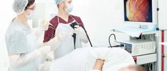
To perform a colonoscopy, special video equipment is used, which is fixed on an elastic probe. The probe is inserted into the patient's intestines. In order to ensure good visibility, a certain volume of air is pumped into the gastrointestinal tract. During the examination, the doctor assesses the condition of the intestines using images that appear on the computer screen. A specialist can notice the changes that have appeared from the inside, which makes it possible to give a real assessment of the structure of the organ’s tissues.
Intestinal ultrasound provides the opportunity to visualize inflammatory processes and neoplasms, the boundaries of the pathological process, and also identify the stage of the disease.
Many patients wonder whether an ultrasound doctor will be able to diagnose intestinal cancer during an examination. The latest generation of ultrasound machines provide this opportunity. After all, the examination is carried out using a special technique. Thanks to the use of an ultrasensitive sensor, it is possible not only to examine the tumor in detail, but also to identify its features.
Both colonoscopy and ultrasound diagnostics provide the opportunity to take high-quality images, which will be used in the future for a more detailed analysis of the pathological process, as well as to assess the dynamics of the treatment.
CT scan
The procedure does not detect all neoplasms. Colonography does not damage the mucous wall. It is possible to study and examine the contours of lesions and the condition of nearby organs. The CT scan procedure is similar to an x-ray. The device produces several frames, performing systematic storage in large quantities. Tomography without colonoscopy is not able to detect cancer in the first stage. For this method, you need to drink a solution or inject the composition. The technique takes longer than an x-ray; the person must remain quietly in a lying position for the duration of the procedure, without moving.
CT bowel
Virtual tomography operates using a special computer program that analyzes CT results and is capable of detecting polyps and growths larger than 1 cm. This method is not used in any center; early detection of diseases with its use is excluded.
How to properly prepare for research
Before performing an ultrasound of the intestines or before a colonoscopy, patients will have to follow certain recommendations of a similar nature:
- a few days before the study, patients need to exclude from their diet any foods that cause increased gas formation;
- approximately 24 hours before the examination, food intake should be minimized and dinner should be replaced with a light snack;
- in the evening or early in the morning you need to empty the intestines of feces. If this cannot be done naturally, then you should definitely resort to a cleansing enema.
- In order to fully cleanse the intestines, absorbent drugs can be very effective, such as:
- "Enterosgel";
- "Smecta";
- "Polysorb".
Before the study, experts also advise taking medications to reduce gas formation. Compliance with all preparatory measures will guarantee a highly informative survey.
Hydrogen test
The procedure takes place over three hours. Every 30 minutes the patient exhales into a special tube. A significant number of bacteria entering the small intestine are being studied. The principle of operation of the method: bacteria do not allow fluid to penetrate into the intestinal mucosa in normal quantities, so defecation is disrupted. When there are widespread symptoms, the indicated method is needed:
- irritable bowel syndrome;
- sugar intolerance;
- the intestines do not absorb foods (cow's milk, some fruits, honey);
- increased accumulation of bacterial flora;
- small production of juice for the function of digesting food;
- symptoms of changed and disturbed microflora (flatulence, diarrhea, constipation);
- assessment of the effectiveness of treatment of intestinal diseases that are associated with atrophy of the intestinal villi lining the walls.
How is ultrasound diagnostics and colonoscopy performed?
Many patients believe that undergoing an ultrasound of the intestine is a study during which a specialist will perform exclusively external manipulations, as is done when examining many organs. An intestinal ultrasound is performed a little differently.
First of all, a special catheter is inserted into the patient through the rectum to a depth of 5 cm. Its diameter is very small and does not exceed 8 mm. With its help, a special liquid is introduced into the intestine - a contrast agent, the purpose of which is to better visualize the walls of the organ.
During the examination, the patient does not experience pain. This diagnostic procedure is not very different from other types of ultrasound examinations.
As for colonoscopy, this is a much more unpleasant and sometimes even quite painful procedure. During the examination, a special device is inserted into the patient's intestines through the anus - an endoscope, which is a flexible tourniquet with a camera at the end.
For a complete examination of the intestines, the specialist will need to insert the endoscope deep enough, which causes pain to the patient. Another significant disadvantage of endoscopy is that it takes much longer to perform than ultrasound diagnostics.
How to detect bowel cancer during examination
In order to identify the symptoms of colon and rectal cancer, which today are one of the most common causes of death from cancer, it is necessary to conduct a thorough examination at the first suspicion.
Often, symptoms of bowel cancer do not appear until the disease has progressed to stage 3 or 4
. Colorectal cancer in later stages may be accompanied by pain in the abdomen and rectum, heavy bleeding from the anus, changes in bowel habits for one week or more, and a feeling of rapid tiredness and tiredness.
The tumor develops in the form of polyps
, most of which are benign, but over time, if they are not removed, they can develop into a malignant tumor, the cells of which will penetrate more and more into the colon and rectum.
People who eat natural, fiber-rich, low-fat foods and exercise regularly can avoid developing this disease.
Is it permissible to perform an ultrasound examination of the abdominal cavity after a colonoscopy?
It is not advisable to perform an ultrasound examination after a colonoscopy. In exceptional cases, specialists can still resort to ultrasound examination, for example, if there is a question about performing a biopsy, removing polyps, or stopping intestinal bleeding. In such cases, ultrasound will help monitor the dynamics of the treatment. In all of the above cases, ultrasound diagnostics are carried out exclusively through the peritoneal cavity.
As a rule, the patient is initially prescribed an ultrasound examination, and only then a colonoscopy. The choice of a specific method depends on the medical history, the patient’s condition and existing complaints.
Which type of diagnosis is better?
It is impossible to unequivocally answer the question of which type of diagnosis is better: intestinal ultrasound or colonoscopy, for the simple reason that these two examination methods simply cannot be compared in terms of effectiveness and significance. They have completely different purposes. Each of the methods can be used only if there are certain indications, and the final decision on prescribing one or another diagnostic method can only be made by a doctor.
However, most often, patients are initially prescribed ultrasound diagnostics, and if it turns out to be uninformative, then after this the patient is sent for a colonoscopy.