A hernia is a protrusion, a release of soft tissue from its physiological position. Each of us has heard or encountered a similar pathology of the spine and intestinal areas. And for some reason this diagnosis does not frighten potential patients of surgeons and neurologists.
The diagnosis of “gastric hernia” is a rare pathology. It frightens a person, although this disease is chronic and before the first symptoms appear, the presence of a protrusion does not bother the gastroenterologist’s patient in any way.
Gastric hernia is a rare disease. It occurs in a chronic form and does not bother a person until a certain point. Symptoms of the disease - heartburn and belching - are not typical.
Treatment is predominantly conservative. In difficult cases, surgical intervention is indicated. After surgery, relapses are unlikely.
How do hernias form?
Hernias occur in the area of “weak spots” of the anterior abdominal wall, under the influence of intra-abdominal pressure. Factors that cause its increase are called producing factors and include: physical activity, cough, childbirth and all those cases when the abdominal press tenses.
“Weak spot” is the area of the abdominal wall where the muscular aponeurotic part is most thinned. This may be the place of muscle attachment to the aponeurosis or physiological openings (inguinal rings, umbilical ring).
An increased risk of hernias is observed in people with predisposing factors: connective tissue weakness syndrome, damage to the nerves innervating the abdominal wall, as well as the presence of postoperative scars.
What does a hernia consist of?
All hernias, regardless of location and size, have a common structure and consist of the following components:
- A hernial orifice is a defect of the anterior abdominal wall, mainly in the aponeurosis. Through them, the internal organs leave the abdomen and end up under the skin. If the hernial orifice is wide, then through it the contents can freely return to the abdominal cavity.
- Hernial sac is a sheet of stretched peritoneum covering organs that extend beyond the abdominal cavity through the hernial orifice. The hernial sac is covered with several membranes, these also include subcutaneous tissue and skin.
- The contents of the hernial sac are the internal organs or their individual parts that extend beyond the abdominal cavity through the hernial orifice.
What will happen if left untreated?
The bulge will gradually increase, will no longer disappear when lying down, and the pain will intensify. The most dangerous complication is strangulation, which can lead to organ necrosis followed by peritonitis, which is a direct threat to life. In case of strangulation, emergency removal of the abdominal hernia is performed.
Important! In this condition, you should not try to correct the defect yourself. It is urgent to call an ambulance.
Other possible complications of hernias:
- inflammation;
- impossibility of reduction;
- coprostasis.
What types of hernias are there?
Depending on the anatomical location, external and internal hernias are distinguished. External ones include:
- Umbilical hernia.
- Inguinal hernia.
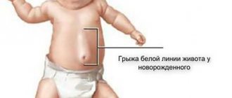
- Hernia of the white line of the abdomen.
- Postoperative hernia.
- Paracolostomy hernia.
- Lumbar hernia.
- Perineal hernia.
- Obturator hernia.
- Hernia of the xiphoid process.
- Sciatic hernia.
Hernias that form inside the abdominal cavity are considered separately. With such hernias, internal organs can be located in pockets of the peritoneum or penetrate into the chest cavity through the openings of the diaphragm.
Depending on the size of the hernia, they are divided into:
- Small: hernial orifice less than 4 cm
- Medium: hernial orifice from 4 to 10 cm
- Large: hernial orifice larger than 10 cm
Hernias are also classified according to the degree of development:
- initial - a small depression is identified in a weak spot of the abdominal wall - a triggering factor for the formation of a hernia;
- canal - internal organs begin to sink into the hernial opening;
- complete - the internal organs have passed through the hernial orifice and are located under the skin.
According to the clinical course, hernias are divided into:
- reducible - the contents of the hernial sac move freely from the abdominal cavity to the hernial sac and back.
- irredeemable.
- strangulated - a condition in which the hernial orifice puts pressure on the structures of the released organ, which leads to disruption of its blood supply and can lead to necrosis. There are: elastic strangulation, fecal strangulation, parietal strangulation, retrograde strangulation, Meckel's diverticulum strangulation, Broca's hernia.
Signs of a gastric hernia
What are the symptoms of a gastric hernia? Signs of disruption of the transition of the esophagus to the stomach:
- heartburn,
- belching,
- feeling of bitterness or acidity in the mouth,
- regurgitation of food
- feeling of food stuck in the lower part of the sternum,
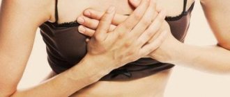
- chest pain during night rest and eating,
- night cough.
Many people are interested: is a hernia on the stomach dangerous? What complications may arise? A hiatal hernia can provoke the appearance of coronary pain due to irritation of the vagus nerve and subsequent spasm of the coronary vessels of the heart. This situation is dangerous due to the occurrence of cardiovascular complications, including myocardial infarction.
What are the symptoms of a hernia?
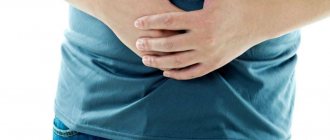
In the initial stages, a hernia may manifest itself as discomfort or slight pain during physical activity at the site of hernia formation. As the aponeurosis thins or the tissues of the anterior abdominal wall weaken, a painless protrusion appears, disappearing when pressure is applied to it. With each new episode of increased intra-abdominal pressure, the hernial orifice will increase in size, and the sac will stretch due to an increase in the volume of contents.
External abdominal hernias that occur without complications are characterized by such general symptoms as: the presence of a hernial protrusion, discomfort in the hernia area, dysfunction of the organs that make up the hernial contents.
All about hernias
go back to the ambulatory surgery department
ALL ABOUT HERNIA
What is a "hernia"? A hernia is a protrusion of abdominal organs under the skin through weak spots in the abdominal wall. The exiting organs are located in the hernial sac formed by the peritoneum (the inner lining of the abdominal wall). The hernial sac can contain almost any organ of the abdominal cavity (intestinal loops, bladder, less often the stomach or part of it), the omentum, and extremely rarely the liver, spleen.
Rice.
Postoperative ventral hernia Why does a hernia form? The abdominal wall, consisting of muscles and aponeuroses, performs a number of functions, one of which is to hold the internal organs in their natural position and counteract the intra-abdominal pressure they create. Under the influence of intra-abdominal pressure, a defect (hernial orifice) can form in the weakest places of the abdominal wall, through which a hernia emerges. Predisposing factors may contribute to this, such as:
- Increases intra-abdominal pressure
1. excessive physical activity
2. severe cough, including chronic (smoker's cough)
3. constipation
4. diseases that cause shortness of breath with difficulty exhaling (bronchial asthma)
- Conditions and diseases associated with the development of connective tissue weakness (obesity, varicose veins of the legs, congenital connective tissue pathologies, hereditary predisposition)
- Patients who have previously been operated on for hernias are also at risk due to predisposing factors
- The formation of a hernia may go unnoticed, or may be accompanied by intense pain. Subsequently, under the influence of the same factors, a gradual increase in the hernia occurs, until most of the abdominal organs exit into the hernial sac.
Who can get a hernia? A hernia can occur in anyone, regardless of gender and age. External signs and symptoms. External manifestations and symptoms of hernias can develop gradually or occur within a short time.
- A feeling of pressure, weakness or pain in the abdomen, groin area or scrotum, which occurs or intensifies with physical activity or straining.
- A visually detectable “bulging”, a bulge in the abdomen, in the groin area, or scrotum, which appears or increases in size during physical activity or straining. Also, in the area of the above formations, a feeling of discomfort or pain may appear during physical activity, coughing, or straining.
If you have any of the above symptoms, you should consult your doctor. The sooner the diagnosis is made and treatment is carried out, the higher the chance of preventing the development of complications, sometimes fatal.
Read more about types of hernias.
Rice. Classification of hernias of the anterior abdominal wall
Lumbar hernia of the anterior abdominal wall A lumbar hernia of the abdomen (Hernia lumbalis) is considered to be a hernia formation on the posterior, lateral walls (in the lumbar region), emerging through various weak points of the lateral abdominal wall. The main anatomical formations through which lumbar hernial formations occur are the Petit triangle and the Greenfelt-Lesgaft gap, aponeurotic fissures. Petit's triangle is bounded posteriorly by the outer edge of the vastus dorsi muscle, anteriorly by the inner edge of the external oblique muscle, and inferiorly by the iliac crest. In the area of Petit's triangle, under the superficial fascia and thin aponeurosis, there is an internal oblique muscle. The Greenfelt-Lesgaft interval is often quadrangular in shape. Its upper border is made up of the lower serratus posterior muscle and the XII rib, medially it is delimited by the longitudinal muscles of the spine, the quadratus lumborum muscle, and the edge of the internal oblique muscle runs in front and below. The shape and size of the gap can vary depending on the length of the XII rib - with a long rib, the Greenfelt-Lesgaft gap is sometimes absent or looks like a gap, and with a short rib it increases in size. Aponeurotic fissures usually form at the site of passage of blood vessels and nerves, but can sometimes appear as a result of rupture or abnormal development of the aponeurosis. Among the causative factors contributing to the occurrence of hernia formation in these areas is weakness of connective tissue and muscle atrophy, inflammatory processes. Hernial protrusions occur on the left more often than on the right; bilateral ones are rare.
Recurrent hernias are a complication of surgical treatment of hernias. The causes of recurrent hernial protrusion may be related to the patient’s lifestyle and structural features of his body.
For example: failure to comply with the terms of the recovery period, when a person begins active physical activity ahead of schedule. With age-related changes and a number of pathological conditions, when tissues can become flabby, change elasticity and structure, which also affects the quality and duration of healing after surgery. Thus, in elderly, exhausted or very obese patients, relapses can be observed regardless of the method of operation and the course of the postoperative period. The main reasons for the formation of recurrent hernias:
- errors related to operational technology
- connective tissue deficiency
- wound infection during or after surgery
- excessive physical exertion, especially soon after surgery
The only treatment for recurrent hernias is surgery. In this case, various methods of hernioplasty using various mesh prostheses are selected.
Rice. Removal of old and deformed graft
Rice. Restoration of the integrity of the inguinal canal and reprosthetic hernioplasty according to Liechtenstein
Why is a hernia dangerous? In addition to the obvious inconvenience associated with the presence of a cosmetic defect, decreased physical activity and ability to work, a hernia carries the risk of developing a number of complications. These include dysfunctions of the organs located in the hernial sac - constipation, urination disorders, and when large volumes of organs leave the abdominal cavity - breathing problems. A serious complication is the development of a strangulated hernia. Strangulation is compression of the hernia in the hernial orifice, resulting in necrosis of the contents of the hernial sac. Incarceration is accompanied by sharp pain in the area of the hernial protrusion. The most dangerous thing with strangulation is the development of intestinal obstruction (a loop of intestine is strangulated) and subsequent peritonitis. This situation requires immediate resolution through surgery. By and large, regardless of which organ is the content of the hernial sac, the final result without appropriate treatment is the same - peritonitis, the only difference is time. Peritonitis - inflammation of the peritoneum - is a serious complication of a large number of diseases, including strangulated hernias; the development of this pathological condition is one of the most difficult problems in surgery. Age, obesity, and the presence of concomitant pathologies further aggravate this situation. Without surgery, there is only one outcome - death. Even if the operation is performed, but more than a day has passed since the onset of the strangulation, up to 30% of patients die in the postoperative period. There is no need to bring such a small problem as a hernia to such a tragic situation.
How is a hernia treated? The only way to treat a hernia in adult patients is to perform surgery (hernia repair).
When should hernia treatment begin? Following from the above, the earlier the operation is performed, the better.
Is it possible to do without surgery? -No. For adult patients with a hernia, the only treatment option is surgery.
Are there any contraindications for surgery? Hernia repair cannot be performed in the presence of severe concomitant pathology, when the operation can only harm and not help. Such cases include: the coming months after myocardial infarction, stroke, and a number of other extremely severe concomitant pathologies. It should be remembered that the presence of chronic diseases is not an absolute contraindication to surgery, but only requires appropriate correction in the preoperative period.
Is it possible to perform other surgical interventions simultaneously with hernia surgery? Yes. Hernia surgery can be supplemented with almost any surgical intervention. Often, especially in older patients, there are several problems that require surgical intervention. In such situations, it is preferable to get by with one operation that combines the removal of the hernia and some other problem. Performing combined operations is a priority method, as it allows you to solve two (or more) problems in one surgical intervention and relieves the patient of psycho-emotional problems associated with the need to undergo several operations.
What types of operations are performed to repair a hernia? Today, more than 300 methods of hernia repair are known - ventral, inguinal, umbilical, femoral, postoperative. But all of them can fundamentally be divided into two groups:
- with plastic surgery using one’s own tissues – the tissue of the abdominal wall around it is used to close the hernial opening
- with plastic surgery using synthetic materials (or plastic surgery “without tension”) - synthetic prostheses made from surgical threads are used to close the hernial opening.
Plastic surgery with one’s own tissues is the oldest group of methods, born in the second half of the 19th century, it is the most extensive and widespread. Its essence is to close the hernial orifice with the patient’s own tissues (muscles, fascia and aponeuroses) in one way or another. The frequency of hernia recurrence after these operations varies from 20% to 70% depending on the condition of the patient’s tissues, the method of hernioplasty and the correctness of its choice. The main disadvantages are severe pain in the first days after surgery due to tissue tension and long periods of physical rehabilitation. Intense physical labor is contraindicated for at least 3 months after surgery.
Methods of plastic surgery “without tension” of the patient’s own tissues have existed since the second half of the 60s of the twentieth century. What distinguishes them from methods of plastic surgery using one’s own tissues is the use of “patches” made of synthetic materials to close the hernial orifice. In recent years, these methods have gained great popularity, which became possible thanks to the creation of advanced synthetic materials and the development of new methods for closing the hernial orifice, which practically guarantee the patient against recurrence of the hernia. The relapse rate does not exceed 1% in specialized clinics, regardless of the type of hernia. Despite the skin incision over the hernia, pain after surgery is minimal, because there is no tension on your own tissues. Intense physical labor is possible a month after the operation, household physical activity is not limited. This allows such operations to be performed on an outpatient basis. A positive point is also the possibility of performing the operation under local or spinal anesthesia, which is especially important for elderly patients and patients with heart and lung diseases. Hernioplasty using the ILLichtenstein method for inguinal hernias has become most widespread due to its reliability and simplicity. It is applicable for any type and size of inguinal hernia.
Rice. Plastic surgery of the inguinal canal with a mesh polypropylene prosthesis according to Liechtenstein.
It is also worth mentioning laparoscopic (through punctures of the anterior abdominal wall) methods of hernia repair. These are operations that are performed under the control of a laparoscope - a device that allows, using a mini-video camera, to eliminate a hernia from the abdominal cavity without making an incision in the skin over the hernia. They were born in the early 80s of the twentieth century with the advent of video technology. In most cases, the abdominal wall defect is closed from the inside of the abdominal cavity with a synthetic mesh prosthesis. The frequency of hernia recurrence after this repair is 2-5%, which is determined by the type of hernia and the preparedness of the surgeons. The important advantages of these methods are low trauma, which means minor pain after surgery, short rehabilitation periods (up to a month for physical labor), as well as the ability to perform bilateral plastic surgery and, if necessary, combined operations in the abdominal cavity through the same punctures in the abdominal wall. Serious disadvantages of this group of methods include the need for general anesthesia (anesthesia), the need to inject gas into the abdominal cavity to create an operative space (dangerous in patients with lung and heart diseases).
Why is a hernia dangerous?
Any hernia is dangerous for the development of complications. The most serious complication of a hernia is strangulation. It occurs when the blood supply to the contents of the hernial sac is disrupted and tissue necrosis occurs.
An equally serious complication is intestinal obstruction. As a result of the prolonged presence of intestinal loops inside the hernial sac, they are compressed, the movement of intestinal contents through them is disrupted, and intestinal obstruction forms.
It is very important to understand that the development of complications can occur suddenly, against the background of complete well-being: on vacation, at the dacha, while traveling - in situations where qualified medical care is difficult to access or not available at all. Treatment of hernia complications requires emergency intervention, and delay can lead to a sharp deterioration in a person’s condition and significantly worsen the prognosis of delayed treatment.
Therefore, it is better to get rid of the hernia before complications develop.
Treatment
Methods of conservative therapy include taking medications that eliminate the symptoms of a hiatal hernia; drugs help reduce the acidity of gastric juice or neutralize it, improve the condition of the mucous membrane, have an anti-inflammatory effect, etc. However, drug therapy has its drawbacks, for example, suppression of gastric juice secretion leads to poor digestion of food. In addition, a decrease in stomach acidity increases the risk of developing a malignant process. Also, conservative therapy requires strict adherence to a diet; it is impossible to eliminate the symptoms of a hiatal hernia without dietary restrictions.
But drug therapy is aimed only at eliminating the signs of the disease; after stopping taking the drugs, the symptoms of a hiatal hernia return. Complete cure is only possible with surgical treatment.
Hernia treatment
It is only possible to completely get rid of a hernia through surgery. There are also conservative methods to alleviate the condition of a hernia, but their use is possible only if there are contraindications to surgery.
Contraindications to elective surgical treatment include severe concomitant diseases, malignant processes in advanced stages, acute diseases and pregnancy. It is important to note that complicated hernias must be operated on urgently for health reasons.
The smaller the size of the hernia, the easier the operation for both the surgeon and the patient. In turn, the treatment of giant hernias represents a major surgical problem and requires the use of non-standard approaches to its elimination.
Hernioplasty (hernia repair) is the name of an operation to “liquidate” a hernia with plastic surgery of a defect in the anterior abdominal wall.
Diagnostics
To prescribe adequate treatment, high-quality diagnosis is important; the diagnosis is made based on the existing symptoms of a hiatal hernia and the results of instrumental examination methods.
- To clarify the size of the hernia, the presence of strictures, ulcers, and assess the motility of the esophagus, an X-ray examination is provided with the introduction of a barium suspension into the esophagus.
- Fibrogastroscopy is one of the main diagnostic methods necessary to assess the condition of the mucous membrane and the degree of pathological changes.
- pH-metry - determination of the level of gastric secretion; esophageal manometry - determination of motility and propulsive ability of the esophagus. Methods are recommended to confirm the need for surgery and select the most effective correction method.
If you have a hiatal hernia, it is important to undergo a comprehensive examination, since the pain that occurs with this pathology may resemble pancreatitis, angina or heart attack. For example, in 1/3 of patients with hiatal hernia, the main symptom, in addition to pain localized in the left side of the chest, is a heart rhythm disturbance resembling extrasystole or paroxysmal tachycardia. Therefore, differential diagnosis is important to avoid wasting time in an attempt to treat a non-existent disease.
To obtain a complete picture of the disease and exclude complications, other examination methods may be needed: ultrasound, computed tomography, ECG, etc. Our clinic offers patients all the necessary modern diagnostic methods.
What are the methods of anterior abdominal wall plastic surgery?
Plastic methods can be tension or non-tension.
Tension is a type of plastic surgery performed using the patient’s own tissues. This method received this name because, in order to eliminate a hernia defect, the tissues must be “tightened” and sewn together. The resulting tension in the tissues can cause pain after surgery and result in a possible relapse. At the present stage of development of medicine, this method of closing hernias defects is significantly inferior to non-tension methods.
Tension-free plastic surgery involves the use of modern mesh prostheses to strengthen the anterior abdominal wall. The prosthesis is a polypropylene network, which, due to its flexibility, strength and high degree of tissue “germination”, has shown its reliability and safety when used in hernia repair. Mesh prostheses come in different sizes, from small ones with a diameter of 5 cm for umbilical hernias, to large ones of 50 x 50 cm for giant incisional hernias. Modern three-dimensional mesh systems make it possible not only to strengthen the hernia defect in the form of a “patch”, but to completely fill it, significantly reducing the risk of relapse. In some situations, a special mesh is installed, the surface of which is coated with a special composition that allows it to safely contact the abdominal organs and avoid the formation of adhesions between them.
The open hernia repair operation consists of several stages:
- Isolation of the hernial sac. A skin incision is made above the hernial protrusion, the hernial sac is freed from the surrounding subcutaneous fatty tissue. The “hernial orifice” is distinguished.
- The hernial sac is opened, the condition of the contents of the hernial sac is assessed, and if there are no complications, the contents are immersed in the abdominal cavity.
- The hernial sac is excised, stitched and plunged into the abdominal cavity.
- The integrity of the anterior abdominal wall is restored (plasty is performed).
Hiatal hernia (HH) - symptoms and treatment
To diagnose hiatal hernia, in addition to detailed questioning of the patient, almost all research methods used in gastroenterology are used. Mandatory diagnostic methods include:
- clinical and radiological examination;
- fibroesophagogastroduodenoscopy (FEGDS);
- esophagotonometry;
- pH-metry of the esophagus and stomach;
- Ultrasound of the abdominal cavity.[12][158]
The leading instrumental methods are considered to be X-ray diagnostics and FEGDS.[8][16]
X-ray diagnostics
Thanks to the X-ray diagnostic method, fundamental research has been carried out on hiatal hernias, classifications have been developed, various forms of this pathology have been studied, and a number of indications and contraindications for various types of treatment of hiatal hernias have been developed.
The modern full name is “Polypositional X-ray diagnostic examination of the esophagus, stomach and duodenum using a liquid suspension of barium sulfate on a troscope.”
This X-ray examination allows you to reliably diagnose various forms of hiatal hernia, including “small” esophageal hernias, identify cardia insufficiency, gastroesophageal reflux, reflux esophagitis, and exclude cardia insufficiency associated with impaired passage of food in the underlying sections of the gastrointestinal tract.
Endoscopic esophagogastroduodenoscopy
In the middle of the 20th century, the latest technologies in endoscopy were developed and widely introduced into clinical practice. They have made it possible to significantly expand the possibilities for diagnosing gastroenterological diseases.
The peculiarity of endoscopic esophagogastroduodenoscopy is:
- the use of flexible fiber optics and the creation of endoscopic devices - fiber gastroscopes;
- high resolution of these devices with the ability to conduct research while visualizing images on the monitor;
- minimally invasive endoscopic diagnostics;
- minimal percentage of complications that arise;
- no need for special preparation of the patient before the start of endoscopic examinations;
- outpatient nature of endoscopic diagnostic methods.
All this allows us to recommend this diagnostic method not only to patients, but also to the population as a whole for medical examination and detection of the disease in the early stages.
Of course, endoscopic diagnosis of hiatal hernia is not an easy procedure, but FEGDS doctors consider it as a screening method indicated for all patients, including people with minimal symptoms of gastroesophageal reflux, dyspepsia or dysphagia (digestive or swallowing disorders), as well as anyone who suffers from digestive diseases. tract.
The main direct and indirect symptoms of hiatal hernia, usually manifested during FEGDS, include:
- reduced distance from the anterior incisors to the cardia;
- reduced length of the abdominal esophagus;
- hernial cavity;
- “second entrance” to the stomach;
- gaping (opening) of the cardia or its incomplete closure;
- prolapse (protrusion) of the gastric mucosa into the esophagus;
- reflux (reverse flow) of stomach contents into the esophagus;
- segmental dilation (expansion) of the esophagus in the area of the ninth segment;
- absent, poorly visualized or blurred Z-line;
- flattened fold of the cardioesophageal junction detected during inversion examination of the cardia;
- a smoothed angle of His, also detected during inversion examination of the cardia.
Most of the listed endoscopic symptoms of the hiatal hernia can be detected through video monitoring during FEGDS, which helps to establish an accurate diagnosis.
Is it possible to operate a hernia without incisions using modern mesh prostheses?
Yes, you can!
At the moment, laparoscopic hernioplasty is the operation of choice for the treatment of various types of hernias. This operation is performed through punctures in the anterior abdominal wall. The surgeon inserts a video camera into the abdominal cavity and, using additionally inserted manipulators, releases the hernial sac from its contents. In the second stage, the surgeon separates the peritoneum of the hernial sac from the tissues of the anterior abdominal wall, dissects it and places a special mesh prosthesis on the hernial orifice. Then, the mesh is reinforced on top with the previously separated peritoneum.
Thanks to this, the prosthesis does not form adhesions with internal organs. This method of plastic surgery avoids tissue tension and greatly reduces the likelihood of relapse. The absence of large incisions on the anterior abdominal wall contributes to a comfortable course of the postoperative period.
How does the postoperative period proceed?
After laparoscopic hernia repair, pain is practically not felt and is therefore easily controlled using tablet painkillers.
The only reminder of the operation was discomfort in the area of small punctures on the anterior abdominal wall.
In uncomplicated cases, discharge from the hospital occurs within 2-3 days. The time frame for returning to work, on average, does not exceed 10-14 days.
Traditional open surgery is characterized by pain in the postoperative period. However, carrying out comprehensive pain relief in a hospital allows you to reduce its severity and feel comfortable throughout the entire recovery period.
After discharge from the hospital, you will be advised to limit physical activity for a period determined by your attending physician based on the complexity of the treatment performed.
My approach to treatment is my own technique
To restore the anatomical relationship in the area of the esophagus and stomach, I use a technique I have improved - a modified Tope fundoplication of 270 degrees, for which a patent has been issued.
During the operation, after reducing the esophagus and upper part of the stomach to a normal level, I sutured the diaphragmatic opening to a normal size, then from the upper 1/3 of the stomach I formed a symmetrical cuff covering the esophagus at 270°. As a result, the anterior-right surface of the esophagus, where the vagus nerve passes, remains free. The cuff, which serves to strengthen the lower esophageal sphincter, will not only prevent the reflux of gastric contents, in contrast to the Nissen method described above, natural protective mechanisms are preserved - the gag reflex and belching.
After partial fundoplication according to Toupet 270 degrees, drinking carbonated drinks, large portions of food and other similar dietary habits will not cause pain or heaviness in the epigastrium. Thanks to improved technology, the number of relapses a year after surgery does not exceed 2%. 5 years after treatment for a hiatal hernia, relapse develops in only 4% of patients.
Who deals with surgical treatment of external abdominal hernias?
You can always contact the clinic of coloproctology and minimally invasive surgery for surgical treatment.
Qualified specialists regularly perform laparoscopic interventions for hernias of the anterior abdominal wall. In some cases, we also use traditional open surgery.
At KKMH, treatment of hernias is carried out both on a paid basis and under the compulsory medical insurance policy.
Sign up for a consultation by phone +7 (499) 11-03-222.