Ultrasound examination of the esophagus is prescribed for accurate diagnosis of diseases of this organ, as well as the entire gastrointestinal tract. Unlike gastroscopy, this diagnostic method is more effective, which makes it possible to detect many dangerous pathologies at an early stage, when they do not make themselves felt.
Introduction: What is Ultrasound?
During this procedure, the doctor uses ultrasound, which is reflected from the sensor by the tissues of the esophagus and then transformed using computer equipment into an image. On the screen of his monitor, the doctor sees all the structures of this organ, as well as its layers: internal (mucous membrane), middle (muscular) and external.
The doctor can also see changes in the mucous membranes of the organ at rest, as well as during swallowing. All data is then processed and used for a diagnostic conclusion. The procedure is completely painless and does not cause discomfort to the patient. It does not replace MRI: today it is the most modern way to determine diseases of the esophagus.
When should an ultrasound be done?
Indications for ultrasound of the esophagus are as follows:
- establishing the causes of heartburn;
- assessment of the condition of the membranes of this organ in order to exclude or confirm the development of esophagitis;
- reflux;
- suspicion of achalasia;
- suspicion of a hernia in the esophageal opening of the diaphragm;
- a state of the body when it is impossible to perform esophagoscopy, or the patient’s refusal to do such a procedure;
- heart or pulmonary failure, as well as damage to the central nervous system;
- diagnosis of organs surrounding the esophagus;
- assessment of the effectiveness of treatment of gastrointestinal diseases.
Decoding the results
What does an abdominal ultrasound show in a child:
- An ultrasound of the liver will help diagnose benign and malignant neoplasms, abscesses, cysts, cirrhosis, and parasites.
- An ultrasound of the spleen reveals an enlargement of this organ, which may indicate various blood diseases.
- Ultrasound of the gallbladder helps to identify cholecystitis, cholelithiasis and its complication - hydrocele, as well as to identify such a common phenomenon among children as dyskinesia of the gallbladder and biliary tract.
- Ultrasound of the pancreas often reveals nonspecific changes in infants - reactive pancreatitis. If it is not treated in time, it becomes acute or chronic, accompanied by abdominal pain, nausea, vomiting and other symptoms.
Normal indicators
Indicators for a newborn:
- the thickness of the right lobe of the liver is on average 50 mm;
- spleen length – from 30 to 50 mm, on average – 40 mm;
- thickness of the pancreas – from 5 to 10 mm, on average – 7 mm;
- The length of the gallbladder is from 12 to 25 mm.
Parents should not interpret an ultrasound of the child’s abdominal cavity on their own. The norm varies for children of different ages - what is the norm for an infant will already be a pathology for a teenager.
How is an ultrasound performed on an adult?
The procedure is determined by the specific structure and location of the organ. Before the examination, the patient must refuse food and water. Alcohol is prohibited the day before. To prevent gases from accumulating in the stomach, a few days before the examination you need to exclude kefir, radishes, legumes, cabbage, jam, and sweets. You can't drink soda.
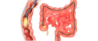
When inserted percutaneously, the sensor moves along the skin above the esophagus. For better contact, a gel is used. Such research is rarely done for adults. More often, an intraesophageal examination is performed with the insertion of a sensor directly into the lumen of the organ.
Ultrasound of the esophagus
Often such research is combined with the introduction of water into the esophagus. This makes it possible to assess gastric motility.
The duration of the procedure is no more than 15 minutes. Discomfort may occur in some cases due to the insertion of an ultrasound probe into the esophagus. In this case, the patient should be in a lateral position. After this procedure, the patient is advised to lie down for a while.
Features of preparation for ultrasound in children
Conducting such a study for a child is preferable to FGDS. The fact is that it is more difficult for children to do FGDS, and their digestive system is more vulnerable.
The child and his parents need to carefully prepare for the procedure. The period of fasting before the procedure is determined by the age of the patient. Thus, newborns can be examined before feeding.
The last meal should be at least three hours before the ultrasound. To eliminate flatulence, carminative drugs are prescribed.
Children between the ages of one and three need to prepare the day before the diagnosis. Fruits and vegetables are excluded, and the child should eat only boiled meat and cereals. Fasting time before the procedure is 4 hours. One and a half hours before an ultrasound scan of the stomach and esophagus, you should not drink water.
Children over three years old need to prepare for the procedure in advance - preferably three days in advance. It is necessary to completely exclude all products that cause increased gas formation. You cannot eat cabbage, sweets, legumes, or dairy products. It is better to give your child fish, boiled meat, and porridge. Before examining the esophagus and stomach, the child must fast for at least six hours. One hour before the ultrasound, you should not drink water.
If the examination of the esophagus, as well as the stomach, is scheduled for the afternoon, a small breakfast is allowed in the morning. However, you need to have breakfast so that there is at least six hours between food and research.
What's next?
With the examination result in hand, the patient returns to the attending physician. He studies the examination protocol and prescribes therapy. If hypoechoic nodes are detected, then the diagnosis should be continued.
Patients should not panic when they read the phrase “hypoechoic formation” in the protocol. This is not a diagnosis, but a description of the density of the structure. In this place, the tissue structure is less dense than around it, and the ultrasound pulse moves more slowly. This means that the doctor will have to determine whether this is a pathology or the norm for this area. Sometimes the results obtained are simply incorrect when performing an ultrasound of the throat and larynx, which does not indicate the presence of serious health problems, but the low qualifications of the sonologist (the specialist who performed the ultrasound).
And one more clarification. Structures with low density may turn out to be cysts. This is a cavity with thin walls, the tissue of which resembles a mucous membrane. There is liquid inside the cavity. However, the term “cyst” is never written in the ultrasound report. Since to establish such a diagnosis it is necessary to perform a biopsy. The procedure can be performed with a special needle or other medical devices.
How is an ultrasound done for children?
The child must be placed on his back. A gel is applied to the location of the esophagus and stomach for better contact of the sensor with the skin. Sometimes it is necessary for a small patient to change his body position.
The sensor is installed on the epigastric region. The doctor constantly changes the angle of inclination so that the image on the screen can be adjusted. Sometimes it may be necessary to change the baby's body.
Sometimes the child is given some water to drink during the procedure. This is done so that the dynamics of fluid movement through the esophagus can be monitored.
An ultrasound probe is not inserted into a child's esophagus . The introduction of an endoscope is also not carried out, which is primarily due to the structural features of the gastrointestinal tract. Normally, the thickness of the wall of the organ under study in children does not exceed several millimeters.
Unfortunately, in the modern world, diseases that in one way or another affect the ENT organs are very common and widespread not only among adult and elderly patients, but also among children. Unfortunately, many people do not attach much importance to such diseases, believing that they can cope with them on their own, not suspecting that a common cold, if not treated, can even develop into an abscess.
A frivolous, neglectful attitude towards one’s own health and similar diseases often lands one in the hospital . And in some cases, if the disease is too advanced, a visual examination is not enough, the doctor has to resort to another diagnosis: an ultrasound examination of the throat or larynx. Being interested in what it shows, the patient learns that such an examination can find out the cause of the patient’s problem and highlight various pathologies and diseases.
Due to the large amount of valuable information necessary for treatment, simplicity, accessibility, widespread use, and low cost, ultrasound remains one of the most popular methods for diagnosing a number of diseases.
When is an ultrasound of the larynx and throat prescribed, how does it work?
This procedure makes it possible to evaluate not only the size and shape, but also the density of the organs being examined. Ultrasound, emitted by a miniature sensor of a special device, penetrates the soft tissues of the body, is reflected from them, and comes back. As a result, the screen displays a complete picture of the area of the body being examined.
The doctor compares the information received with data accepted as the norm and assesses the patient’s condition. If we are talking about the throat, then the front part of the neck is examined, where the esophagus with the trachea and larynx are located.
- Most often, with complaints in this area, ultrasound can identify problems such as:
- Various inflammatory processes in different stages.
- Benign formations, malignant tumors (cysts, such unpleasant phenomena as polyps, cancerous tumors).
- The presence of foreign objects that should not be in the examined organs.
- Diseases associated with an organ such as the thyroid gland.
- Ultrasounds are also prescribed when the doctor suspects the presence of any pathologies in the structure of the trachea or esophagus (narrowing, curvature, enlargement of the lumens).
- Any scars, papillomas, fibromas, other formations.
ultrasound of the throat
Typically, an ultrasound is prescribed when the patient complains of certain problems and ailments. Most often these include the following symptoms:
- Any discomfort or pain, as well as a “lump in the throat”, that is, difficulty swallowing.
- Changes in timbre (the appearance of hoarseness, hoarseness in the voice).
- If the patient suddenly has a feeling as if the skin on the throat is too tight.
- In cases where the patient feels as if there is a foreign object in his esophagus.
- If the thyroid gland enlarges.
- If the lymph nodes in the neck are enlarged.
- If masses are felt on the neck, or if the doctor suspects the presence of a tumor.
- The patient complains of a prolonged cough that cannot be treated and has nothing to do with the condition of the lungs or bronchi.
If a person experiences at least one of these symptoms, there is no need to put off the ultrasound and wonder what exactly this procedure shows. You need to see a therapist, and he will refer you to a specialist. To begin with, you should go to an otolaryngologist, take the tests he prescribed, and carry out all the recommended studies.
If it turns out that the problem comes from the thyroid gland, the patient is sent to an endocrinologist. You should not choose on your own: the therapist will make this choice better than the patient. If an ultrasound is prescribed, you do not have to prepare in any way, there are no restrictions, and you can drink in any quantity.
The examination is carried out as follows:
- When an ultrasound of the throat or larynx is performed, the patient exposes the neck area, removes all jewelry from it, and lies down on the couch.
- The doctor carefully spreads a special gel on his neck, making it easier for the device’s sensor to move along it.
- The sensor slides over the skin, emits ultrasonic waves, which are reflected from body tissues and received back by the device.
- The specialist sees images of the areas of interest on the screen, enters all the data into a special protocol, deciphers the results obtained, and hands the card to the patient. The ultrasound procedure itself takes on average about twenty minutes.
After the examination, it is necessary to begin treatment as quickly as possible. If nothing is revealed, and the unpleasant sensations remain, the patient should go to a neurologist or psychotherapist.
Particularly alarming are complaints such as rapid weight loss, constant fatigue, fatigue, enlarged nodes, purulent discharge (despite the fact that the person is not currently suffering from colds), pain during swallowing movements, accompanied by an increase in temperature, and a change in voice , as well as fainting and frequent dizziness.
These signs indicate the possible development of diseases not only of the larynx, as well as the thyroid gland or ligaments, but also cancerous tumors. It should be noted that ultrasound is used not only to diagnose diseases or confirm existing diagnoses, but also to monitor the patient’s recovery process. The examination has no contraindications, except for the presence of wounds or other injuries on the patient’s neck (as they can distort the results obtained).
If the patient is taking antibiotics, the doctor may recommend temporarily stopping them if he suspects the development of an inflammatory process in the throat or larynx. As for the population groups that are most often prescribed such an ultrasound, these are men aged about sixty years.
What do the statistics show in connection with this survey? That the percentage of throat and larynx diseases in men is significantly higher than in women. This is due not only to smoking and drinking alcoholic beverages, frequent neglect of inflammatory processes, but also to working in various hazardous enterprises.
What diseases does this ultrasound detect?
Diagnosis of the condition of the throat and larynx can identify or, conversely, exclude the following pathologies:
- Various tumors, their location, boundaries.
- Hyperthyroidism.
- Cysts.
- The presence of a foreign body in the throat, its exact location.
- Purulent abscesses of various nature.
- Consequences of neglected, untreated inflammatory, purulent processes in the larynx.
- Injuries of the larynx, its deformations after accidents.
Multiple nodes and cystic formations in the enlarged right lobe of the thyroid gland.
All hypoechoic areas are examined by the doctor with double attention. Most attention is paid, of course, to the possibility of developing malignant tumors. Moreover, they are quite widespread and difficult to detect. This is due to the fact that at an early stage the formations are very small in size and do not expand beyond the mucous membrane, and therefore people do not know that they are sick for quite a long time.
If the thyroid gland is the focus of attention, then the doctor looks at its echogenicity and acoustic density, which are expressed on the device screen as darkening against the general background of the image. The echogenicity of the gland can be normal, decreased, increased, or completely absent.
If an ultrasound shows that there are no structural changes in the tissues of the thyroid gland, then everything is normal and there is nothing to worry about. If hypoechoic areas are detected, then the development of malignant formations can be suspected. Hyperechoic zones are areas of increased density and indicate the proliferation of connective tissue.
What to do next?
If, during an ultrasound, a specialist identifies nodes with hypoechoic areas, he will certainly refer the patient for a number of additional examinations. This can be a biopsy, radiography or computed tomography, as well as fibrolaryngoscopy, which shows in more detail what exactly is going on in the patient’s body. At the same time, of course, various blood tests are prescribed, a treatment plan is developed, and instructions are given to the patient.
Fibrolaryngoscopy procedure
Quite often, doctors again prescribe an ultrasound and, under its control, take an aspiration puncture. The received materials are sent for research (histological). To confirm the result, a repeat puncture will be required. If, as a result of a survey using this method, the result is positive, then its reliability is almost one hundred percent.
Conclusion
This ultrasound remains one of the most informative, reliable and accessible methods of examining a patient. Thanks to constant improvements in devices and techniques, it is not inferior to the latest diagnostics. The main thing is to see a doctor as early as possible and not think about whether the necessary examination is being done at the nearest hospital, but to quickly find means and ways to conduct an ultrasound of the throat. The chances of successful treatment, and sometimes the patient’s very life, depend on this. As for the cost of the procedure, it usually fluctuates around five hundred rubles.
What pathologies does ultrasound detect?
With this examination, many pathologies of the gastrointestinal tract can be diagnosed. Protrusion of the diaphragm opening. This is a very common disease diagnosed using ultrasound. Ultrasound also detects the backward movement of stomach contents.
- Esophageal-gastric reflux.
- Achalasia cardia is a pathology characterized by a significant change in the passage of food through the esophagus. On ultrasound, a cone-shaped or cylindrical formation in the esophagus is noticeable. The abdominal section of the organ narrows.
- Esophageal carcinoma. For this disease, thickening of the walls of the organ, uneven contours, as well as deviations in the structure of the walls.
- Narrowing of the output section.
- Polyps.
- Stomach cancer or lymphoma.
- Pyloric stenosis (congenital or acquired).
- Varicose veins of the esophagus or stomach.
- Esophageal atresia.
- Diverticula.
- Benign formations.
Stomach cancer on ultrasound image