Indications
- suspicion of an ulcerative process;
- detection of a neoplasm;
- protrusions or other deformations of the gastric walls;
- inflammatory processes in the stomach;
- state of dysphagia (functional impairment of swallowing);
- abdominal pain;
- severe constant heartburn;
- sudden involuntary release of gas into the oral cavity from the stomach or esophagus with a sour odor;
- the appearance of scarlet blood in the stool;
- decrease in red blood cells;
- sudden weight loss without objective reasons;
- pain in the epigastric region;
- detection of mucous and purulent discharge, as well as blood impurities, in feces;
- chronic increase in intervals between acts of defecation, hardening of stools, feeling of incomplete bowel movement;
- frequent diarrhea with changes in stool color (black, tar-like);
- rapid weight loss not due to dieting.
X-rays of the large and small intestines are necessarily indicated for suspected congenital malformations, oncopathology, polyposis, saccular protrusions of the intestinal wall, granulomatous inflammation with segmental damage to different parts of the digestive tract, chronic colitis and enterocolitis.
Contraindications
X-ray of the stomach with barium is contraindicated in the following cases: disturbances in the functioning of the hematopoietic system, a pathological condition associated with clouding of the lens of the eye, affecting visual acuity, oncopathology of the bronchopulmonary system, carrying a child at any stage, endocrine pathologies of the thyroid gland. There are available for X-ray examination of the intestine the following contraindications:
- intestinal perforation;
- unconsciousness of the patient;
- general serious condition of the patient;
- nonspecific ulcerative colitis;
- a combination of segmental or total dilatation of the colon against the background of signs of systemic toxicity;
- severe diseases of the cardiovascular system;
- complete disruption of the passage of contents through the intestines;
- internal bleeding;
- severe pain in the epigastric region;
- pregnancy.
Pros and cons of fluoroscopy
The positive aspects of the procedure include the fact that X-ray examination of the esophagus is almost harmless and does not require special preparation. The specialist receives the data in real time and can not only localize the pathology and assess the degree of its development, but also draw a conclusion about the functioning of the esophagus as a whole.
Meanwhile, the examination is carried out using ionizing radiation. Therefore, the patient receives a certain amount of radiation (0.2-0.5 mSv at a rate of 5 mSv per year). Because of this, the procedure is not recommended for pregnant women, children and patients in serious condition.
Preparation
In order for the procedure to be successful and give reliable results, it is necessary to properly prepare for an X-ray of the stomach with barium. This is a mandatory condition, if not met, the radiologist may refuse to perform the diagnosis. Preparation for an x-ray of the stomach should begin 3 days before the scheduled examination and includes the following. It is necessary to adhere to a certain diet, which involves excluding from the diet foods that cause increased gas formation (legumes, sauerkraut, black bread, fresh fruits and vegetables, whole milk). On these days, you need to eat low-fat food, steamed or boiled/baked. 12 hours before the scheduled x-ray examination, you must completely abstain from food. The procedure is performed exclusively on an empty stomach. Some patients are advised to cleanse the intestines with an enema and gastric lavage. Drinking alcohol and smoking before the examination is strictly prohibited. Immediately before the procedure in the X-ray room, the patient takes off clothes with metal fittings, jewelry, removable dentures, etc., which can negatively affect the quality of the images. An X-ray of the small intestine will also be most effective if the patient prepares for it correctly. The subject must adhere to a strict diet three days before the procedure, which will prevent flatulence and fermentation in the intestines. The doctor who sends the patient for such an examination, as a rule, explains what needs to be excluded from the diet and gives the patient a reminder. If the patient suffers from chronic constipation, then he will need to use laxatives and try to drink more clean water without gas. Colon cleansing must be done in the evening the day before the scheduled x-ray. This is done in the classical way using a series of enemas, which are also repeated on the day of the examination or using special pharmaceutical preparations - microenemas. The day before the examination, you should completely give up your usual food. Allowed to drink are broths, herbal teas, clear fruit juices. Before the procedure, it is recommended to refrain from smoking and alcohol for at least 7 days.
Where to get an X-ray of the gastrointestinal tract
Every clinic provides a similar service. It does not require special equipment, although private clinics have digital devices that provide quick results on electronic media. An X-ray of the stomach and duodenum can be taken at a public clinic, hospital, or clinic.
Private medical institutions also conduct X-ray examinations of the gastrointestinal tract. The price for an x-ray will be 40-50 dollars, fluoroscopy is priced separately - from 45 to 65 dollars. The price includes contrast agent and diagnostics. In some private diagnostic centers you will have to pay separately for decoding the results.
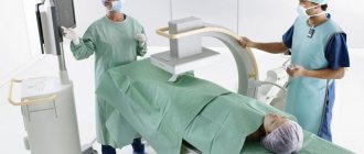
Carrying out
An X-ray of the esophagus and stomach begins with a general image of the abdominal cavity. After this, the patient is asked to drink prepared barium sulfate. The initial targeted photograph is taken after two sips of the drug. At this moment, the relief of the walls of the esophagus with barium is determined. Then the patient is allowed to finish the rest of the drug. During the examination, the doctor can press on the patient's abdomen to promote better distribution of the contrast. During the process, various pictures are taken in different positions - lying on the back as standard or with the pelvis elevated at an angle of 45°, lying on the side, standing. At the same time, at the command of the radiologist, the patient must hold his breath. Modern X-ray rooms are equipped with a special table that rotates while taking pictures. As a rule, to examine the upper part of the gastrointestinal tract, it is enough for an adult to take 250–300 ml of dissolved barium sulfate (in some cases, the dose may be increased). If radiography is prescribed for a child, then the required amount of suspension is calculated based on the age category. For children, as a rule, 100 ml of barium paste is enough.
Bowel examination
X-ray of the small intestine with barium is performed in stages. The procedure begins with the patient drinking 0.5 liters of barium suspension. If the study is carried out with double contrast, the drug enters the body through a special tube that is inserted into the patient’s mouth. Along with the contrast, air or inert gas is supplied. After this, wait at least 2 hours - during this time the barium has time to reach the small intestine. As contrast fills the small intestine, the radiologist takes a series of pictures, asking the patient to assume different body positions. And after relieving themselves, they take the last control shot. After filling them with contrast, the diagnostician examines different segments of the small intestine in detail on the monitor for half an hour. The contrast procedure allows you to evaluate intestinal motility and its mucous membrane. While the contrast agent is still present in small quantities, the relief of the inner wall of the intestines is examined, and when there is a lot of contrast, the shape, size, contours and functionality of the intestines are assessed. With maximum filling of barium, it is possible to identify inflamed segments, ulcerative processes and identify neoplasms. If the passage of barium is disrupted, then the radiologist gently presses on the anterior wall of the peritoneum to distribute it evenly. During a procedure with contrast, as the substance is distributed in the intestinal lumen, an experienced radiologist can draw conclusions about the presence of pathological processes. If the barium suspension is distributed in the form of flakes, then this is a clear sign of impaired absorption. And if the contrast fills the lumen unevenly, this may indicate oncopathology.
What does a gastrointestinal x-ray show?
An examination of the digestive organs is necessary to assess their condition, functioning, detect problems and eliminate them. Today there are several technologies for this: endoscopy, MRI, computed tomography, angiography.
The very first research technique was x-ray (x-ray) - radiation diagnostics of the internal structure of the body. With its help, one-time images of the stomach and duodenum are obtained.
The result obtained is recorded on film, from which the doctor will evaluate the condition of the internal organs. This procedure is also called gastrography.
Content:
- What does a gastrointestinal x-ray show?
- Indications for examination
- Contraindications to the procedure
- How to prepare for an x-ray
- How does this happen
- Where to get an X-ray of the gastrointestinal tract
- Possible complications
- Alternative examination methods
Often, images alone are not enough to fully study the anatomy of the digestive tract. In such cases, radiography is combined with fluoroscopy.
The latter is carried out using a special amplifier - a contrast agent. In laboratory conditions, a special barium-based solution is prepared, which does not transmit x-rays.
The patient drinks this cocktail during the procedure, as a result the doctor can observe in real time the patency of the esophagus, the peristalsis of the stomach, and have a good look at its internal walls and folds.
As a rule, these two procedures are carried out in combination. Radiography provides images, and fluoroscopy allows you to see the movement of internal organs and record data on electronic media. This makes it possible to study in detail the structure of the walls of the esophagus, duodenum, and stomach without invasive intervention.
Radiation diagnostics remains the most accessible method of examining a patient; it can be carried out in every hospital. The success of this process depends on the experience and skill of the radiologist.
But this technique also has a significant drawback: during the examination, the patient receives a certain dose of radiation, especially during fluoroscopy. Therefore, doctors resort to it only in extreme cases. The doctor must first carry out all non-radiation examination methods and only if they do not give the desired result, prescribe an x-ray.
X-ray of the gastrointestinal tract shows:
- diseases of the esophagus: diverticulum (protrusion of the mucous membrane), tumors, narrowing, varicose veins;
- foreign bodies in the digestive tract;
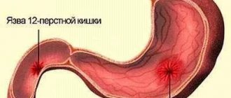
- pathologies of the duodenum: ulcers, cancer, spastic narrowing;
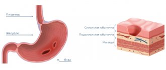
- stomach diseases: gastritis, cancer, ulcers, impaired absorption and weak peristalsis, etc.;
- other abdominal organs are not the main target of diagnosis, but their contours are still visible on the image. Based on the image, the doctor can notice problems in other organs.
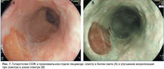
In some cases, the patient has to undergo several procedures at once, since each type of diagnosis shows different areas. For example, endoscopy reflects the condition of the internal mucous membrane and allows you to take material for a biopsy. And on the x-ray you can see the outer part of the organs, a little internal, tumors and neoplasms, narrowing of the esophagus are visible.
results
- abnormalities in the structure of the digestive tract;
- acute expansion or narrowing of the lumens of the stomach/esophagus;
- malformations of certain organs of the gastrointestinal tract;
- hypertonicity/hypotonicity of the muscular wall of the stomach;
- tumors, papillomas, foreign bodies;
- reduction or radial arrangement of shell folding;
- cicatricial changes at the site of tumors, ulcers, chemical burns;
- pathological narrowing;
- saccular protrusions and elasticity of the walls;
- the introduction of one section of the intestine into another with the possible development of gastrointestinal obstruction;
- intestinal motor function;
- the presence of ulcerative and inflammatory processes;
- tumors, polyposis.
An examination can show how the bauhinium valve functions. This is the structure that separates the small and large intestines and is responsible for passing food between them. If it has pathological changes, then the food gets back access, and this poses a danger to the patient’s life.