Questioning
Dysphagia (difficulty swallowing) with solid foods is usually observed with carcinoma, benign stricture, esophageal membrane; when eating solid and liquid food - indicates a violation of motility of the esophagus (achalasia, diffuse spasm of the esophagus and other motility disorders), damage to the esophagus by caustic alkalis or acids (burn of the esophagus), severe peptic esophagitis, infectious lesions of the esophagus (HIV infection, cytomegalo- and herpes viruses, fungal infections), diffuse spasm of the esophagus
Heartburn and pain behind the sternum when food passes through the esophagus are typical symptoms of cancer and reflux esophagitis.
Preparation period
Before an x-ray of the esophagus, the patient should undergo simple but mandatory preparation. You should find out how best to prepare for the procedure from the specialist who prescribed the examination. However, there are a number of general recommendations.
3 days before the procedure, you should adjust your diet by eliminating foods that promote gas formation. On the eve of the examination, at lunchtime, it is recommended to take castor oil. In the evening and immediately before x-rays of the esophagus, cleansing enemas are prescribed.
X-ray examination of the esophagus
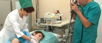
X-ray examination currently remains the main indicative diagnostic method. With various rotations of the patient around the vertical axis, a contrast X-ray examination is performed with an aqueous suspension of barium sulfate, and if perforation is suspected, with a water-soluble contrast. Pay attention to the nature of the contours, peristalsis, relief of the mucous membrane, and the function of the esophageal sphincters. Examination of the patient in a horizontal position with the foot end of the X-ray table raised allows us to identify dysfunction of the lower esophageal sphincter (reflux of contrast material from the stomach into the esophagus).
Measures to prepare for the study
Even children can have an ultrasound.
Preparation for the procedure will include the need to adhere to a strict diet for several days before the ultrasound. The ban also applies to smoking and drinking alcohol, since they can irritate the mucous membrane of the esophagus and stomach, and this distorts the results of the study.
On the day of the examination, eating is prohibited. The exception is half a glass of tea with a small cracker. Dinner the day before should take place no later than 6-7 pm.
In addition, before the ultrasound, the patient is prohibited from consuming foods that promote gas formation in the stomach and intestines:
- Rye bread;
- fresh vegetables and fruits;
- carbonated drinks;
- peas and cabbage dishes.
For children, preparations for an ultrasound also include dietary restrictions, but taking into account the diet recommended for children. So, if an infant is going to undergo an ultrasound, you can bring him for examination at any time, but it is better to do this immediately before feeding or at least 2 hours after the last. This will increase the reliability of the results.
Our equipment
In our hospital, diagnostics of the digestive tract using MRI and CT methods is carried out by experienced specialists using modern digital tomographs from Philips, (Netherlands).
MRI of the gastrointestinal tract is performed on an Ingenia 1.5 Tesla magnetic resonance imaging scanner , which provides highly informative images of the internal organs of the abdominal cavity and retroperitoneal space, vessels and soft tissues, specifically examined areas. Speeds up examination by 40% compared to other tomographs.
CT of the gastrointestinal tract is performed on an Ingenuity Elite 128-slice computed tomograph, which is capable of visualizing small details, which is important in the diagnosis of malignant neoplasms. Equipped with iMR technology, which improves image quality by 60-80% while reducing radiation exposure.
If you need to undergo a CT or MRI of the gastrointestinal tract in Moscow , contact the Clinical Hospital on Yauza. You will receive expert-level research in the shortest possible time.
Diagnostics
SIGN UP FOR A CONSULTATION +7 (812) 951-7-951
Diagnosis of esophageal cancer should be comprehensive and based on data from clinical, radiological, endoscopic, ultrasound and morphological methods of examining the patient. In some cases, to clarify the extent of the process and preoperatively determine the stage of the process, a computed tomographic study is required.
Laboratory and functional research methods are of great and sometimes decisive importance in assessing a patient’s curability. The final stage of diagnosis when deciding on the extent of esophageal cancer should be revision laparotomy with intraoperative ultrasound examination of the liver and areas of regional lymph nodes below the diaphragm. In a number of patients, intraoperative ultrasound diagnostics should also be used at the stage of thoracotomy to resolve the issue of interest in the tumor process of the organs surrounding the esophagus and assess the resectability of the process. X-ray methods should be used to study the esophagus, stomach (while maintaining the patency of the esophagus) and lungs (necessarily with a tomographic examination of the mediastinum at the level of the roots of the lungs). According to indications, X-ray examinations of other organs are performed. The objectives of x-ray examination are: - identification of tumor lesions of the organ; - determination of the extent of the tumor lesion, its localization in accordance with the segmental division of the esophagus; - identification of narrowing of the lumen of the esophagus, suprastenotic expansion; - identification of ulceration or fistula in the tumor area with a clear description of their size and depth; - determination of the depth of tumor invasion of the esophageal wall and the severity of the peri-esophageal soft tissue component of the tumor; - identification of concomitant diseases of the esophagus (diverticula, hiatal hernia, reflux esophagitis, achalasia, esophageal and cardiospasm). The main radiological symptoms of esophageal cancer are: rigidity of the walls and disruption of the relief of the mucous membrane in the area of the tumor lesion, narrowing of the lumen of the organ and suprastenotic expansion. X-ray diagnosis of advanced esophageal cancer, as a rule, does not cause difficulties. It is much more difficult to diagnose small tumors, up to 3 cm. In these cases, there can be both X-ray negative tumors and cases with an underestimated extent of 0.5 to 1 cm compared to the true extent of the process. When X-ray detection of a tumor of the esophagus, it is possible to determine the form of tumor growth - predominantly exophytic growth form, predominantly endophytic or mixed. More accurately, the growth pattern of small tumors is usually determined using endoscopic diagnostic methods. The latter include esophagoscopy, gastroscopy (if it is possible to pass an endoscope through a narrowed section of the esophagus), tracheobronchoscopy and laparoscopy. The objectives of esophagoscopy are: - identification of tumor lesions of the esophagus and its macroscopic picture; - identification of possible inflammatory and non-tumor changes in the mucous membrane of the esophagus; - determination of the upper and, if possible, lower boundaries of the lesion; - establishing the presence of a circular lesion of the walls of the esophagus; -taking a biopsy to establish morphological confirmation of the tumor process and its histological structure; - assessment of the immediate effect of radiation or chemo-radiation therapy in the case of combined or radiation treatment; - assessment of the possibility of using photodynamic therapy or laser tumor destruction (in cases of small tumors of the esophagus that do not grow into the muscle layer of the wall). - detection of the presence of early esophageal cancer using diagnostic drugs of the hematoporphyrin series. During endoscopic examination, the following types of tumor lesions are distinguished: 1. Tumors with predominantly exophytic growth - polypoid, papillomatous, large-tuberous, saucer-shaped cancer. 2. Tumors with predominantly endophytic growth - focal flat infiltrate, ulcerative-infiltrative cancer, infiltrative-stenotic cancer. If possible, gastroscopy is performed, the purpose of which is to determine the condition of the gastric mucosa, the presence or absence of tumor transfer to the stomach and additional tumor growths in the stomach. The purpose of tracheobronchoscopy is: - assessment of the condition of the mucous membrane of the bronchial tree; - identification of narrowing of the lumen of the trachea and bronchi and the degree of its severity due to germination or compression from the outside by metastatic lymph nodes; - determination of the distance to the carina (tracheal bifurcation); - identification of the primary multiplicity of the tumor process (damage to the bronchial tree by synchronous lung tumors). If a narrowing of the lumen of the trachea or bronchi is detected due to compression by the tumor by more than 1/3, the resectability of the tumor is very doubtful and most often the operation in these cases is limited to a trial thoracotomy. Ultrasound examination is performed for the purpose of: - identifying metastatic focal liver lesions; - identification of enlarged lymph nodes in regional areas below the diaphragm and accessible areas of the mediastinum; - assessment of the intramural spread of the tumor (in cases where it is possible to pass a transesophageal ultrasound probe beyond the area of narrowing of the esophagus. When the tumor is localized in the lower parts of the esophagus, in order to exclude metastatic liver damage, if there is suspicion based on clinical, ultrasound and computed tomography, laparoscopy is performed. In the latter years, thanks to the improvement of ultrasound, endoscopic methods and computed tomography, it has become possible to take biopsy material for morphological confirmation of the tumor specificity of the identified changes.Morphological diagnosis of esophageal cancer is a mandatory element of the examination and includes cytological and histological methods for studying material obtained during esophagoscopy, tracheobronchoscopy, laparoscopy, puncture biopsy, as well as in the study of surgical material (tumor specimen with the esophagus and regional lymph nodes).The final stage of diagnosis is revision laparotomy, which can be an independent stage in surgical or combined treatment in combination with esophageal resection or gastrostomy. A comprehensive assessment of the functional state and reserves of the body according to functional studies is mandatory. These should include studies of the cardiovascular system, external respiration function with determination of the contribution to gas exchange of each lung, since operations on the esophagus are most often accompanied by thoracotomy with one-lung ventilation. Stage IV pulmonary ventilation insufficiency is considered an absolute contraindication to surgery. Differential diagnosis Esophageal cancer is differentiated from cardiospasm, cicatricial narrowing of the esophagus, esophageal ulcer and ulcerative esophagitis, benign tumors of the esophagus, varicose veins of the esophagus, diverticula of the esophagus, compression of the esophagus from the outside by tumors of the mediastinum, scars after mediastinitis, abnormally located vessels in the mediastinum, etc. in the differential diagnosis of esophageal cancer with other diseases is the morphological diagnostic method. The detection of malignant cells in a biopsy specimen clearly indicates the presence of esophageal cancer. However, the absence of malignant cells in the obtained material does not mean the absence of esophageal cancer. Only a repeated negative answer along with dynamic observation can make it possible to make a relatively favorable judgment.
Share link:
Laboratory research methods
- Blood tests
Carrying out blood tests is one of the important laboratory methods for diagnosing gastrointestinal problems; it allows one to assess the general condition of the body, characterize the functional state of individual organs, the degree of structural (morphological) tissue damage and prescribe optimal treatment.
- Clinical stool examination
The coprogram is important in diagnosing damage to the intestines, pancreas, biliary system and, indirectly, the stomach. The consistency of stool, smell, color, presence and amount of undigested food residues (muscle fibers, fiber, starch, fat, etc.), and the presence of mucus and blood in the stool are subject to assessment.
- Gastric juice studies
Allows you to study the functional and morphological state of the gastric mucosa, as well as roughly assess its evacuation function. Various probe and probe-free procedures are most widely used to characterize gastric secretion.
- Duodenal sounding
In diseases of the liver and biliary tract, the study of duodenal contents (the contents of the duodenum) is important, since one of its constituent parts is bile. Based on data from duodenal contents, bile secretion can be judged with a high degree of accuracy.
In addition, thanks to this method, you can learn more about the function of the pancreas.
- Biopsy
A biopsy is a diagnostic procedure during which any suspicious cells or tissues are taken intravitally for further microscopic examination. A biopsy is important to exclude oncological pathologies.