Gastric adenoma (adenomatous polyp) is a benign neoplasm of glandular epithelial cells with signs of dysplasia. Macroscopically, it is an outgrowth of the mucous membrane on a thin stalk or on a wide base. Microscopically, chaotic proliferation of the epithelium with numerous morphological changes (nuclear polymorphism, signs of proliferation, etc.) is determined.
- Reasons for development
- Symptoms
- Classification of gastric adenoma
- Diagnostics
- Treatment of gastric adenoma
- Nutrition for gastric adenoma
- Prevention
Gastric adenomas are a serious pathology. The risk of their degeneration into cancer is extremely high - 40-50%. Therefore, timely detection and treatment of adenomatous formations is considered one of the priorities of modern gastroenterology.
General information
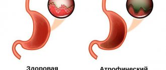
Hyperplasia is a pathological process characterized by increased growth of any tissue due to excessive cell division.
Changes can be detected in almost any organ, be it the mammary glands, uterus, or adrenal glands. However, most often the pathological process affects the mucous wall of the stomach. Hyperplasia is classified as a dangerous process, because rapid cell growth and increased division can cause neoplasms.
In some cases, with hyperplasia, not only an increase in the number of cells is observed, but also their structural restructuring, which inevitably leads to the start of the oncological process. What is characteristic is that the process of cell making during hyperplasia is no different from ordinary cell division. The only difference is that the number of cells during hyperplasia increases sharply. Pronounced structural abnormalities appear only in advanced cases.
Hyperplasia leads to the proliferation of the mucous membrane, which can be either focal or diffuse in nature.
Diagnostics
Endoscopic examination of the stomach (FGDS) allows diagnosing pathology and determining the histotype of the tumor. Using gastroscopy, you can study in detail the condition of the organ mucosa, detect adenomas even smaller than 5 mm, evaluate the structure of each neoplasm and take histological material for analysis.
Diagnostics also includes a survey and examination of the patient, fluoroscopy of the stomach with contrast, general clinical tests, laboratory tests and other techniques. If a malignant process is confirmed, further examination and treatment is carried out by an oncologist.
Classification
It is customary to distinguish several types of hyperplasia. The classification is based on the type of cells involved in the pathological process, as well as the location of the pathological focus.
Focal gastric hyperplasia
Focal hyperplasia is characterized by damage to a clearly defined area of the gastric mucosa. It is considered a precursor to polyposis . The affected area can have different sizes and shapes. Most often, a small growth with a characteristic modified structure is detected. Such lesions lend themselves well to coloring and stand out against the background of unchanged, healthy tissue. It is this property that has important diagnostic significance.
The focal form is characterized by the appearance of both one lesion and several modified areas. Such lesions may have a specific stalk or look like a small tubercle. Often in the literature you can find another name - warty hyperplasia . Focal hyperplasia very often develops after exposure to various damaging factors that provoke erosions and ulcers.
Lymphoid hyperplasia of the stomach
Lymphoid hyperplasia is characterized by an increase in the number of lymphocytic cells. Most often, such processes are observed after infectious diseases that can activate immune system . But in some cases, the proliferation of lymphoid tissue is the result of pathological changes occurring in the nodes themselves.
Under the epithelial layer in the mucous membrane there is a huge number of lymphatic vessels with nodules, the pathology of which forms hyperplasia of lymphoid tissue .
Lymphofollicular hyperplasia
The pathology is diagnosed in people of various age groups, regardless of place of residence and food preferences. Lymphofollicular hyperplasia is characterized by increased cell division in the lymphatic system located in the mucosal wall.
The disease develops against the background of various inflammatory processes in the stomach. Predisposing factors include Helicobacter pylori and frequent consumption of a variety of carcinogenic food additives. Chronic stress is of no small importance .
Hyperplasia of the integumentary epithelium of the stomach
The mucous wall of the stomach is covered with columnar, single-layer epithelium, which can hyperplasia under the influence of certain factors. Hyperplasia of the gastric mucosa is considered a dangerous pathology due to the high risk of malignancy.
As a result of hyperplasia, not only the proliferation of the epithelium occurs, but also certain structural changes are formed. Cytological examination clearly demonstrates an increase in cell volume, displacement of the nucleus towards the base, as well as the accumulation of mucin in the cytoplasm.
Hyperplasia of the antrum of the stomach
Quite often, hyperplastic changes are recorded in the antrum of the stomach. This phenomenon is explained by the fact that this section is the closing one and it is from here that the food bolus enters the intestinal lumen. The antrum accounts for one-third of the entire stomach. The closing section of the stomach is most often exposed to various diseases, because experiences maximum loads.
The functional responsibilities of the antral stomach are based on grinding incoming food and further pushing the food bolus into the lumen of the duodenum. Most often, it is in the antrum of the stomach that various forms of hyperplasia are diagnosed. The most common cause of hyperplasia is chronic gastritis .
Glandular hyperplasia
Hyperplastic changes can also affect cells that perform the functions of glands. In this case, polyp-like formations are formed on the mucous membrane, the body of which is represented by glandular cells. The growths can be oval or round in shape. Compared to other forms of hyperplasia, glandular hyperplasia is extremely rare.
Foveal hyperplasia
Also known as regenerative polyposis . What is foveal hyperplasia? The pathology is characterized by the proliferation and thickening of the folds of the gastric mucosa. Most often, the cause of the disease is excessive use of drugs from the NSAID group. This form of hyperplasia is characterized by a clear clinical picture.
Follicular hyperplasia
In the medical literature it is also found as lymphofollicular. The mechanism of development of the pathology is the accumulation of multiple lymphocytes in the follicles, which leads to thickening and proliferation of tissue. The most common provoking factor is gastritis . Follicular hyperplasia is considered an extremely dangerous form due to the high risk of malignancy.
Treatment of gastric adenoma
A diagnosed gastric adenoma requires active medical tactics, since in most cases it transforms into cancer, and here the tubular form is especially dangerous. The choice of treatment mainly depends on the histological structure and size of the tumor.
Papillary and mixed variants in some cases allow for expectant management (small size, presence of medical contraindications to surgery, etc.). If the adenomatous polyp in the stomach is not removed, the patient undergoes gastroscopy annually. If signs of malignancy, rapid tumor growth, or complications appear, the question of surgical intervention is raised.
Tubular adenomas are always subject to surgical treatment. A gastroscope is used to remove small polyps on a thin stalk that do not have signs of atypia. A loop is placed over the mucous growth and it is removed using electrocoagulation. This method is considered less traumatic and less dangerous for the patient. The gastric mucosa recovers in 2-8 weeks. 2-3 months after surgery, a control endoscopy is performed to confirm the absence of relapse.
If the adenoma has a wide base, the tumor has a large diameter, or there are several large polyps, laparoscopic access is used. If malignant cells are detected in the adenomatous formation, gastric resection or other radical operations can be performed, taking into account the indications and stage of the process. Surgical interventions can also be performed using an “open” approach through an incision in the abdominal wall.
Causes
The causes of hyperplasia, including those with damage to the stomach, have not been sufficiently studied to date. However, it is customary to identify several predisposing factors:
- changes in the nervous regulation of the stomach;
- various infectious lesions, including Helicobacter pylori;
- changes in hormonal regulation;
- the presence of inflammatory processes;
- negative impact of carcinogenic substances;
- hereditary, genetic predisposition;
- changes in the secretory functioning of the stomach;
- the presence of ulcerative defects, gastritis, erosions.
Breast hyperplasia
The proliferation of cells in a woman’s breast (hyperplasia of the glands and milk ducts) is a fairly common disease recently. Most often, mammary hyperplasia is diagnosed at the age of 20 years. There are typical and atypical breast hyperplasia. Both of these forms of the disease are dangerous because, if not diagnosed early, they can lead to breast cancer.
How does this hyperplasia manifest itself? Symptoms that you should pay attention to:
- chest pain;
- lumps in the chest;
- nipple discharge;
- menstrual irregularities;
- mood changes: depression, dysphoria, lability.
Glandular hyperplasia, unless it is a malignant process, can be successfully treated with medication.
Symptoms
It is quite difficult to identify the disease at the very initial stage, since patients are almost completely free of any symptoms. An increase in the number of cells does not cause discomfort. Pain syndrome does not appear even when polyps begin to grow. When they grow excessively, there are difficulties in swallowing food, in some cases severe pain and severe bleeding .
As the pathology progresses, patients begin to complain about disturbances in the functioning of the stomach, and problems with digestion are noted. Main symptoms:
- heartburn;
- pain that occurs immediately after eating or during prolonged fasting; pain can be short-term or permanent;
- constipation;
- increased gas formation, flatulence ;
- vomiting, nausea;
- belching with sour contents;
- anemia;
- dizziness;
- feeling of aching throughout the body;
- refusal to eat.
Hyperplastic processes in the uterus
Hyperplasia in gynecology is a fairly common disease. Endometrial hyperplasia is a disease of the uterine body in which changes in the mucous membranes and glands of the uterine lining are observed. In other words, endometrial hyperplasia or uterine hyperplasia is an overgrowth of the endometrium that causes it to become thicker than normal.
The endometrium is the inner layer of the uterus, equipped with blood vessels, which is constantly renewed. It is to the endometrium that the embryo is attached, so the condition of this tissue is very important for a woman’s reproductive health.
Uterine hyperplasia can be glandular and glandular-cystic. Adenomyosis hyperplasia and atypical hyperplasia are also distinguished. A separate pathological growth of the walls of the uterus (hyperplasia) - polyp.
Why does uterine hyperplasia occur? The reasons for the growth of the endometrium are the following:
- ovarian tumors;
- lack of ovulation;
- pathologies of the adrenal cortex;
- pathology of the pituitary gland;
- incorrect use of hormonal contraceptives;
- advanced chronic inflammation of the endometrium.
Hyperplasia most often occurs due to hormonal imbalance. The causes of the disease lie in a violation of the ratio of estrogen and progesterone. In addition, uterine hyperplasia can occur due to metabolic disorders - obesity, decreased glucose tolerance, increased cholesterol levels.
Hyperplasia of the uterine mucosa is in most cases a benign process, but if the disease is neglected, atypical hyperplasia may be detected.
Atypical hyperplasia is a proliferation of the uterine mucosa, in which altered glands with atypical cells are observed in large numbers. This is the first sign that endometrial tissue hyperplasia is degenerating into cancer (adenocarcinoma).
Adenomyosis is a hyperplasia in which the endometrium grows deep into the inner layers of the uterus. Adenomyosis is internal hyperplasia, causing a deterioration of immunity, a decrease in hormone levels and the functionality of the uterus. In addition, adenomyosis is a hyperplasia that often causes infertility. This occurs due to the fact that the endometrium becomes loose, and the embryo cannot gain a foothold in it.
Another manifestation of the disease hyperplasia is a polyp on the walls of the uterus. A polyp is the same growth of cells, but it is distinguished from other hyperplastic processes by the presence of a stalk, with the help of which it is attached to the tissues of the uterus.
Most often, such hyperplasia is benign - the polyp only in advanced cases degenerates into a malignant formation. Exclusively surgical treatment involves such hyperplasia - the polyp or polyps, regardless of their nature (benign or malignant), are cut off from the walls of the uterus.
Hyperplasia often appears during menopause. This is understandable, because during this period a woman experiences constant fluctuations in hormone levels and ovarian function deteriorates. Such hyperplasia is treated by curettage of the uterine cavity, cervix and subsequent administration of gestagens.
How does uterine hyperplasia manifest? Symptoms of growth of the endometrium of the uterus, which should raise suspicion:
- menstrual irregularities;
- dysfunctional prolonged bleeding from the uterus that occurs during menstruation or between menstruation. It can be moderate or abundant;
- intermenstrual bleeding;
- pain in the lower abdomen;
- infertility.
We can say that hyperplasia is completely curable. Treatment of women of reproductive age, unless we are talking about the formation of polyps or a precancerous condition of uterine tissue, is limited to taking hormones and performing restorative therapy. In other cases, a woman is indicated for complex treatment: curettage of the uterine cavities and hormonal therapy.
Another type of cell proliferation that gynecologists often encounter in their practice is placental hyperplasia (enlargement of the placenta in size), a disease that occurs in pregnant women and requires immediate identification of the cause and treatment. The placenta ensures normal intrauterine development of the fetus, so placental hyperplasia can cause developmental delays in the child, the appearance of hemodynamic disorders, polyhydramnios or oligohydramnios, and premature birth.
Placental hyperplasia develops due to:
- severe anemia;
- diabetes mellitus;
- Rhesus conflict;
- syphilis, ureaplasmosis, toxoplasmosis, mycoplasmosis, chlamydia and other infections.
Placental hyperplasia is treated with medication - a woman can be prescribed Actovegin, Curantil, Essentiale Forte. In addition, placental hyperplasia requires a thorough diagnosis and identification of the root cause of the disease, hyperplasia in a pregnant woman, which mainly determines the course of treatment.
Hyperplastic processes of the prostate
Hyperplasia of the prostate epithelium is a benign process. Adenoma or BPH (benign prostatic hyperplasia) is also called prostatic hyperplasia.
Prostatic epithelial hyperplasia is a pathological proliferation of tissue and the formation of nodules in the prostate. Subsequently, hyperplasia can lead to obstruction (obstruction) of the urinary tract.
Prostate hyperplasia is a fairly common disease among men. Why does such hyperplasia develop? The causes of this disease are hormonal age-related changes and impaired testosterone metabolism.
Such hyperplasia is easily treatable. Treatment of a timely identified disease can only be medicinal - hormonal or herbal preparations can be prescribed. In later stages, prostatic hyperplasia is treated exclusively with surgery.
In the early stages, prostatic hyperplasia appears only on ultrasound - in the form of microscopic nodules. In later stages, the patient may feel discomfort or pain in the prostate area, and problems with urination may begin.
Adrenal hyperplasia
Adrenal cortical hyperplasia is a congenital disease in which a person’s production of the hormone cortisol is impaired. This hyperplasia is known for various manifestations. Symptoms vary depending on which genes are affected. Oligomenorrhea, acne, increased fatigue, infertility, and hirsutism may occur. In addition, adrenal hyperplasia can be noticeable in laboratory indicators:
- slight increase in testosterone levels;
- moderate increase in DHEA sulfate levels;
- an increase in the level of the cortisol precursor hormone – 17-hydroxyprogesterone.
Adrenal hyperplasia is diagnosed during examination by an endocrinologist: after a blood test for hormone levels, examination of the patient, and collection of anamnesis.
Liver hyperplasia
Hyperplasia of liver tissue can occur due to vascular thrombosis, preoperative embolization, and pathological proliferation of cells of unknown origin. For these reasons, focal liver hyperplasia develops.
In addition, a rather rare disease is identified separately - focal nodular hyperplasia of the liver, which is a benign tumor without a capsule.
Hyperplasia of liver tissue is subject to surgical treatment in most cases. The exception is focal liver hyperplasia, since it is a benign process. The tumor is kept under observation and only if necessary, a biopsy is performed or surgery is performed.
Complications
Any tumor in the stomach is characterized by complications:
- pain syndrome,
- ulceration with acute or chronic bleeding,
- perforation of the organ wall with the development of peritonitis.
In addition, with lymphoma, there may be a decrease in the specific gamma globulin IgG produced by normal immune cells, which contributes to frequent infections and inflammatory processes. The deficiency is compensated by intravenous administration of immunoglobulin.
In all cases, before starting chemotherapy, prevention of tumor lysis syndrome, which occurs when malignant cells are highly sensitive to drugs, is carried out.