Gastric adenoma (adenomatous polyp) is a benign neoplasm of glandular epithelial cells with signs of dysplasia. Macroscopically, it is an outgrowth of the mucous membrane on a thin stalk or on a wide base. Microscopically, chaotic proliferation of the epithelium with numerous morphological changes (nuclear polymorphism, signs of proliferation, etc.) is determined.
- Reasons for development
- Symptoms
- Classification of gastric adenoma
- Diagnostics
- Treatment of gastric adenoma
- Nutrition for gastric adenoma
- Prevention
Gastric adenomas are a serious pathology. The risk of their degeneration into cancer is extremely high - 40-50%. Therefore, timely detection and treatment of adenomatous formations is considered one of the priorities of modern gastroenterology.
Reasons for development
Most often, adenomatous polyps of the stomach develop due to atrophic gastritis or gastritis with low acidity. Also, any chronic diseases of the digestive system (erosions, peptic ulcers, etc.) can serve as a background for the appearance of benign tumors.
The main reasons leading to the formation of adenomas are the following:
- “invasion” of sections of intestinal epithelium into the lining of the gastric mucosa - intestinal metaplasia;
- excessive cellular proliferation against the background of chronic inflammation with subsequent hyperplasia of epithelial tissue;
- damage to the inner lining of the stomach by waste products of microorganisms Helicobacter pulori, which reduce the acidity of digestive juice and the protective properties of secreted mucus;
- systematic use of proton pump inhibitors, which, by suppressing the production of hydrochloric acid, thereby increase the production of gastrin, which stimulates cell division.
According to statistics, men get sick twice as often as women. Pathology usually develops at the age of 40-60 years. Some people have a genetic predisposition to developing benign tumors of the gastrointestinal tract. Excessive smoking, consumption of rough food and strong alcoholic drinks, and taking certain medications also contribute to the appearance of adenomas.
Pathology of the uterine cavity
- Endometrial pathology
Based on the above information, the pathology of the uterine cavity can be conditionally divided into several subgroups:- Endometrial hyperplasia is a pathological growth of the endometrium, sometimes uneven, focal. Individual areas of growth are called “polyps.” Treatment of endometrial hyperplasia is surgical, followed by conservative therapy (if necessary).
Intrauterine synechiae (adhesions, adhesions), which form after intrauterine interventions (abortions), with long-term wearing of spirals or after inflammation of the endometrium (endometritis);
- Malignant diseases of the endometrium (adenocarcinoma);
- Remains of the fertilized egg after curettage of the uterine cavity;
- Foreign bodies in the uterine cavity (intrauterine devices or threads after surgery).
- Myometrial pathology
- Submucous uterine fibroids located in the uterine cavity;
- Interstitial uterine fibroids, presenting to the uterine cavity and deforming it;
- Adenomyosis (internal endometriosis) - the presence of which can be confirmed by hysteroscopy.
- Uterine malformations
- in particular, intrauterine septums
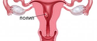
The main types of pathology of the uterine cavity are presented in Figure 2.
Figure 2. Main types of pathology of the uterine cavity
Symptoms
Gastric adenoma is often asymptomatic and becomes an “accidental” finding during endoscopic examination of the gastrointestinal tract. Since neoplasms are formed predominantly against the background of atrophic gastritis (less often they are associated with other gastric pathologies), the patient will experience a clinical picture characteristic of this disease:
- decreased appetite;
- belching;
- heartburn;
- nausea;
- vomiting after eating rough, “heavy”, fatty, spicy food;
- discomfort, heaviness and pain in the epigastric region;
- a feeling of fullness in the stomach, even after eating a small portion of food.
When polyps grow and their size increases to 2 cm or more, the following symptoms of pathology may appear:
- Constant aching pain in the stomach, depending on food intake, associated with the presence of inflammation in the mucous membranes of the gastrointestinal tract.
- Signs of internal bleeding that occur when the adenoma is damaged or ulcerated (tarry stools, vomiting blood, anemia).
- Symptoms associated with chronic blood loss are weight loss, fatigue, loss of strength, fatigue, dizziness, tachycardia, etc.
An adenomatous polyp of the stomach can transform into adenocarcinoma at any time. As a rule, when a tumor becomes malignant, a person’s condition worsens.
Adenomas can lead to the development of serious complications. When the pedicle feeding the polyp is twisted, vascular thrombosis and tissue necrosis occur. The patient develops intense pain in the stomach, body temperature rises and other signs of intoxication of the body are also added. If an adenomatous formation is infringed, the movement of food through the digestive tract stops and sharp cramping pain appears in the epigastric region. Both conditions require emergency medical attention.
Why does glandular hyperplasia of the uterus occur in menopausal women?
The main cause of any type of hyperplasia is hormonal imbalance, so this disease is especially common in menopausal patients.
Normally, after ovulation, a woman produces gestagen hormones, which prevent tissue proliferation and prepare the body for the onset of menstruation. If this does not happen, the body continues to produce estrogens, which cause mucosal growth.
In the cramped uterine cavity, glandular cells have nowhere to grow, so they begin to grow “into themselves,” forming deformed convoluted glands with cystic contents (secretion). As a result, the uterine layers thicken and are rejected, the process is accompanied by bleeding. During this period, you can observe blood with clots consisting of immature cells of the inner uterine layer.
Pathology can be provoked by inflammation in the uterus, myomas and fibroids, sudden refusal of hormonal contraception, surgical abortions and curettage, late menopause.
Classification of gastric adenoma
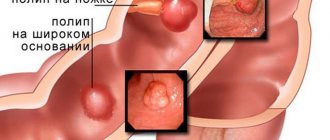
An adenomatous polyp can be located on any part of the organ. More often, gastric adenoma is localized in the antrum and in the pyloric region (the place of transition to the duodenum).
Gastric adenomas are divided into three groups according to histological type:
- Villous (villous, papillary) type of adenoma. It is the most favorable in terms of further prognosis. The tumor typically has a wide base and a characteristic “hairy” appearance. It consists of many villi and connective tissue elements with a small amount of glandular epithelium (no more than 25%).
- Mixed form of adenoma. It has a more pronounced degree of dysplasia compared to the previous type. The tumor contains villous and tubular structures in varying proportions. The share of papillary tissue accounts for from 25 to 75%).
- Tubular adenoma. It is the most common type. The tumor has clear boundaries, is crimson in color, and consists of epithelial glands. Under a microscope, numerous altered, branching tubes are visible, longer and wider than normal. This adenoma is characterized by slow growth and a high degree of malignancy.
Clinicians divide adenomatous polyps of the stomach into benign, transitional types and malignant (cancer).
Why is it dangerous?
Such a diagnosis can be dangerous for a woman’s health. That is why you should pay attention to the symptoms that arise and immediately consult a doctor for medical help.
Risks and dangers associated with hyperplastic processes:
- Infertility. if the nature of the endometrium is disrupted, the fertilized egg simply cannot be implanted into the wall of the uterus. It is for this reason that women cannot get pregnant for months and years. And the reason turns out to be this problem.
- Hyperplasia can cause excessive bleeding. As a result, severe forms of anemia (a drop in hemoglobin levels) may develop. With the development of anemia, the entire body, all organs and systems suffer from a lack of oxygen (hypoxia).
- Some forms of endometrial hyperplasia are precancerous diseases. If you ignore this process, malignancy and growth of a malignant tumor may occur.
Diagnostics
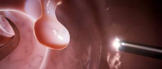
Endoscopic examination of the stomach (FGDS) allows diagnosing pathology and determining the histotype of the tumor. Using gastroscopy, you can study in detail the condition of the organ mucosa, detect adenomas even smaller than 5 mm, evaluate the structure of each neoplasm and take histological material for analysis.
Diagnostics also includes a survey and examination of the patient, fluoroscopy of the stomach with contrast, general clinical tests, laboratory tests and other techniques. If a malignant process is confirmed, further examination and treatment is carried out by an oncologist.
Endometrial hyperplasia
Let's consider one of the most common pathologies of the uterine cavity - endometrial hyperplasia.
Endometrial hyperplasia is a process characterized by inadequate and non-invasive proliferation (growth) of endometrial glands with varying quality of the underlying stroma. Recently, great importance in the development of this process has been attached to inadequate excessive growth of blood vessels, i.e. excessive angiogenesis. In the future, when creating drugs that can block these processes, i.e. have antiangiogenic properties, endometrial hyperplasia and treatment will be a very easy task for the doctor.
Treatment of gastric adenoma
A diagnosed gastric adenoma requires active medical tactics, since in most cases it transforms into cancer, and here the tubular form is especially dangerous. The choice of treatment mainly depends on the histological structure and size of the tumor.
Papillary and mixed variants in some cases allow for expectant management (small size, presence of medical contraindications to surgery, etc.). If the adenomatous polyp in the stomach is not removed, the patient undergoes gastroscopy annually. If signs of malignancy, rapid tumor growth, or complications appear, the question of surgical intervention is raised.
Tubular adenomas are always subject to surgical treatment. A gastroscope is used to remove small polyps on a thin stalk that do not have signs of atypia. A loop is placed over the mucous growth and it is removed using electrocoagulation. This method is considered less traumatic and less dangerous for the patient. The gastric mucosa recovers in 2-8 weeks. 2-3 months after surgery, a control endoscopy is performed to confirm the absence of relapse.
If the adenoma has a wide base, the tumor has a large diameter, or there are several large polyps, laparoscopic access is used. If malignant cells are detected in the adenomatous formation, gastric resection or other radical operations can be performed, taking into account the indications and stage of the process. Surgical interventions can also be performed using an “open” approach through an incision in the abdominal wall.
Intrauterine synechiae
Intrauterine synechiae are adhesions in the uterine cavity, which very often interfere with the implantation of a fertilized egg, which leads to infertility. The causes of intrauterine synechiae can be inflammatory diseases of the pelvic organs, as well as intrauterine interventions (abortion, curettage for a missed pregnancy, surgical interventions on the uterus).
The presence of intrauterine synechiae is safe for life, and can manifest itself as scanty menstruation or its absence. However, if pregnancy issues arise, intrauterine adhesions can be a serious problem, especially with common forms and severe damage to the uterus./p>
Nutrition for gastric adenoma
The diet for adenomatous polyps in the stomach is prescribed by a gastroenterologist, taking into account the background pathology (peptic ulcer, gastritis, etc.). To prevent the growth of adenomas and their recurrence after surgery, it is recommended to adhere to the following rules:
- “Heavy”, fatty, spicy and any dishes that can injure the gastric mucosa or cause irritation are excluded from the menu. The same goes for drinks.
- The consumption of “junk” food, fast food, processed foods, etc. is minimized.
- You should not overeat, it is advisable to eat in small portions in compliance with the principles of rational nutrition.
Any error in diet can aggravate the course of the disease.
Our doctors
Fedorova Elena Vladimirovna
Surgeon, Candidate of Medical Sciences, doctor of the highest category
22 years of experience
Make an appointment
Prokhorov Yuri Anatolievich
Surgeon, head of the surgical service of CELT, candidate of medical sciences, doctor of the highest category
33 years of experience
Make an appointment
Lutsevich Oleg Emmanuilovich
Chief Surgeon of CELT, Honored Doctor of the Russian Federation, Chief Specialist of the Moscow Department of Health in Endosurgery and Endoscopy, Corresponding Member of the Russian Academy of Sciences, Head of the Department of Faculty Surgery No. 1 of the State Budgetary Educational Institution BPO MGMSU, Doctor of Medical Sciences, Doctor of the Highest Category, Professor
43 years of experience
Make an appointment
Gordeev Sergey Alexandrovich
Surgeon, Candidate of Medical Sciences, doctor of the highest category
42 years of experience
Make an appointment
What else to read:
- Causes of autoimmune gastritis and its prevention In the modern world of numerous super-cool gadgets, lack of exercise and the increasing share of fast food and genetically modified products, it is very difficult not only to remain healthy throughout life, but also to simply be born......
- Symptoms and methods of treatment of reflux gastritis Contents of the article: 1 Types 2 Symptoms 3 Treatment 3.1 Diet 3.2 Physiotherapy treatment 3.3 Herbal medicine Reflux gastritis or GERD is a common disease. Reflux gastritis usually develops as a result of insufficient functioning of the gastric sphincters (cardiac and pyloric),……
- Treatment of superficial gastritis: features for different types of disease Contents of the article:1 Causes and pathogenesis2 Classification3 Signs3.1 Increased acidity3.2 Decreased acidity4 Stages5 Features of the course of the disease6 Diagnosis7 Therapy and its features7.1 Various acidity8 Diet Superficial gastritis is one of......