A foreign body of the esophagus is food stuck in the lumen of the organ or some inedible object swallowed accidentally or on purpose. Children, who make up approximately 12% of patients with this problem 1, most often swallow coins, coin-cell batteries, and magnetic balls from construction sets. In adults, the “assortment” is more diverse: bones, dentures, packages with psychotropic substances, nails, needles. The exact statistics of the frequency of foreign bodies entering is unknown, since according to rough estimates, 80% of swallowed objects leave the body naturally. Among patients presenting with complaints characteristic of a foreign body in the esophagus, the foreign object itself is found in approximately 40% of cases. In the rest, only the damage to the mucous membrane caused by it is detected.
Throat bone (fish bone)
Among cases of foreign bodies of the upper respiratory tract in the practice of an otorhinolaryngologist, fish bones are the most common. The peak demand for removal of fish bones occurs in the summer months, when the diet contains a lot of freshly caught river fish. Samara is no exception, as it is located on the Volga River. Removing and pushing through fish bones is done at home with a crust of bread. Most often, small, thin bones—ribs—get stuck. The bone becomes lodged in the upper respiratory and digestive tracts at the time of ingestion. The most favorite places for bone fixation in the pharynx are the palatine tonsils, lingual tonsil, lateral ridges, posterior palatine arches, and pyriform sinuses. The palatine tonsils become a target for fish bones, since they actively accompany the bolus of food at the moment of swallowing. The lingual tonsil suffers for the same reasons. The tissue of the palatine and lingual tonsils is represented by lymphadenoid tissue, which is very loose and easily threaded onto a thin fish bone. Concomitant pathology in the form of chronic tonsillitis with hypertrophy of the tonsils increases the risk of bone entering the tissue. In the case where the bone is stuck in the upper parts of the pharynx and is in the line of sight, removing the fish bone in such a situation is not difficult. The situation with bone fixation in the lower parts of the pharynx requires the participation of a specialist. It is extremely difficult to remove such a bone without the help of an otolaryngologist. Complications of pharyngeal trauma caused by fish bones are rare. This form of sore throat is classified as traumatic; if the bone remains in the tonsil tissue for a long time, paratonsillitis can develop, which will end in a peritonsillar abscess. Acute pharyngitis, lateropharyngeal abscess, mediastinitis, phlegmon of the pharynx, neck, sepsis, laryngeal stenosis as a complication are quite rare. Removal of fish bones in Samara is performed by ENT doctors at Outpatient Center No. 1. First medical aid. During pharyngoscopy, you should carefully examine the palatine tonsils, moving away the palatine arches; with indirect laryngoscopy, you should carefully examine the root of the tongue, the vallecula of the tongue, and pear-shaped pouches. Finger examination is allowed. The foreign body is removed with a forceps or tweezers under visual control, after which it is recommended to rinse the oropharynx with an antiseptic solution and adhere to a gentle diet. If the foreign bodies are located in a different location in the pharynx, the patient should be urgently hospitalized in the otorhinolaryngology department. Specialized assistance. Foreign bodies of the lingual tonsil, vallecula of the tongue root and pyriform pouches are removed during indirect laryngoscopy in adults and direct hypopharyngoscopy in children using a laryngeal forceps or forceps. Anti-inflammatory therapy is prescribed. If a foreign body is not detected in the pharynx, and the pain syndrome persists, it is necessary to exclude a foreign body of the esophagus. For this purpose, fibrohypopharyngoscopy and esophagoscopy are performed.
How to give first aid to a child
First aid on the spot must be provided quickly and decisively. The child should be thrown in a prone position over the knee or the back of a chair, and then struck between the shoulder blades with an open palm. If a child inhales a round object, you can lift the baby by the legs, head down, and hit him several times with an open palm between the shoulder blades. The swallowed object will fall out or dislodge, allowing the baby to breathe.
If a child has inhaled coins or batteries, this should not be done. Flat objects will not be able to slip into the narrow glottis in the right direction. In children of the first years of life, you can use a chest shake to try to change the position of the foreign body so that the child can breathe and wait for the doctor to arrive. For this purpose, you can apply several short blows with your palm between the shoulder blades or put the baby on his back on the floor or other hard surface, lift his chin up, and with two fingers sharply press in the area between the navel and the angle formed by the ribs in an upward direction.
An older child should be placed on his lap, with his back to himself. Place the fist of one hand on the upper abdomen, cover it with the palm of the other hand and press sharply (this must be repeated up to five times).
If the child loses consciousness, quickly turn him onto his right side. Sometimes, during loss of consciousness, the muscles relax and impacts between the shoulder blades “knock out” the foreign body from the trachea. Before the ambulance arrives, cardiopulmonary resuscitation must be performed.
If a child who has inhaled a foreign body is breathing and coughing, do not pat him on the back. Call emergency services and, while waiting, help your child cough up the object.
In any case, even if you managed to get rid of the foreign body before the ambulance arrived, the baby must be shown to a doctor to prevent injury to the mucous membrane of the respiratory tract and its further infection.
Foreign bodies of the pharynx
Causes.
They are usually localized in the oropharynx and laryngopharynx, where they enter with food, sometimes during manipulations in the mouth (pin, needle, toothpick). The most common foreign body in the pharynx is a fish bone, which penetrates into the loose tissue of the palatine, lingual tonsils, and into the vallecula of the root of the tongue. Less often, foreign bodies (coin, meat bone) are fixed in pear-shaped pockets. Foreign bodies enter the nasopharynx from the nasal cavity (needle), or from the lower parts of the pharynx during vomiting. This occurs more often in children and the elderly.
Symptoms.
Pain in the throat when swallowing with irradiation into the ear (stabbing with a fish bone), discomfort in the projection of a foreign body, sometimes hypersalivation, vomiting, difficulty swallowing.
Complications.
Bleeding, acute pharyngitis, lateropharyngeal abscess, mediastinitis, phlegmon of the pharynx, neck, sepsis, laryngeal stenosis.
First medical aid.
During pharyngoscopy, you should carefully examine the palatine tonsils, moving away the palatine arches; with indirect laryngoscopy, you should carefully examine the root of the tongue, the vallecula of the tongue, and pear-shaped pouches. Finger examination is allowed.
The foreign body is removed with a forceps or tweezers under visual control, after which it is recommended to rinse the oropharynx with an antiseptic solution and adhere to a gentle diet. If the foreign bodies are located in a different location in the pharynx, the patient should be urgently hospitalized in the otorhinolaryngology department.
Specialized assistance.
Foreign bodies of the lingual tonsil, vallecula of the tongue root and pyriform pouches are removed during indirect laryngoscopy in adults and direct hypopharyngoscopy in children using a laryngeal forceps or forceps. Anti-inflammatory therapy is prescribed. If a foreign body is not detected in the pharynx, and the pain syndrome persists, it is necessary to exclude a foreign body of the esophagus. For this purpose, fibrohypopharyngoscopy and esophagoscopy are performed.
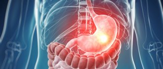
Classification of esophageal foreign bodies
Foreign bodies can be organic or inorganic in nature. Among organic foreign bodies, trichobezoars stand out separately - accumulations of hair swallowed by a patient (most often with an unstable psyche). Among inorganic batteries, batteries are especially dangerous - under the influence of a weak electric charge or alkali, necrosis of the wall of the esophagus can occur literally in a matter of hours. By shape, foreign bodies of the esophagus can be round, piercing, cutting, cylindrical, irregular in shape, and so on. By their ability to absorb X-rays: radiopaque and radiopaque non-opaque.
Foreign bodies of the esophagus
Causes.
Hasty eating, missing teeth, inadequate dentures, decreased pharyngeal reflex, alcohol intoxication, cicatricial narrowing of the esophagus. Foreign bodies usually get stuck in the area of physiological narrowings, most often at the level of the first thoracic vertebra.
Symptoms.
The onset of the disease is sudden and associated with food intake. Characterized by pain in the throat or behind the sternum with irradiation to the back, interscapular area, dysphagia, aphagia, drooling, general weakness, malaise, pain on palpation of the neck (on the left), aggravated by tapping on the spine, possibly forced positioning of the head.
When a foreign body is localized in the area of the first physiological narrowing of the esophagus, the head is tilted forward, down, the patient holds it motionless, and turns the whole body. When a foreign body is localized in the thoracic esophagus, the patient is in a semi-bent position (“carrying posture”).
Indirect laryngoscopy reveals swelling, hyperemia of the mucous membrane in the area of the aryepiglottic folds, arytenoid cartilages, and accumulation of saliva in the pyriform pouch (usually the left one). Vomiting and coughing are possible. A large foreign body can cause difficulty breathing through the larynx.
Complications.
Perforation of the esophagus, periesophagitis, mediastinitis, bleeding from the great vessels.
First medical aid.
. Immediate evacuation to hospital. Attempts to push a foreign body by swallowing bread crusts or using bougies are prohibited.
Specialized assistance
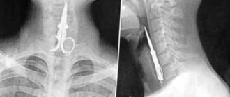
provided by otorhinolaryngologists together with endoscopists. To do this, indirect laryngoscopy and x-ray examination of the cervical spine in two projections (according to G.M. Zemtsov) are performed, which makes it possible to detect the shadow of a foreign body, indirect signs of a non-contrast foreign body of the esophagus or damage to its walls.
These symptoms are:
- straightening of the cervical spine due to tension in the scalene muscles;
- expansion of the prevertebral space;
- the presence of a symptom of an air “arrow” - an accumulation of air released from the stomach below the level of a foreign body, the pointed end of the “arrow” indicating the location of the foreign body;
- striped clearings in the prevertebral space are a sign of air penetration into the retroesophageal tissue or the development of putrefactive inflammation with the formation of gas.
Fibroesophagoscopy is also performed for diagnostic and therapeutic purposes. If it is impossible to remove a strangulated foreign body of the esophagus during esophagoscopy, an esophagotomy is performed. Anti-inflammatory therapy is prescribed.
O.V. Vozgoment
PGMA named after.
acad. E.A. Wagner, Perm Based on the results of an expert assessment of the quality of medical care for 2 children with damage to the esophagus caused by foreign bodies and complicated by mediastinitis and tracheoesophageal fistula, the deficiencies in intensive care and anesthesia, which in one case contributed to an unfavorable outcome, in the other - to prolongation, are analyzed. length of hospitalization. It is concluded that in the treatment of such patients, the most important problems are the choice of surgical and anesthetic tactics, as well as adequate antibacterial, detoxification therapy, respiratory and nutritional support, taking into account the specific clinical situation. Key words: esophagus, damage, mediastinitis, tracheoesophageal fistula, respiratory support, intensive care, examination.
Information about the author: Oleg Vasilievich Vozgoment – Candidate of Medical Sciences, Associate Professor of the Department of Anesthesiology of Reanimatology, Perm State Medical Academy named after. acad. E.A. Wagner
OV Vozgoment
EAVagner Perm SMA
The paper assesses in the expert way intensive care in two children with esophagus damage caused by foreign bodies, complicated with mediastinitis and tracheoesophageal fistula. Shortcomings of intensive care and anesthesia that in 1st case promoted adverse outcome and prolonged hospitalization in the 2nd case have been discussed. The author concluded that in these patients the choice of anesthetic and surgical tactics were of the most importance, as well as proper antibacterial agents, detoxification, respiratory and nutritional support; specifications of every case should have been taken into account.
Keywords: esophagus, damage, mediastinitis, tracheoesophageal fistula, respiratory support, intensive care, expert assessment.
===
Mechanical damage to the esophagus is one of the most severe injuries, often resulting in death. The incidence of esophageal damage due to foreign bodies varies widely from 30 to 77% [1, 3, 10]. In children, damage to the esophagus is observed relatively rarely, occurring mainly in connection with chemical burns or perforation of the organ wall (foreign bodies, instruments). Most often, esophageal foreign bodies occur in children under two years of age [2, 6, 8]. Mortality due to esophageal foreign bodies has remained stable for decades at about 1% [6,9]. However, in the most severe cases, when deep perforations occur, complicated by mediastinitis, pleurisy, mortality increases to 40–45% [1,9]. The cause of unfavorable outcomes in most cases is septic complications, but it can also be shortcomings in the anesthetic management, as exemplified by the following clinical observation.
Patient N., 1.5 years old, was treated in the surgical department of the children's hospital from 13.08. to 14.10., including from 24.08. to 9.09. - in the ICU. Diagnosis: Foreign body in/3 of the esophagus (battery), standing for a long time. Complications: Fibrinous esophagitis. Tracheoesophageal fistula. Associated: Obstructive bronchitis. Moderate anemia.
10.08. The child swallowed a round battery. At the initial visit to the place of residence, X-rays of the chest and abdominal cavity were not taken. 13.08. referred to the ENT department. A foreign body in the larynx was diagnosed by X-ray. During esophagoscopy - a foreign body in the upper third of the esophagus. Bedsore of the esophagus. It was not possible to remove the battery due to the risk of perforation of the esophagus. He was transferred for further treatment to a children's surgical clinic, where upon admission an emergency fibroesophagogastroduodenoscopy (FEGDS) was performed. Foreign body (20×2mm button cell) in the third third of the esophagus, tightly fixed, fibrin around, swollen mucosa, food debris. The foreign body has been removed.
The condition remained serious, febrile temperature. The child was fed and given water, and there was no vomiting. Choking was observed during feeding. From 23.08. negative dynamics – shortness of breath and acrocyanosis increased. Fasting, parenteral nutrition, and change of antibiotics were prescribed. A CPV on the left and a nasogastric tube were installed. The condition has stabilized. 24.08. R-scopy diagnosed a tracheoesophageal fistula. Transferred to ICU. 27.08. Kader gastrostomy was performed. Subsequently, tube feeding was carried out. The condition is closer to moderate severity, the volume of gastrostomy feeding has been increased to 50 ml x 6 r/day. 9.09. transferred to the surgical department, where enteral feeding through a gastrostomy tube was increased to 150 ml × 6 r/day. There was a 3-day episode of hyperthermia up to 39°C. 28.09. discharged for outpatient treatment with a recommendation to return for a planned operation in 10–14 days. However, 29.09. is again hospitalized in the surgical department due to a sharp increase in cough, accompanied by the discharge of a significant amount of mucous sputum and the urge to vomit. Prepared for radical surgical intervention - elimination of tracheoesophageal fistula. 14.10. Under endotracheal combined anesthesia (ataralgesia + epidural anesthesia), an operation was performed - posterolateral right thoracomediastinotomy. During the operation, during an attempt to suture the upper defect of the esophagus, the child experienced a “sudden” cessation of blood circulation. Resuscitation efforts within 30 minutes were unsuccessful. Biological death was declared.
Main diagnosis: Foreign body of the esophagus (electric battery).
Complications: Chemical burn of the upper third of the esophagus. Fibrinous esophagitis. Necrosis of the wall of the esophagus and trachea against the background of limited mediastinitis. Formed wide tracheo-bronchial fistula. Right-sided focal confluent aspiration pneumonia. Condition after gastrostomy placement on August 27. Condition after extrapleural thoracomediastinotomy from 14.10. Division of tracheoesophageal fistula. Suturing of the tracheal wall defect and attempts to suturing the defect in the esophagus. OSSN III Art. Brain swelling. Herniation of the medulla oblongata? Pulmonary embolism?
Pathologist. main diagnosis: Foreign body of the esophagus (electric battery).
Complications: Chemical burn of the esophagus in the upper third. Fibrinous esophagitis. Focal necrosis of the wall of the esophagus and trachea with the formation of a wide tracheo-esophageal fistula, limited mediastinitis. Condition after gastrostomy placement on August 27. Catarrhal tracheobronchitis with signs of chronicity of the process. Bilateral focal leukocyte-desquamative pneumonia. Accidental transformation of the thymus, phases 3–4. Condition after extrapleural thoracomediastinotomy; separation of tracheoesophageal fistula; suturing the tracheal wall defect and attempting to suture the defect in the esophagus from 10/14. Swelling of the brain with the formation of a groove of herniation of the inferior tonsils of the cerebellum into the foramen magnum. Small hemorrhages in the epidural tissue of the spinal canal, in the pia mater. Liquid blood in the cavities of the heart. Venous congestion of internal organs. Dystrophic changes in parenchymal organs.
Associated complications. ARVI – desquamative bronchitis.
Pathological epicrisis. 14.10. During radical surgical removal of a tracheoesophageal fistula, the patient developed acute cardiovascular failure (ACF) and cerebral edema, which led to death during the operation.
A comment. In this case, the immediate cause of circulatory arrest in patient N. during surgery was most likely not primary OSHF, but hypoventilation of the lungs during mechanical ventilation during endotracheal anesthesia. The operation lasted a long time, about 4 hours. The tracheal fistula, which was quite large in size, was treated and sutured for a long time. During this manipulation, which naturally increased the size of the opening in the trachea, the depressurization of the respiratory circuit became more significant compared to the initial situation, when the esophagus was inflated during mechanical ventilation. The result was expulsion of air during inspiration, a decrease in tidal volume, and the development of ventilatory or hypercapnic respiratory failure. Ventilation DN is characterized by hypercapnia, which in this case led to cerebral edema and brain herniation [4, 5, 7]. Hypoxia also occurred, which forced us to switch to 100% oxygen inhalation and turn off nitrous oxide, although it was not diagnosed by pulse oximetry. 100% oxygen could further maintain sufficient oxygenation. Such a situation should have been predicted and measures taken to prevent it. During depressurization, it was possible to use either single-lung ventilation, or high-frequency mechanical ventilation, or, as a last resort, increase the tidal volume.
The following example of successful treatment of such a pathology.
Patient S., 1.5 years old, was treated in the surgical department of the children's hospital from 20.05. until 24.05. regarding ulcerative esophagitis, healing stage. Condition after removal of a foreign body of the esophagus. He was admitted to the hospital with complaints of difficulty swallowing solid foods and vomiting after eating. Mom did not exclude the possibility of swallowing a foreign body. 20.05. Esophagoscopy was performed. In the region A foreign body was found in the esophagus - a coin, which was removed. During a control examination, linear mucosal defects up to 1.5 cm in length were found on the lateral walls of the esophagus, at a distance of 15 cm from the incisors. The bottom of the defect was made of dirty gray fibrin. 24.05. – control FEGDS revealed the same mucosal defects, covered with yellow fibrin. Conclusion: Ulcerative esophagitis, healing stage. Condition after removal of a foreign body of the esophagus. Treatment was carried out and prescribed: table 16, amikacin, fat-hormonal mixture. 24.05. the patient was discharged for outpatient treatment under the supervision of a surgeon at the place of residence with a recommendation for a control FEGDS after 1 month, which was not performed.
22.08. An emergency medical team was called to the child due to drug poisoning (phenazepam). According to his mother, he ate an unknown number of pills. On examination, the child is lethargic and drowsy. The stomach was lavaged “until clean waters were obtained.” During rinsing, the child began to scream and became agitated, which is why he was taken to the emergency department of the surgical hospital. The condition upon admission was severe, agitated, heart rate – 160/min, blood pressure – 120/80 mm Hg. Art., BH – 30 per minute. He was hospitalized in the intensive care ward (ICU) of the somatic department, where subcutaneous emphysema was soon discovered in the chest area, spreading to the neck. An ultrasound scan revealed air in the mediastinum. Transferred to the intensive care unit (ICU) with a diagnosis of Pneumomediastinum. FEGDS revealed a rupture of the esophagus in the i/o, 2 cm long (from the incisors 8–10 cm), 1 cm wide, covered with mucus. There are no signs of ongoing bleeding. On the R-gram from 08/22. severe subcutaneous emphysema in the neck area. Pneumomediastinum. Antibacterial therapy, infusion therapy, forced diuresis, and fractional administration of fluid through a tube were prescribed. Due to stabilization of the condition, reduction of subcutaneous emphysema on August 24. the patient was transferred to the ICU with the recommendation of fractional meals from August 26. through a tube and parenteral nutrition. The child's condition was assessed as moderate. Fever up to febrile levels. 25.05. In violation of the recommendations, the gastric tube was removed and oral feeding was started. 27.08 consulted with a surgeon. The control R-gram shows pneumohydrothorax on the right. Transferred back to the ICU. 27.08. A pleural puncture was performed and 65 ml of clear yellow exudate was obtained. X-ray picture of perforation of the esophagus, mediastinitis, pleurisy on the right. On FEGDS from 28.08. – rupture of the esophagus in the i/z. 28.08. Thoracentesis was installed on the right.
Due to negative dynamics and lack of effect from conservative therapy, August 29. An operation was performed - cervical mediastinotomy on the right, suturing the esophageal rupture, drainage of the mediastinum. 30.08. Discharge of purulent effusion from the mediastinum was noted. From 31.08. They started feeding the child through a tube. 2.09. The thoracentesis tube was removed. From 4.09. tube feeding was prescribed. 11.09. FEGDS revealed incompetence of the sutures on the esophagus. The R-gram shows positive dynamics in the lungs and mediastinum. An esophageal fistula has formed, which by 25.09. closed, as determined by control R-graphy and FEGDS. In addition to surgical intervention, the patient received treatment: respiratory support, until 3.09. was on a ventilator, 10.09. was extubated, rational antibacterial therapy was carried out, corrective infusion therapy, red blood cell transfusion, combined nutrition - tube and parenteral, general metabolic drugs, symptomatic treatment. The result was recovery. 4.10. The patient was discharged home under the supervision of a pediatrician and surgeon. At FEGDS from 6.11. no pathology was detected.
A comment. Despite the favorable outcome, an expert study was carried out, since the mother complained about poor quality medical care provided by the EMS doctor. In her opinion, the actions of the doctor who performed gastric lavage were the cause of this serious complication. In this case, indeed, we have an example of a serious complication of a seemingly simple procedure of gastric lavage. However, the emergency physician is not to blame for the occurrence of perforation of the esophagus, and in this case we should not be talking about perforation, but about rupture of the esophagus. Gastric lavage, which was carried out according to absolute indications, or rather, the insertion of a probe into the stomach, was naturally accompanied by gagging, and, consequently, an increase in intraesophageal pressure. This, apparently, led to a rupture of the esophagus at the site of its defect, possibly a thinning of the wall, which was formed as a result of a former bedsore (ulcerative process) caused by the presence of a foreign body. Damage to the esophagus by the probe could not have occurred because the manipulation as described was performed without technical difficulties. What is obvious is the lack of attention to the child on the part of the parents, due to whose negligence the child swallows a coin and then medications within 2 months. It is also noteworthy that in the medical record that was entered upon admission to the surgical hospital, there is no information in the anamnestic data about the episode of swallowing a coin, which would help to correctly interpret the clinical situation. Therefore, we can assume that they were not known to the EMS doctor. Thus, it can be considered that the main drawback in providing care to patient S. was the early removal of the gastric tube in the intensive care unit, which led to an increase in the inflammatory process in the mediastinum and the need for surgery. Perhaps early placement of a gastrostomy, as recommended by many authors [2, 8, 10] in such cases, could prevent exacerbation of the process in the mediastinum, but the chosen tactics were deliberate and based on the rapid improvement and stabilization of the patient’s condition immediately after admission to the hospital. The complication that developed in the patient is classified as severe, accompanied by high mortality. In this case, obviously, only the professionalism of the doctors who provided assistance helped the patient stay alive. The treatment defect that was allowed, namely the early removal of the probe, caused the length of treatment for the patient and, fortunately, did not affect the results of treatment.
The above clinical observations indicate the extreme severity of mechanical damage to the esophagus, especially those penetrating into the mediastinum with the subsequent development of mediastinitis, pleurisy, pneumomediastinum, tracheoesophageal fistula, which is accompanied by the occurrence of life-threatening syndromes requiring intensive care, and in some cases, surgical intervention. The most important problems in the treatment of such patients are the choice of surgical tactics, as well as adequate antibacterial, detoxification therapy, respiratory and nutritional support, taking into account the specific clinical situation. It should also be remembered that there is a high anesthetic risk when performing operations for damage to the esophagus, which in certain cases requires the use of special anesthetic techniques.
Foreign bodies in the respiratory tract
Causes.
Aspiration of liquid or obstruction by particles of food or soil during a sudden deep breath, falling, crying, fright, talking, laughing. This is facilitated by the distraction of the victim’s attention while eating, the habit of holding foreign objects in the mouth, a decrease in the laryngeal-pharyngeal reflex, wearing removable dentures, alcohol intoxication, lack of consciousness due to traumatic brain injury, or poisoning.
Foreign bodies of the bronchi (88%) are more common, less common are the trachea (8.8%) and larynx (3.2%). The clinical picture depends on the nature, shape and level of presence of the foreign body in the respiratory tract.
Foreign bodies of the larynx
Symptoms.
When foreign bodies cover more than half of the glottis (live fish, a piece of food, adenoid tissue), fulminant stenosis develops: suffocation occurs, the voice disappears, and consciousness is lost.
Sharp and thin metal foreign bodies (pins, sewing needles, fish bones) initially do not cause severe breathing problems. A convulsive cough occurs, accompanied by sudden difficulty breathing, voice disorder, vomiting, and pain in the larynx are possible. During laryngoscopy, a foreign body can be detected that has stuck into the area of the arytenoid cartilage and aryepiglottic fold. The addition of mucosal edema causes an increase in inspiratory dyspnea.
Complications.
A foreign body obstructing the lumen of the larynx causes fulminant stenosis, and without proper assistance in the next few minutes leads to death. If the larynx is not completely obstructed by a foreign object, acute laryngeal stenosis develops in the coming hours.
First medical aid.
At the IV (terminal) stage of laryngeal stenosis, a conicotomy or cricoconicotomy is performed; at the III (decompensated) stage of stenosis, an urgent tracheostomy is performed. Dehydrating, diuretic, antihistamine, and corticosteroid drugs are administered. The victim is immediately evacuated to the ENT department.
Specialized assistance
consists of immediate removal of the foreign body during indirect (in children) or direct fibrolaryngoscopy with the participation of an endoscopist and an anesthesiologist, carrying out anti-edematous, anti-inflammatory, symptomatic therapy.
Tracheal foreign bodies
Symptoms.
Foreign bodies (nuts, beans, peas, watermelon seeds), carried away by the inhaled air, can pass through the glottis and become fixed on the mucous membrane of the trachea. This leads to paroxysmal convulsive dry cough, difficulty breathing, chest pain, vomiting during coughing attacks. Symptoms of “balloting” or “popping” of a foreign object in the trachea are detected. The addition of mucosal edema leads to inspiratory dyspnea.
Complications.
Foreign bodies capable of swelling (bean seeds), in combination with reactive edema of the tracheal mucosa, can lead to its stenosis, especially in young children, and the development of tracheitis.
First medical aid.
Prescribe sedatives, dehydrating, antihistamines, corticosteroids, antibiotics, oxygen inhalations. For decompensated stenosis, tracheostomy is performed.
Specialized assistance
consists of urgent removal of a foreign body during upper tracheoscopy under anesthesia using muscle relaxants. If it is impossible to remove a swollen foreign body through the glottis, a tracheostomy is performed on a bronchoscopic tube and removed through an incision in the trachea. Prescribe anti-inflammatory, decongestant, symptomatic therapy.