How do foreign bodies get into the stomach?
- Swallowing - children under 2-3 years old have a habit of tasting everything, and they can involuntarily swallow their toy. Therefore, parents always need to follow the golden rule - do not give the child objects that can entirely (or parts thereof) fit in the mouth.
- “professionals” - some types of professions, such as seamstress, shoemaker, hairdresser, etc. Due to the bad habit of holding needles, pins, hairpins, nails in the mouth, they can be swallowed.
- Another category of “swallowers” are mentally ill people, prisoners (with the aim of ending up in a medical unit), smugglers (transportation of prohibited substances in the stomach cavity).
- negligence - any mentally healthy adult can swallow a fruit pit, fish bone, etc.
Swallowed items may include buttons, spoons, forks, nails, needles, coins, etc.
- Medical error - during surgical interventions, surgical material or instruments may remain in the stomach - suture material, ligatures, tampon, clamp.
- Gunshot wound where the bullet remains inside the stomach
- Contents of fistulas - during a long-term inflammatory process, for example, in the gall bladder, a passage can form between the stomach and the gall bladder - a fistula, through which a gallstone can penetrate into the stomach
- Bezoars are an extremely rare pathology that occurs when digestive function is impaired, when substrates that enter the stomach are not digested and do not move through the digestive tract, but remain in the lumen of the stomach.
Foreign bodies of the gastrointestinal tract
Surgeon Nikolaeva O.S. Surgical department No. 1
1. How common are foreign bodies in the gastrointestinal tract?
Every year, millions of foreign bodies enter the gastrointestinal tract, either the stomach or rectum, and every year from 1,500 to 2,750 people die from this. However, only approximately 10-20% of patients with foreign bodies require any treatment; in other cases, foreign bodies pass through the entire gastrointestinal tract unimpeded.
2. Are there groups at risk of increased risk of foreign bodies entering the gastrointestinal tract? Approximately 80% of all patients with gastrointestinal foreign bodies are children, but rectal foreign bodies almost always occur in adults. Other high-risk groups include people with mental disorders, patients in psychiatric hospitals and people who abuse alcohol and sedative-hypnotics. An increased risk of foreign bodies entering the gastrointestinal tract exists in older people who have poor-quality dentures, and in old people with weakened criticism of their behavior due to drug therapy, senile dementia, and also in the presence of dysphagia due to stroke. Intentional insertion of foreign bodies into the gastrointestinal tract has been described in people smuggling illegal drugs, narcotics, jewelry, or other valuable items.
3. In what parts of the gastrointestinal tract are there obstacles for the passage of foreign bodies? Along the entire gastrointestinal tract there are several areas of anatomical as well as physiological narrowing of the lumen where foreign bodies can get stuck. Natural narrowings of the esophagus include:
- area of the cricopharyngeus muscle (t. cricopharyngeus) in the proximal esophagus,
- external compression of the esophagus in its middle third by the aortic arch and the left main bronchus
- the area of the lower esophageal sphincter above the esophagogastric junction.
Other areas of the upper gastrointestinal tract where foreign bodies may be obstructed include the pyloric sphincter at the gastroduodenal junction, the C-loop of the duodenum, and the ligament of Treitz.
Further, obstacles to the passage of foreign bodies can arise in the area of the ileocecal valve at the transition of the small intestine to the large intestine, in the area of the hepatic and splenic flexures of the colon, the Houston valves in the rectum, as well as in the area of the sphincters of the anal canal.
In addition, numerous anatomical and physiological narrowings of the lumen of the digestive tract due to congenital and acquired diseases (strictures, stenoses, tumors, etc.) can create obstacles to the passage of foreign bodies.
4. What foreign bodies most often enter the gastrointestinal tract?
Although the list of foreign bodies that enter the gastrointestinal tract is as extensive as the human imagination, children most often swallow coins. In most adults, dense, unchewed pieces of meat may become stuck above the esophageal stricture. More than half of the foreign bodies in the rectum are accidentally lost various sexual stimulants.
5. What are the characteristic clinical manifestations of foreign bodies entering the gastrointestinal tract? In adults, the onset of clinical symptoms is associated with food intake (accidentally swallowed a bone or piece of meat), foreign bodies, or certain foods. In children, clinical manifestations may occur at a later date, up to several months after a foreign body enters the gastrointestinal tract. Patients with foreign bodies in the rectum can have a wide variety of reasons: from the accidental entry of various foreign bodies into the rectum to the special introduction of certain objects for therapeutic purposes.
6. Can sharp foreign bodies that enter the gastrointestinal tract perforate the intestine? Yes they can. However, perforation of the intestine by foreign bodies is quite rare, rather an exception.
Sharp objects (pins, needles, nails and toothpicks) can of course cause some anxiety for both the patient and the doctor. However, it is important to realize that 70-90% of the time these items pass through the gastrointestinal tract without complications. There are two factors that allow foreign bodies to pass safely through the gastrointestinal tract. Foreign bodies usually pass along the central axis of the intestinal lumen. In addition, reflex relaxation of the muscles of the intestinal wall and slowing down of intestinal peristalsis leads to the fact that sharp objects in the intestinal lumen unfold in such a way that they move forward with their blunt end. In the colon, foreign objects are usually found in the center of stool bolus, which further protects the colon wall from damage.
7. Why is it so important to know what kind of foreign body has entered the gastrointestinal tract? How quickly after foreign bodies enter the gastrointestinal tract must they be removed? Any object whose entry into the gastrointestinal tract carries a high risk of complications should be removed from the gastrointestinal tract endoscopically, the sooner the better. Small batteries must be removed immediately as they cause severe damage to the lining of the esophagus. If sharp objects can be reached with an endoscope, they should also be removed immediately. Long objects (more than 6 cm) should be removed as soon as they can be found. Finally, large solid objects lodged in the esophagus prevent the patient from swallowing persistent saliva and should therefore be removed promptly to reduce the risk of aspiration.
8. Clinical symptoms of complications that occur when foreign bodies enter the gastrointestinal tract.
The presence of symptoms of respiratory distress suggests that the foreign body is lodged either in the lower parts of the pharynx, or in the trachea, or in the pyriform sinus (sinus pyriformis), or in Zenker's diverticulum. Sharp objects can perforate the intestinal wall, obstruct the intestinal lumen, or, penetrating the esophagus or intestinal wall, manifest as pain in the neck, chest, or abdomen, causing a clinical picture ranging from mild discomfort to acute abdomen. Damage to the esophageal wall may result in hematemesis, fever, tachycardia, neck swelling, and crepitus. Profuse hematemesis may occur due to the formation of an aortic-esophageal fistula. When the esophagus is blocked, excessive salivation, inability to swallow saliva, and recurrent vomiting occur. Abdominal bloating and increased intestinal motility suggest the development of intestinal obstruction. With perforation of the wall of the gastrointestinal tract, intestinal peristalsis can be either weakened or completely absent, while abdominal pain and protective tension in the muscles of the abdominal wall develop during palpation. The occurrence of an intestinal-aortic fistula after an acute foreign body enters the gastrointestinal tract leads to the development of severe bleeding.
9. How can foreign bodies from the gastrointestinal tract be removed?
In most cases, foreign bodies pass through the entire gastrointestinal tract and exit the rectum on their own, without any intervention, but in some cases they need to be removed. Almost all foreign bodies that are detected during fiberoscopy can be removed using an endoscope by an experienced endoscopist. In addition, various other techniques have been used to remove foreign bodies with varying success, although sometimes serious complications develop. In cases where there is reason to suspect intestinal perforation or the development of other serious complications, urgent consultation with a surgeon is necessary.
It's easier to prevent than to treat...
Symptoms
Any foreign body will cause discomfort.
- Pain. Stitching pain occurs when sharp objects enter the stomach, for example, a fish bone or a fork. Aching pain occurs when blunt objects enter the stomach, the size of which does not allow them to exit naturally. The pain may worsen after eating.
- nausea, vomiting, as a consequence of the body’s reflex ridding of a foreign object.
- Young children may experience weakness, decreased appetite, and moodiness.
- symptoms associated with the occurrence of complications - insertion of a pointed object into the wall of the stomach or perforation of the stomach wall - acute pain, vomiting blood, fever, chills, cold sweat, etc.
Preparing for surgery
Endoscopic removal of foreign bodies is classified as an emergency intervention. Therefore, no special preparation is required.
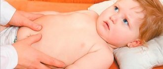
The procedure is performed on an empty stomach (the fasting break should be 4-6 hours). Before minimally invasive manipulation, the child is examined by an endoscopist to determine therapeutic tactics. If removal of a foreign object is planned under sedation (medicated sleep), then electrocardiography is performed and a consultation with an anesthesiologist-resuscitator is organized.
Depending on the age and general condition of the child, the procedure can be performed both on an outpatient and inpatient basis.
Diagnostics
A thorough history taking comes to the forefront of the diagnostic search. This way, parents themselves could notice that their child had swallowed a toy, indicate the size of this object, thereby speeding up the work of doctors.
X-ray examination can help in diagnosis, which will allow you to see a foreign body on an x-ray as a contrast object.
Fibrogastroscopy helps not only to see the swallowed object, but also to remove the foreign body.
Operation
Endoscopic removal is indicated if the foreign body is lodged in the esophagus, stomach, initial part of the small intestine, or exit part of the large intestine. The middle sections of the intestine are inaccessible for inspection.
Minimally invasive intervention in pediatric practice is performed under anesthesia. At the first stage, the surgeon carefully examines the lumen of the organ and determines the presence of a foreign body and the tissue reaction to its presence. After this, the foreign object is carefully grabbed by an endoscopic instrument and removed from the digestive tract under visual control. If the foreign body has piercing ends or a traumatic surface, then immediately after grasping it, an outer tube is put on the endoscope. This helps protect the walls of the digestive tract from damage.
The duration of endoscopic intervention is determined by the complexity of the clinical case. On average it ranges from 10 to 30 minutes.
When to ask for help?
Medical help should be sought if a child has swallowed a dangerous object: large, sharp or toxic (see list). A specialist is also required if the child cannot swallow, has the urge to vomit, has abdominal pain, blood in the vomit or stool, has developed a severe cough, or has a deteriorating general condition. Consultation is required if the foreign body has not been passed in stool within three days.
Adults are advised to seek emergency care in case of chest pain, acute abdominal pain, difficulty swallowing or difficulty breathing, bloody vomiting (or vomiting “coffee grounds”), or blood in the stool.
Removal of foreign bodies from the gastrointestinal tract in SM-Doctor
- Experienced doctors.
Endoscopists at our Center constantly improve their professionalism in accordance with the latest advances in medicine. This allows you to remove even small foreign bodies as gently and at the same time effectively.
- Modern equipment.
The latest generation of endoscopic equipment is characterized by particularly thin and flexible conductors. Such endoscopes easily penetrate even hard-to-reach places and carefully remove stuck objects.
- Endoscopy without pain and fear.
If there are indications and the desire of the patient, endoscopic examination and removal of foreign bodies from the digestive tract is carried out under sedation (medicated sleep). This provides comfort for the child and a more thorough examination of the lumen of the digestive tract.
- Efficiency.
Removal of foreign bodies from the gastrointestinal tract is carried out as quickly as possible after consultation with specialized specialists. The center provides emergency assistance in full from 9 to 21!
- Safety.
All manipulations are performed under visual control. An enlarged image is displayed on the screen of the endoscopic stand for visual assessment and identification of even minimal deviations from the norm.
First aid
A victim with a foreign body in the esophagus can be given first aid until doctors arrive or before going to the hospital. A strong cough helps expel the foreign body from the esophagus. If a person is unable to cough, the Red Cross's five-and-five approach can be used: five firm pressures on the abdomen (Heimlich maneuvers) can be alternated with five firm taps on the back. This strategy is designed to help people who are choking, but the Mayo Clinic recommends using it if a foreign body is suspected in the esophagus.
- Five claps on the back. You need to stand on the side of the injured adult; you can pick up the child. For support, the hand is placed under the victim's chest. He needs to lean forward so that his body is parallel to the ground. You need to deliver five abrupt blows with your palm to the back.
- Five presses on the stomach. The person providing assistance stands behind the victim, wraps his arms around him and sharply presses on his stomach. This technique is contraindicated in children under one year of age.
If assistance is provided by one person, he should perform the indicated techniques before calling an ambulance. If there are more people present, one of them should call doctors in parallel with the start of first aid.
Ultrasound diagnostics of gastric phytobezoars. Clinical observation
Ultrasound machine RS85
Revolutionary changes in expert diagnostics.
Impeccable image quality, lightning-fast operating speed, a new generation of visualization technologies and quantitative analysis of ultrasound scanning data.
Introduction
Bezoars (from the French - bezoard) are foreign bodies that form in the stomach itself as a result of the entry into it, primarily with food, of components that are not digested, but accumulate and form a foreign body. There are several types of bezoars: phyto-, tricho-, shellac- (from dyes, shellac, resin), hemato- (from blood clots), sebo- (from some types of fat, goat fat), pseudo- and polybezoars [1, 2]. Phytobezoars are the most common, accounting for up to 80% of all bezoars. They are formed from plant fiber, seed skins or seeds of persimmons, wild plums, cherries, grapes, figs, dates, and bird cherry. According to some reports, bezoars can be formed due to the proliferation of Candida fungi in the stomach. The rate of formation of a phytobezoar depends on its organic nature and ranges from 1-5 days to tens of years. Very quickly, phytobezoars are formed from unripe persimmons, which contain large quantities of resinous and astringent substances that glue food debris, primarily plant matter, into a compact mass.
Depending on the duration of their existence, bezoars can have a consistency from soft to rocky density, be single or multiple, ranging in size from a few millimeters to 10 cm or more.
Trichobezoars (the disease was first described in 1912 by V.M. Mysh), formed when wool or hair gets into the stomach, are found, as a rule, among hairdressers, as well as in people with an unbalanced psyche, suffering from an irresistible desire to bite hair.
Bezoars do not have clear clinical symptoms; the main diagnostic methods are x-ray and endoscopic.
The experience gained in recent years in successful ultrasound diagnosis of various pathological conditions of the upper digestive tract [3-5] makes it possible to diagnose new diseases of the esophagus, stomach, and duodenum, which was previously considered impossible.
In this message we would like to share our experience of ultrasound diagnostics of gastric phytobezoar.
Description of observation
Patient K., born in 1932, turned to the local doctor of the clinic with complaints of a feeling of heaviness in the epigastric region that had appeared in the last 1-2 months, which intensified after eating. Life history without any features.
On examination: condition is satisfactory. Normal nutrition. The skin and visible mucous membranes are clean and of normal color. The cardiovascular system and lungs are within the age norm. Pulse 80 beats per minute, rhythmic, satisfactory filling. A/D = 130/80 mm Hg. The tongue is moist and covered with a gray coating. The abdomen is soft and painless on palpation in all parts. Noteworthy is the presence in the left upper quadrant of the abdomen, closer to the midline, of an oval-shaped tight-elastic consistency of a painless, displaceable formation up to 15 cm long.
With a suspected tumor of the abdominal cavity, the patient was sent for an ultrasound examination, in which: the liver is not enlarged in size, its contour is even, the parenchyma is homogeneous. No focal pathology was identified. Intra- and extrahepatic bile ducts, veins, and portal vein are not dilated. The gallbladder is medium in size, its wall is compacted, the contents are homogeneous. The pancreas is of normal size, the structure is homogeneous. The Wirsung duct is not visualized. The cross-sectional diameter of the digestive tract at the level of the esophageal opening of the diaphragm is 14.8 mm, the wall thickness is 2.5 mm. In the left hypochondrium at the level of the left parasternal line, a hyperechoic formation 53.6 x 94.9 mm is visualized, giving behind an acoustic shadow (Fig. 1). The spleen has normal size and structure. The splenic vein is not dilated. The kidneys are usually located, not enlarged, the chest is not enlarged, and no stones are identified. The abdominal aorta is of normal diameter, its wall is compacted. No free fluid is detected in the abdominal cavity.
Rice. 1.
Sonographic picture of a bezoar of the stomach.
A)
Cross section.
b)
Longitudinal section.
To resolve the issue of the organ affiliation of the identified formation, the patient was asked to drink 500 ml of boiled water. After filling the stomach with water (Fig. 2), a hyperechoic oval-shaped formation 53.7 x 117.1 mm is visualized in its cavity, giving behind an acoustic shadow. The thickness of the stomach wall does not exceed 5.3 mm, peristalsis is not observed.
Rice. 2.
Sonographic picture of a bezoar after filling the stomach with water.
A)
Cross section.
b)
Longitudinal section.
Conclusion: foreign body (bezoar) of the stomach. Signs of a hiatal hernia. For further examination, it is recommended to perform endoscopy and fluoroscopy of the stomach.
During the questioning of the patient, it was clarified that for many years, being a big fan of persimmons, every winter she ate 1-2 fruits almost every day. This year, on one day, she ate 1.5 kg of persimmon.
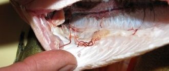
An endoscopic examination performed the next day revealed the following: the endoscope was inserted freely. The esophagus is not changed, the cardia closes completely. The visible gastric mucosa is pink, with small focal hyperemia in the antrum, peristalsis is uniform. The duodenal bulb is not deformed, its mucous membrane is pasty. In the stomach, a large bezoar is detected (Fig. 3), occupying almost the entire stomach cavity, tapering towards the lower third of the body, and is dense upon palpation. During the study, it was not possible to destroy it.
Rice. 3.
Endoscopic picture of a bezoar of the stomach.
Conclusion: bezoar of the stomach. Gastritis. Duodenitis.
X-ray showed the following picture: the act of swallowing was not impaired. The esophagus and cardia are freely passable. There is an axial unfixed hiatal hernia. The stomach is usually located. On an empty stomach it contains a small amount of liquid and mucus, has an elongated shape and sags. The stomach cavity contains a foreign body - a bezoar - about 4 x 11 cm, slightly tapering downward (Fig. 4). Against the background of the bezoar, the relief of the mucous membrane of the body and the angle of the stomach cannot be traced. In the antrum there are folds of mucous membrane of medium caliber, elastic. Stomach motility is preserved. Peristalsis can be traced only in the antrum of the stomach. Evacuation is timely. The compression is painless. The bulb and descending parts of the duodenum are without features. Conclusion: axial unfixed hiatal hernia. Stomach bezoar.
Rice. 4.
X-ray of a bezoar of the stomach.
To resolve the issue of the nature of treatment, a patient diagnosed with gastric phytobezoar was sent to the surgical department of the hospital.
Conclusion
This observation proves the possibility of ultrasound diagnosis of foreign bodies of the stomach, in particular, bezoars (phytobezoars) and the fact that echography can claim to be not only an auxiliary, but also, on a par with x-ray, the main method of diagnosing this pathological condition.
Having identified a space-occupying formation, it could not be ruled out that it belonged to the large intestine, and therefore additional research was required after filling the stomach with water. Carrying out an echography of the abdominal organs and having received data on the presence of a hyperechoic formation in the area of the projection of the stomach, which gives an acoustic shadow, the ultrasound doctor must indicate in his conclusion the possible presence of a gastric bezoar.
Literature
- Surgical diseases / Ed. M.I. Cousin. - M.: Medicine, 1986. - 564 p.
- Handbook of gastroenterology / Ed. V.Kh. Vasilenko. M.: Medicine, 1976. - P.64 - 65.
- Burkov S.G., Arutyunov A.G., Gurova N.Yu. Duodenal diverticulum simulating a pancreatic cyst // Sonoace International, 2001. - Vol. 8. - pp. 34 - 37.
- Clinical guide to ultrasound diagnostics / Ed. V.V. Mitkova. IV volume. - M.: Vidar, 1997. - 388 p.
- Berstad A., Hausken T., Gilja OH Ultrasonography of the human stomach // Scand. J. Gastroent., 1996. - Vol. 220 Suppl. —Pp. 25-82.
Ultrasound machine RS85
Revolutionary changes in expert diagnostics.
Impeccable image quality, lightning-fast operating speed, a new generation of visualization technologies and quantitative analysis of ultrasound scanning data.
What we swallow
As expected, children swallow foreign objects more often than adults. These objects can be classified according to various criteria, for example, according to size, material (detection during X-ray examination depends on this). They are also divided according to the degree of danger.
Less dangerous foreign bodies, according to Australian pediatricians:
- Small stones (pebbles).
- fruit seeds,
- Lost teeth.
More dangerous:
- Button type batteries. They can lead to burns of the mucous membrane of the esophagus and stomach. Before arriving at the hospital emergency room, the child should be given honey; it can prevent burns.
- Objects longer than 6 centimeters or thicker than 2.5 centimeters.
- Items made of lead.
- Magnets (especially when there is more than one). They can be attracted to one another, causing intestinal obstruction or perforation.
- Sharp objects (needles, pins, push pins, nails, fingernails, toothpicks, etc.).
Not only children swallow and inhale foreign objects; this also happens to adults. The “Top 3” foreign bodies in the esophagus and throat in adults look like this:
- Fish bone (9-45%).
- Bone (any except fish) (8-40%).
- Denture (4-18%).
Prevention of foreign body ingestion
For children, Australian pediatricians recommend:
- Eat only while sitting.
- Teach children to chew properly.
- Cut, grind hard foods for children under 5 years of age.
- Keep small objects out of the reach of children.
- Make sure that small pieces cannot be broken off from toys.
- Keep batteries and devices that can be removed from children.
Canadian doctors give the following advice to adults:
- Food needs to be cut into small pieces.
- You need to eat slowly, chew food thoroughly, and swallow little by little.
- There is no need to laugh and talk while eating.
- Don't eat while you're doing something else (like driving).
- Do not put nails, pins or other sharp objects in your mouth (do not insert them between your lips or teeth).
- Reduce alcohol consumption with meals.