The course of esophagitis
Esophagitis - occurs due to frequent consumption of spicy and hot foods.
Esophagitis belongs to the category of fairly common diseases. A pathological process occurs as a result of systematic irritation.
The culprit of the disease is the person himself, who often consumes excessively hot or spicy foods, as well as alcoholic beverages.
Esophagitis can also accompany other infectious diseases. The cause of the development of the disease is often injuries to the esophagus. During inflammatory processes in the tonsils and maxillary sinuses, this pathology can also appear.
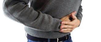
With esophagitis, the patient experiences a constant burning sensation in the chest area. Patients also complain that they often have the feeling that a bolus of food is moving down the esophagus. This disease is characterized by increased secretion of saliva. With esophagitis, pain occurs in the throat area.
To treat esophagitis, it is necessary to use a gentle diet. Patients are recommended to eat small meals, which means six meals a day. Food should be moderately heated. During the acute phase, the patient is advised to fast daily.
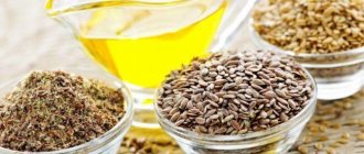
During the remission stage, a gradual subsidence of the inflammatory process is observed. During this period, the patient is allowed to consume pureed vegetable soups, warm milk, and liquid porridges. Patients are also recommended to take sea buckthorn or olive oil on an empty stomach. The patient should regularly drink still mineral water. For esophagitis, drug treatment is also indicated.
The choice of certain medications is made by the doctor depending on the characteristics of the disease.
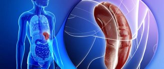
Diseases of the esophagus
The most common esophageal disease is gastroesophageal reflux disease (GERD). The main manifestations of GERD are heartburn, belching of air or food, nausea, vomiting, an unpleasant sour or bitter taste in the mouth in the morning, and difficulty swallowing. Sometimes patients complain of burning pain, discomfort in the epigastric region or behind the lower third of the sternum, which in some cases can be difficult to distinguish from manifestations of serious cardiovascular diseases. The listed symptoms often appear after eating, are associated with changes in body position and occur when bending forward, in a lying position. Patients may also be bothered by extraesophageal manifestations (masks of the disease), such as pain and a feeling of a lump in the throat, hoarseness, cough, destruction of tooth enamel, and excessive salivation. According to modern concepts, the main mechanism for the development of GERD is considered to be a violation of the motor activity of the esophagus and stomach. Between the stomach and the esophagus there is a lower esophageal sphincter, consisting of circular muscle fibers; in a healthy person, this valve closes tightly, preventing the contents of the stomach from flowing back into the esophagus. In patients with GERD, there is a decrease in the tone of the lower esophageal sphincter and a weakening of the esophagus' ability to quickly evacuate contents back into the stomach. As a result, the acidic contents of the stomach enter the esophagus (gastroesophageal reflux), contributing to the development of inflammation of the esophageal mucosa. In the case of prolonged contact in the absence of adequate treatment, erosions and ulcers develop at the site of inflammation, leading in some cases to scarring or bleeding, which may require surgical intervention. The risk of precancerous changes in the structure of the mucous membrane – Barrett’s esophagus – increases. Bile acids, enzymes, bicarbonates, which are part of the contents of the duodenum, also have a strong damaging effect on the esophageal mucosa. When these substances are thrown into the stomach (duodenogastric reflux), their reverse movement into the esophagus can be observed. The main causes of motility disorders of the esophagus and stomach are dietary errors (overeating, spicy and fatty foods, alcohol, coffee, chocolate, carbonated drinks), hiatal hernia, taking medications (non-steroidal anti-inflammatory drugs, calcium channel blockers, nitrates, psychotropic drugs, progesterone, aminophylline, etc.), obesity, neuroses, smoking, wearing tight belts, pregnancy.
In addition to motor disorders, the causes of inflammation of the esophagus can be infectious diseases (influenza, herpes, diphtheria, etc.), candidiasis (with immunodeficiency), trauma, burns.
share information
0
Social buttons for Joomla
Esophageal regurgitation
There are many different diseases of the esophagus.
Regurgitation is a pathological process in which food spontaneously moves out of the esophagus.
With the development of this disease, acidic contents are thrown from the stomach into the esophagus.
This is why many patients complain of a sour or bitter taste in the mouth.
If a patient has a narrowing or obstruction of the esophagus, this leads to the reverse movement of liquid, which has no taste and is characterized by the presence of pieces of undigested food and mucus.
Rejection of food from the stomach is usually observed 10-15 minutes after eating. In most cases, the person chews food and swallows it again. The process of treating a pathological process is directly influenced by the cause of its occurrence.
In the absence of a physical cause, the patient is prescribed Metoclopropamide, Cerucal, Reglan, as well as Cisapride, Coordinax. With the help of these pharmaceutical drugs, contractions of the esophagus are stimulated. Also, therapy for the disease can be carried out using auto-training.
Regurgitation is an unpleasant disease of the esophagus that can be treated with medication.
Publications in the media
Damage to the esophagus is a violation of the integrity of its wall. Classification • Internal (closed), arising from the mucous membrane • External (open), arising from the connective tissue membrane or peritoneum. Etiology • Iatrogenic: •• FEGDS •• Bougienage •• Cardiodilation •• Nasogastric intubation of the gastrointestinal tract •• Tracheostomy •• Tracheal intubation •• Surgeries on the chest, neck and abdomen • Foreign bodies • Diseases of the esophagus leading to perforation of its wall (tumors , ulcers, chemical burns, etc.) • Ruptures of the esophagus after vomiting (75% of cases), straining and coughing: •• Mallory-Weiss syndrome •• Spontaneous rupture of the esophagus (Boerhaave syndrome) • Impaired coordination of the upper and lower esophageal sphincters in as a result of alcohol intoxication, diseases of the central nervous system • Damage •• Cold steel •• Firearms •• With closed injuries to the body.
Pathological anatomy • Incomplete damage to the esophagus within one or more membranes, but not the entire wall of the organ • Complete damage to the esophagus throughout the entire depth of the organ wall •• Damage to the cervical esophagus - peri- or retroesophageal purulent-necrotic phlegmon of the neck develops •• Damage to the thoracic esophagus - mediastinitis, with damage to the pleura - pleurisy, pericardium - pericarditis •• Damage to the abdominal esophagus - peritonitis. Clinical picture • Pain along the esophagus • Sensation of a foreign body in the esophagus • Hypersalivation • Vomiting with blood • Subcutaneous emphysema • Salivation through the wound. Diagnostics • Survey radiography of the abdominal organs allows us to identify emphysema of the mediastinum or the tissue of the neck, hydropneumothorax, pneumoperitoneum • X-ray with a water-soluble contrast agent in various positions of the body (on the back, side, abdomen) allows us to determine the location and size of the defect • FEGDS with a rigid esophagoscope under anesthesia.
TREATMENT Surgical treatment • Radical surgery - elimination of a defect in the wall of the esophagus (no later than 12-24 hours from the moment of injury) • Palliative operations - wide drainage of the peri-esophageal tissue using one or another access: •• cervical lateral mediastinotomy •• transthoracic mediastinotomy, simultaneous drainage of the mediastinum and pleural cavity with four drainages • Gastrostomy for feeding the patient in the postoperative period. Conservative treatment only complements surgical treatment • Complete exclusion of enteral nutrition • Drug correction of homeostasis disorders • Antibiotic therapy with targeted action.
ICD-10 • K22.3 Perforation of the esophagus
Causes and course of the disease
Very often, solutions of acids or caustic alkalis become the cause of a burn of the esophagus. Depending on the volume of liquid drunk, its concentration and viscosity, the depth of damage may vary and corresponds to the following degrees of burns:
- I degree – damage to the surface layer of the epithelium occurs;
- II degree - first there is a lesion, and then necrosis of the deep layers of the mucous membrane and submucosal layer;
- III degree – necrosis of almost all layers of the esophageal wall.
Pathological changes that develop during a burn of the esophagus are divided into four stages.
At the first stage, the mucous membrane at the burn site looks swollen and loosened, and sharp hyperemia (redness) is observed.
At the second stage, necrosis of the esophageal wall occurs, which is rejected in some areas. As a result, ulcers of varying depths are formed: the superficial ones quickly epithelialize, and the deep ones heal with the formation of granulations.
At the third stage, the emerging connective tissue gradually forms scars and wrinkles. Depending on the degree of burn, scars can be superficial, deep, or sclerotic (very dense in consistency). The whole process is accompanied by a narrowing of the lumen of the esophagus.
At the last, fourth stage, deep scars may form in several places, which then shrink, quite often involving the surrounding tissue in the process. Fusion and deformation of the esophagus are observed. The duration of the scarring and narrowing stages can range from 2 weeks to several years.
How to cleanse the esophagus from mouth to stomach: yoga practice – Om Activ
In hataha yoga, there is a whole section dedicated to cleansing practices - shatkarama. We will introduce you to each of these practices and give detailed recommendations on them. Here you can get acquainted with Shank Prakshalana - the practice of cleansing the intestines.
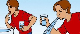
Kunjal Kriya (vaman-dhauti) is a method of cleansing the esophagus from the stomach to the oral cavity.
Your task is to fill your stomach to capacity with warm water and artificially induce vomiting for health and therapeutic purposes.
The word "kunjala" means "elephant". The name of the practice came from observing an elephant bathing, which draws water with its trunk and then spews it out in a fountain. "Vamana" means "inside", "middle".
The biggest obstacle to doing this practice is not understanding what exactly needs to happen. Typically, vomiting is the last attempt of the stomach to get rid of unclean or heavy food, associated with unpleasant sensations. During practice, water is evacuated simply, without excessive spasms and discomfort.
Benefit
● removal of decay products and internal poisons; ● prevention and treatment of gastric prolapse; ● increased secretion of gastric juice; ● improvement of digestion and absorption of food; ● cure chronic gastritis, colitis, constipation; ● removal of excess bile from the stomach; ● getting rid of acne, blackheads, rashes and eczema, including allergic ones; ● prevention and treatment of asthma, since relaxing the abdomen, stimulating nerve reflexes in the lungs and reducing the pressure of the diaphragm on them, reduces the frequency and severity of attacks; ● help in curing diabetes; ● loss of excess weight; ● elimination of cough and bad breath; ● diaphragm training.
Ayurvedicists add to this list: curing diseases of the teeth, chest, constipation, rickets, tonsillitis, night blindness, improving heart function, preventing bile spillage.
Preparation
Prepare 1-2 liters of water at body temperature. You can add sea salt to the water (one teaspoon per liter of water), but this is not necessary.
Procedure
- 1Squat down (kagasana) or stand over a sink or any other container.
- 2Drink 4-6 glasses of water quickly. You should feel like you are “puffed up” to capacity.
- 3Drink another glass as hard as you can. The amount of water you drink depends on the build, height, and weight of the person. A full one will need more water.
- 4If you drank water while standing, lean forward, and if sitting, raise your lower back so that your upper torso is parallel to the ground.
- 5Take a deep breath and draw in your stomach.
- 6Open your mouth as wide as possible, insert your index and middle fingers into it and begin to smoothly move them along the surface of the tongue in the direction from the root of the tongue to the tip, press harder on the root of the tongue near the small uvula. This will almost immediately cause an eruption of water from the stomach.
- 7 During the eruption of water, remove your fingers from your mouth. Try not to strain your body.
- Pink color of water may indicate a peptic ulcer.
- 8After the first portion of water has erupted, insert your fingers into your mouth again, press on your tongue and induce vomiting.
- 9Repeat the procedure until all the water comes out of the stomach and pressing on the root of the tongue or tickling it no longer causes vomiting.
- During the last eruptions, yellowish or greenish water may come out, and a bitter sensation may appear in the mouth due to the removal of excess bile from the stomach.
- 10 Finally, you can rinse your mouth, brush your teeth and rinse your nose.
Nutrition
After practice, refrain from eating for 30 minutes.
After half an hour, eat, for example, rice with ghee or any other porridge with butter to lubricate the stomach well. Avoid sour juices, sour fruits and milk during your first meal after practice - they can cause stomach upset.
Recommendations
- Do not practice before going out into the cold.
Kunjal Kriya removes mucus from the upper part of the esophagus and stomach and requires energy both to perform the practice itself and to restore the mucous membrane.
Contraindications: abdominal hernia, stomach ulcer, high blood pressure, heart disease.
- 1Perform the practice on an empty stomach, early in the morning or at least 3-4 hours after eating.
- 2Before performing the procedure, wash your hands thoroughly and, if possible, trim your nails short. You can use gloves.
- 3If you are unable to induce a gag reflex, insert your fingers deeper and press harder on the root of your tongue.
- 4If it still doesn’t work, don’t worry—it’s probably due to an untrained diaphragm. The water will come out in the usual way.
- 5Yoga master Swami Satyananda Saraswati advises healthy people to cleanse their stomach at least once a week.
- 6Do stomach cleansing after bowel cleansing.
(9758)
comments powered by HyperComments
Capsule endoscopy in Russia Medtronic & Covidien. Given Imaging
Trauma should be considered a violation of the integrity of the wall of the esophagus due to the sudden impact of a physical factor. Traumatic ruptures of the esophagus are rare, but deadly. Knife, gunshot, and stab wounds of the esophagus from the outside often affect the cervical spine; in car accidents or falls from a great height, the thoracic esophagus is sometimes ruptured. Typically, such injuries are accompanied by injuries to adjacent organs - blood vessels, lungs, heart, spine, which further worsens the prognosis. According to forensic reports, the esophagus is damaged by thoracic or thoracoabdominal injuries in 2.6% of cases.
Cause of esophageal injury
Based on this criterion, all mechanical damage to the esophagus is divided into damage by foreign bodies, instruments, spontaneous, hydraulic and pneumatic ruptures, damage by compressed air, gunshot and stab wounds, blunt injuries; neck, chest and abdomen.
The above classification answers many questions facing the problem of clinical description of mechanical damage to the esophagus. Based on the origin of the injury, all injuries to the esophagus are divided into external and internal. External injuries include injuries to the esophagus, which can occur in its cervical, thoracic and abdominal sections. As follows from the above classification, these injuries are divided into isolated and combined.
Injuries to the esophagus
Isolated wounds of the esophagus (stabbed, cut) are rare; they are more often combined with damage to neighboring tissues and organs. Gunshot wounds of the esophagus are especially severe.
Injuries to the cervical esophagus
If the cervical esophagus is damaged, the trachea, thyroid gland, large vessels, recurrent nerve, and spinal cord may simultaneously be injured.
Symptoms of esophageal injury
Symptoms of esophageal injury are as follows: pain when swallowing, discharge of saliva, blood and food from the wound when eating. Subcutaneous emphysema can often occur when the wound channel communicates with the larynx or cervical trachea. Any injury to the esophagus poses a serious risk of infectious and purulent complications, which are usually caused by anaerobic infection. Often esophagitis develops within 24 hours after injury, periesophagitis develops on the 2nd day, and mediastinitis on the 3rd day. The latter often develops as a result of purulent leakage. These complications are accompanied by swelling in the neck and smoothness of its relief, serous-bloody, then purulent discharge from the wound, sharp pain in the throat and neck when turning the head, which intensifies when throwing the head back. This causes a forced flexion position in the cervical spine. Body temperature reaches 39°C, the resulting septic condition is manifested by severe chills, pale skin, and cardiac dysfunction. The patient's general condition is progressively deteriorating.
When the thoracic esophagus is injured, injuries to the heart, lungs, large vessels of the mediastinum, trachea and bronchi can occur, which in most cases lead to either the immediate death of the victim or severe delayed complications with the same fatal outcome. If the patient is conscious, he complains of chest pain when swallowing, flexion, and especially when extending the thoracic spine. In a soporous state, bloody vomiting may occur. In case of damage to the esophagus, combined with damage to the trachea or bronchi, a severe syndrome of mediastinal emphysema develops with compression of the lungs, heart and aorta. The phenomena of mediastinitis, pleurisy, pericarditis develop rapidly, usually ending in death.
Injuries to the abdominal esophagus can be combined with injuries to the stomach, parenchymal abdominal organs, and large vessels. With such wounds, in addition to general pain, signs of peritonitis, internal bleeding, and intestinal obstruction develop.
Morphological changes in esophageal perforations
The dynamics of these changes go through several stages.
The stage of serous inflammation is characterized by rapidly increasing traumatic edema of loose peri-esophageal tissue, emphysema of the tissues of the neck and mediastinum. A complication of mediastinal emphysema can be rupture of the mediastinal pleura.
The stage of fibropurulent inflammation occurs 6-8 hours after injury: the edges of the esophageal wound are covered with fibrin and infiltrated with leukocytes. A reactive effusion of a hemorrhagic nature forms in the pleural cavity corresponding to the side of the wound. Often, primary or secondary pneumothorax develops. The peptic factor, which occurs when gastric juice enters the mediastinum, enhances necrotic and lytic processes in the mediastinal tissue and contributes to a more rapid course of mediastinitis. As for emphysema, with a favorable course of the postoperative period it usually resolves within 8-10 days and does not noticeably affect the further course of the process.
The stage of purulent exhaustion and late complications is characterized, according to the cited authors, by the so-called purulent-resorptive fever and wound exhaustion. At this stage, 7-8 days after perforation, purulent leaks spread, resulting in the formation of secondary pleural empyema, purulent pericarditis, and abscess formation of lung tissue. Such patients die from arrosive bleeding from large vessels of the mediastinum, resulting from the strong fibrinolytic effect of purulent exudate. Late complications of the pathological condition under consideration include purulent-fibrinous pericarditis, which occurs with perforations of the lower third of the esophagus, as well as in cases where the false passage passes in close proximity to the pericardium.
The stage of reparation (healing) usually occurs after opening the abscess, emptying and draining it, especially if the purulent focus is limited or encysted.
Closed esophageal injuries
Closed injuries of the esophagus are very rare and occur with severe bruises and compression of the chest and abdominal cavity as a result of road accidents, falls from a height, or in the workplace due to non-compliance with safety precautions among moving units. Closed injuries of the esophagus can be combined with ruptures of the liver, spleen, stomach, colon, abdominal aorta, which sharply worsens the general condition of the patient and often leads to death at the scene from massive internal bleeding and traumatic shock. The reparative stage lasts from 3 weeks to 3 months and depends not so much on the size of the abscess cavity in the peri-esophageal tissue, but on the size of the esophageal wall, since only after the flow of esophageal contents into the mediastinum has stopped can recovery occur.
The esophageal defect is closed by secondary intention. Unsutured defects larger than 1.5 cm are replaced by scar tissue, which subsequently results in deformations of the esophagus and the formation of diverticula with their inherent dysfunction.
Classification of mechanical injuries of the esophagus
Localization of injury
By level: cervical, thoracic, abdominal sections of the esophagus and their combination.
Injuries to the cervical esophagus are the most common and occur as a result of foreign body wedging or an unsuccessful attempt to remove them. With bougienage, damage to the esophagus is localized in the thoracic esophagus, and with cardiodilation - in the supradiaphragmatic and abdominal sections. The most dangerous manipulation is “blind” bougienage of the esophagus, in which multiple perforations often occur due to loss of elasticity of its wall. According to the involvement of the walls in the pathological process: anterior, posterior, right, left, their combinations, circular damage. The anterior wall is relatively rarely damaged. Foreign bodies most often injure the side walls. Instrumental ruptures of the cervical esophagus are often located on the posterior wall, and of the thoracic esophagus - on the right wall. Hydraulic ruptures are observed on the right wall of the middle third of the thoracic esophagus, spontaneous ones - in the lower third of this section and more often on the left. Circular injuries, characterized by avulsions of the esophagus, occur with blunt trauma to the chest and abdomen.
Depth of injury
- Non-penetrating injuries (abrasions, scalp ruptures of the mucous membrane and submucosal layer, submucosal hematomas) are the most common type of esophageal injury and are associated with foreign bodies or rough manipulation of instruments. Penetrating injuries (perforations, penetrating wounds) can be caused by the same mechanism as non-penetrating injuries, or by gunshot wounds. Depending on the mechanism, injuries can be isolated or combined with damage to neighboring organs and anatomical formations. Mechanism of injury
- Stab, cut, laceration, gunshot wounds, bedsores with perforation, combined.
- Damage by foreign bodies is most often a puncture wound and much less often a cut wound, resulting from a double-edged wound wedging into the esophagus. Instrumental injuries look like lacerations, and intraoperative injuries look like linear wounds with smooth edges.
Condition of the esophageal wall
- A scarred wall affected by varicose veins, a deep chemical burn, or a cancerous tumor.
The identification of this classification feature is of great practical importance, since the course of the injury and surgical tactics largely depend on: the previous condition of the walls of the esophagus. In particular, purulent complications with a rupture of a scarred esophagus develop later than with a rupture of an unchanged wall. In addition, the esophagus, with pronounced cicatricial changes, is a functionally inferior organ that has lost elasticity and pliability - such important qualities for the safe performance of instrumental manipulations. With food varicose veins, there is a risk of profuse bleeding, and if the wall of the esophagus is damaged by a cancerous tumor, there is a significant probability of its perforation during esophagoscopy with a rigid esophagoscope.
Associated damage
- Perforation of the esophageal wall in a complex manner without damage to neighboring organs.
These injuries concern only the esophagus and occur when it is perforated by foreign bodies, balloon probes, an esophagoscope, a bougie, a biopsy hook, an endotracheal tube, a gastric tube and is always accompanied by the appearance of a so-called false tract of varying length with the destruction of the peri-esophageal tissue of the neck or mediastinum. Perforation of the esophageal wall with damage to the mediastinal pleura.
Such damage can be localized on the right, seva or be bilateral. They can be combined with damage to the tracheobroichial tree and large vessels.
Diagnosis of esophageal injury
Diagnosis of esophageal injury is an extremely important stage in measures to cure this injury. The factor of early diagnosis with the establishment of the cause, size and depth of damage to the esophagus is extremely important, since the nature of medical care depends on this. The following sequence in carrying out diagnostic measures is generally accepted: survey fluoroscopy of the neck and posterior mediastinum, X-ray methods of examination with contrast, diagnostic esophagoscopy, puncture of the pleural cavity. The results of these studies, as well as anamnesis, assessment of the circumstances that led to esophageal injury syndrome and the nature of the clinical course allow for differential diagnosis both between different types of esophageal injuries, and between the latter and other forms of esophageal diseases.
During plain X-ray, air bubbles are visible in the peri-esophageal tissue; this phenomenon is called deep emphysema. Damage to the pleura is indicated by the presence of pneumo- and hydrothorax.
When conducting X-ray studies with contrast, some thoracic surgeons and radiologists prefer oil-based iodine-containing contrast agents. However, with a narrow perforation path, the oil solution, due to its viscosity, does not always penetrate into it, which makes it impossible to diagnose damage. In addition, these drugs, when in contact with the mediastinal tissue, are firmly fixed to it, and it is much more difficult to wash them off than a suspension of barium sulfate. The most acceptable are two- and three-iodine-containing water-soluble compounds, which are widely used in the diagnosis of esophageal ruptures. They do not irritate mediastinal tissue and, having low viscosity, penetrate well even into small wound defects. As noted by B.D. Komarov et al. (1981), these contrast agents are quickly absorbed, which makes them indispensable for obstruction of the esophagus and suspected presence of esophageal-respiratory fistulas, they provide a bactericidal effect and can be repeatedly used for dynamic monitoring of the healing process of the damaged area in the postoperative period.
When using X-ray methods of examination with contrast, it is possible to detect damage to the mucous membrane, exit of the contrast agent beyond the contour of the esophagus, determination of the position, direction and size of the false passage, its relationship to the lumen of the esophagus, mediastinal pleura, diaphragm, retroperitoneal space. All this is crucial when choosing treatment tactics.
Diagnostic esophagoscopy for injuries of the esophagus is not as widespread as x-ray examination. The reasons for this are as follows: esophagoscopy cannot always be performed due to the severity of the patient’s condition; after this manipulation there is always a deterioration in the condition. These obstacles are eliminated using intratracheal anesthesia with muscle relaxation, during which a thorough and calm examination of the esophagus along its entire length is possible and an accurate determination of the location, size and depth of the damage is possible. Diagnostic esophagoscopy has not only diagnostic, but also therapeutic value, since with its help it is possible to remove blood and other masses accumulated in the mediastinum from the false tract, as well as to insert a feeding probe into the stomach.
Puncture of the pleural cavity is an integral part of preoperative preparation and as a therapeutic and diagnostic measure. Its role increases with late diagnosis of esophageal perforation. The discovery of admixtures of food particles and gastric juice in the punctate confirms this diagnosis.
Differential diagnosis of mechanical injuries of the esophagus
In differential diagnosis, it should be borne in mind that in case of open trauma to the neck and chest, the diagnosis of damage to the esophagus is established during primary surgical treatment: with intraoperative trauma, damage to the esophagus is usually discovered during surgery (manipulation - probing, esophagoscopy with a rigid esophagoscope); Damage to the esophagus due to a closed injury to the chest or abdomen can only be diagnosed radiographically, since the clinical picture is dominated by signs of traumatic shock.
When the thoracic esophagus is ruptured, the resulting symptoms of esophageal injury may resemble many acute diseases of the cardiovascular system, respiratory system and chest wall, the occurrence of which is accompanied by severe pain (myocardial infarction, dissecting aortic aneurysm, pleuropneumonia, spontaneous pneumothorax, intercostal neuralgia).
A closed chest injury with a rupture of the esophagus, according to the clinical picture, has a certain similarity with a rupture of the diaphragm. As clinical practice shows, due to the fact that the physical examination data (tachycardia, hypotension, hydro- and pneumothorax), as well as the further course of the process (increasing intoxication, increased body temperature, soporous and comatose state) do not have specific signs of damage to the esophagus, Differential diagnosis in case of traumatic rupture cannot be carried out with a sufficiently high probability with most of the above diseases. However, as B.D. Komarov et al. point out. (1981), a clear history (vomiting due to spontaneous and hydraulic ruptures, foreign bodies or endoscopic manipulations) makes it possible to suspect damage to the esophagus. This suspicion can only be confirmed or refuted by performing an X-ray examination of the patient, but if this examination does not provide a clear answer to the condition of the esophageal wall, then esophagoscopy is performed.
A rupture of the lower third of the thoracic esophagus and the abdominal esophagus is manifested by symptoms very similar to those of perforation of the hollow organs of the abdominal cavity, in particular a perforated gastric ulcer.
According to B.D. Komarov et al. (1981), differential diagnosis for ruptures of the esophagus should be carried out not only with diseases such as pulmonary embolism and strangulated diaphragmatic hernia, but also with acute diseases of the abdominal organs (perforation of a hollow organ, acute pancreatitis and cholecystitis, thrombosis of mesenteric vessels).
In the differential diagnosis of esophageal injuries, one should keep in mind some similarities with Hammen's syndrome, which occurs in a woman in labor during pushing: subcutaneous emphysema, pneumothorax, shortness of breath, cyanosis, blood circulation disorders, pain, extracardiac murmurs synchronous with heart contractions. X-ray shows air in the mediastinum.
Against the background of primary symptoms associated with rupture of the esophagus, significant difficulties arise in the differential diagnosis of acute mediastinitis due to trauma to the esophagus from chronic sclerosing mediastinitis, which is a consequence of long-term inflammatory processes in the chest cavity and mediastinum (nonspecific pneumonia, bronchiectasis, pneumoconiosis, etc.) and is characterized by diffuse infiltration of the mediastinum, against the background of which foci of calcification can be identified radiographically. These lesions can simulate leakage of contrast agent beyond the contours of the esophagus, if due attention is not paid to them during plain radioscopy of the mediastinum.
How to examine?
X-ray of the esophagus
Endoscopy of the esophagus
Esophageal examination
Esophagogastroduodenoscopy
Esophagoscopy
Treatment of esophageal injury
Treatment of esophageal injury is divided into non-operative and surgical. When determining treatment tactics and choosing its method, the cause of the injury, its mechanism, morphological characteristics of the damaged tissues, localization, condition of the peri-esophageal tissue and the period elapsed since the injury to the esophagus are taken into account.
As a rule, non-operative treatment of esophageal trauma is indicated for patients with non-penetrating injuries of the esophagus, with perforations of the esophagus by a foreign body and with instrumental injuries of the esophagus.
In case of non-penetrating injury to the esophagus, the need for hospitalization and non-operative treatment arises when multiple and deep abrasions of the mucous membrane and submucosal layer are detected during esophagoscopy and x-ray examination, accompanied by swelling of the peri-esophageal tissue of the neck and mediastinal tissue. According to B.D. Komarov et al. (1981), with superficial abrasions of the mucous membrane without significant swelling of the peri-esophageal tissue, patients can undergo outpatient treatment, which in the vast majority of cases leads to recovery. They are recommended to eat gentle warm food, mucous decoctions, take beaten raw egg whites, drink small portions of decoctions of St. John's wort, medicinal chamomile and other herbs that have antiseptic properties and are not capable of irritating the mucous membrane. With this form of treatment at home, the patient should be informed about the possible appearance of signs of complications of an existing injury (increased pain, difficulty swallowing, chills, rise in body temperature). If they occur, you should immediately consult a doctor. As noted by the above authors, but according to their observations, in 1.8-2% of 372 patients with non-penetrating injuries of the esophagus, abscesses formed in the peri-esophageal tissue immediately adjacent to the area of non-penetrating damage after 5-6 days.
When the esophagus is perforated by a foreign body that penetrates the peri-esophageal tissue, an inflammatory process always occurs in this area, which on the 1st day after the injury is limited to a small area adjacent to the site of the damaged esophageal wall. The use of massive doses of antibiotics during this period leads in most cases to limiting inflammation and then to recovery. Indications for drainage of a limited abscess formed during antibacterial therapy occurred only in 5-8% of cases. Adequate drainage of the abscess also leads to recovery.
The presence of a foreign body in the lumen of the damaged esophagus causes massive infection of the peri-esophageal tissues and the development of phlegmonous (often putrefactive) inflammation. Attempts at non-operative treatment of such patients are erroneous, since if surgical intervention is delayed, diffuse mediastinitis develops with unpredictable consequences.
In case of instrumental injuries of the esophagus, non-operative treatment of esophageal injury is possible only if there is an effective outflow of purulent discharge from the damage zone into the lumen of the esophagus, when the rupture of its wall is no more than 1-1.5 cm and is not accompanied by damage to surrounding organs and the mediastinal pleura, and a false passage in tissue of the neck or mediastinum does not exceed 2 cm. In case of instrumental ruptures of the scarred wall of the esophagus, in which the false passage does not exceed 3 cm, non-operative treatment is also possible, since sclerotic changes in the peri-esophageal tissue accompanying sclerosis of the esophagus prevent the spread of the inflammatory process.
Typically, non-operative treatment of esophageal trauma and related indications is carried out either in a surgical thoracic or in an ENT department, especially if an uncomplicated (non-penetrating) foreign body was removed in the latter, leaving behind damage requiring only non-operative treatment.
Methodologically, non-operative treatment of esophageal injury, carried out according to appropriate indications in an inpatient setting, consists of massive antibiotic therapy and restriction or exclusion of oral nutrition for a certain period.
For non-penetrating injuries of the esophagus that do not require complete exclusion of oral nutrition, along with antibiotics, a solution of penicillin (1 million units in 200 ml of water) or a solution of furatsilin 1:5000 is prescribed per os, the purpose of which is to wash deep abrasions and scalp wounds from fibrin, pus and food debris.
In case of penetrating injuries of the esophagus, the dose of antibiotics is increased to the maximum possible, oral nutrition is excluded until the defect in the esophageal wall is healed. The tactics for managing a patient with such damage to the esophagus, according to the recommendations of B.D. Komarov et al., should be as follows. If it is assumed that healing will occur within a week, which usually occurs with puncture wounds with a foreign body, instrumental injuries up to 5-8 mm with a false stroke of the same length, then patients can be managed on full parenteral nutrition during this period. In such cases, patients should receive 2000-2500 ml of various solutions, including 800 ml of a 10% glucose solution with insulin (16 units), 400 ml of a 10% solution of Aminosol or Aminon, 400 ml of a balanced solution of electrolytes and vitamins. The deficiency of amino acids is compensated for by intravenous administration of Amnoplasmal E.
If healing of the esophageal injury is expected to take a long time, for example, in the presence of a bedsore of a scarred esophageal wall, an instrumental rupture larger than 1 cm in size with a false tract of the same length, then patients should immediately be transferred to tube feeding. For this, only thin silicone probes are used, which can remain in the esophagus for up to 4 months without causing irritation of the mucous membrane and without causing any discomfort to the patient. Feeding is carried out through a funnel or using a syringe to rinse the cavities with products of a creamy consistency, including pureed meat and boiled vegetables, broths, and fermented milk products. After feeding, the tube should be rinsed by passing 100-150 ml of boiled water at room temperature. In case of extensive destruction of the esophagus, requiring reconstructive surgical interventions, the patient is fed through a gastrostomy tube.
Treatment of esophageal injuries that cannot be treated nonoperatively consists of emergency surgery, which is performed by a specialist in cervical surgery, a thoracic surgeon, or an abdominal surgeon, depending on the level of injury. In case of severe injuries, the esophagus is exposed in the neck, mediastinotomy or laparotomy and diaphragmotomy are performed. When the cervical esophagus is injured, the wound of its wall is sutured, leaving the remaining tissues of the wound unsutured, and the wound cavity is drained. After the operation, the patient is placed on a bed with the head end down to prevent the contents of the wound, including inflammatory exudate (pus), from flowing into the mediastinum. Nutrition is carried out through a tube inserted through the nose; in particularly severe cases, a gastrostomy tube is applied. Drinking and eating are prohibited for 3 days. Antibiotics are prescribed.
With the development of mediastinitis, pleurisy or peritonitis, mediastinotomy, pleurotomy and laparotomy are indicated, which are performed by appropriate specialists in the relevant departments.
source: https://ilive.com.ua