Home » Health » Diagram of the location of human internal organs in the body of a man and a woman
The human body is the most perfect organic structure on Earth. The coordinated functioning of all organs makes it possible to carry out the life process.
Blood is pumped through the vessels by the heart, oxygen is processed into carbon dioxide by the lungs, brain activity controls all physiology. Thus, we can move, think, exist.
Having studied the structure of our body , we can come to the conclusion that it is similar to other creatures living on the planet. This is no accident. It is assumed that humans owe their origin to other mammals.
Research has confirmed that our physiology is similar to other animals, this is confirmed by the structure of an individual cell.
The features of the external structure can be seen with your own eyes. Our body consists of:
- The head, on which the sensory organs of the face are located - eyes, nose, ears, upper and lower jaws, food intake and speech communication occur through the mouth. The nose provides the functions of smell, the ears provide the organs of hearing.
- The neck, which connects the head to the body.
- The torso, including the chest and back.
- Upper and lower extremities – arms and legs. The feet provide stability for a person, and the hands provide the necessary manipulations.
Human anatomy includes systems consisting of individual internal organs that support the life process.
Let's list them:
- The musculoskeletal system allows us to move and ensures upright posture. Includes the skeleton, muscle structure, ligaments and joints. The location of the internal muscles ensures that the necessary functions are performed. The basis of the skeleton is the spine. Its disc numbers are from 1 to 24. The ribs form the chest.
- The digestive system is the most complex structure of all. During the digestion of food, it maintains the necessary energy balance. It begins with the nasopharynx and ends with the intestines.
- Respiratory system - includes the lungs and respiratory tract. Enriches our blood by converting oxygen into carbon dioxide.
- Transport functions are provided by the cardiovascular system. It allows blood to move through vessels, the arrangement of which ensures blood supply to the entire body.
- The nervous system regulates all physiological processes. It includes two types of brain: the brain and the spinal cord. In addition, there are nerve endings consisting of individual cells.
- Metabolism is regulated by the endocrine system.
- The pelvic organs are combined into the reproductive and urinary structures. It varies depending on gender. Provides reproduction and removes waste substances.
- The skin system protects the internal environment from aggressive environmental influences.
Let's take a closer look at the structure of the internal organs of our body.
The essence and features of MRI of the abdominal cavity
Magnetic resonance imaging is a non-invasive method, safe for health and very effective. A special device - a tomograph - works on the principle of creating a magnetic field. Its impact causes a response from hydrogen atoms located in the cells of the human body. These changes are recorded by the device in the form of layer-by-layer images, which are transferred to a computer, processed in a program and converted into three-dimensional images.
In fact, the tomograph is a large magnet. After turning it off, the hydrogen atoms return to their normal form, so there are no consequences of the procedure.
Unlike computed tomography and x-rays, MRI does not involve exposure of the body to harmful ionizing radiation. Thanks to this property, the procedure can be carried out with any frequency. The device helps to obtain an image of the abdominal organs, conduct analysis at the required depth, and identify signs and causes of diseases.
Systems and organs studied
Which organs are studied during the examination depends on the reasons for its conduct, existing symptoms, and preliminary diagnosis. MRI shows structural features, condition, pathologies:
- The pancreas along with the ducts;
- Gallbladder, its ducts;
- Blood vessels;
- Intestines;
- Adrenal glands and kidneys;
- Liver;
- Lymph nodes;
- Spleen;
- Soft tissues.
The doctor may order a study for a comprehensive check of the abdominal organs or indicate in the direction of which of them requires a detailed study.
Functions of the organ
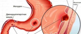
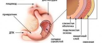
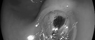
The main purpose of the stomach is to digest ingested food, and saliva, pancreatic juice and other secretions from the digestive tract contribute to this. After chewing, which occurs in the mouth, it goes through the esophagus to the stomach, where, under the influence of mechanical (peristaltic) muscle movement and the chemical action of hydrochloric acid, bile and other enzymes, it is converted into chyme. This mushy mass rises through the digestive tract to continue assimilation of essential nutrients and the resulting waste that cannot be used by the human body.
In the photo you can see a cross-section of the stomach
A secondary, but no less important function of the stomach is protective. Along with food, pathogenic bacteria also enter the body. Hydrochloric acid minimizes their number, that is, destroys them.
Indications for the procedure
MRI of the abdominal cavity is recommended in case of suspected dysfunction of organs located in this area. This examination is recommended for:
- Pain in the abdomen, hypochondrium, stomach, which occurs regularly or is constantly present;
- Problems with urination - increased frequency, changes in the volume and color of urine, discomfort when emptying the bladder;
- Detecting blood in the urine;
- Regular nausea or vomiting;
- Yellowing of the skin and mucous membranes;
- Changed stool consistency;
- Sudden sharp weight loss;
- Increase in organ size;
- Severe swelling of the limbs.
An MRI of the abdominal cavity is performed if there is suspicion of internal bleeding, injury, or the presence of foreign objects. The examination is performed before or after surgery to identify postoperative complications. Tomography allows you to evaluate the effectiveness of the treatment and, if necessary, adjust it.
If cancer is suspected and neoplasms are detected using other methods, MRI is prescribed to clarify the location of the tumor, its size and boundaries, nature, extent and characteristics of the blood supply.
Tomography helps to identify small tumors of unknown localization, diagnose metastases in the lymph nodes, and causes of liver enlargement of unknown etiology. Examination of the bile ducts shows the presence of narrowings, polyps, and blood supply disorders, including atherosclerosis of the arteries and vascular thrombosis.
External genitalia
1 – pubis; 2 – foreskin of the clitoris; 3 – head of the clitoris; 4 - labia minora; 5 - external opening of the urethra; 6 - hymen (is the border between the external and internal genital organs); 7 - Bartholin gland; 8 - anus; 9 — entrance to the vagina; 10 - labia majora.
Pubis
The pubis is an elevation located in front and slightly above the pubic joint, covered with hair, the upper limit of which grows horizontally (in men, hair growth extends upward along the midline).
Clitoris
The clitoris is a small (up to 1-1.5 cm), but very sensitive and important organ, consisting mainly of the corpus cavernosum. The male penis has a similar structure. The cavernous body has voids filled with circulating blood. During sexual arousal, these voids are intensely filled with blood, and the clitoris becomes enlarged and thickened—an erection. The corpus cavernosum is not capable of contracting like blood vessels, so traumatic injury to the clitoris is dangerous due to excessive bleeding.
Labia minora
The labia minora (LGB) are two folds of skin between the labia majora and the opening of the vagina. In front, connecting, they form the foreskin of the clitoris. Normally, the labia minora protrude slightly beyond the boundaries of the major lips, their color varies from pale pink to dark brown in the posterior regions. MPGs have a large number of vessels and nerve endings and are an ergenic zone; during sexual arousal, they increase in size due to blood flow.
PGMs are variable in shape and size, and are often asymmetrical. If the shape and size of the labia minora cause physical or mental discomfort, surgical correction of their size and the shape of the labia minora are performed.
sex slit
The genital fissure is the space between the labia majora and labia minora.
Labia majora
The labia majora (LGB) are two pronounced longitudinal folds of skin located on the sides of the genital slit. Anteriorly, the BPG converge into the anterior commissure, located above the clitoris. Behind, narrowing and converging one on the other, the BPG pass into the posterior commissure. The skin of the outer surface of the GPG has hair and contains sweat and sebaceous glands. The thickness of the labia majora contains blood vessels, nerves and Bartholin glands. On the inside they are covered with thin pink skin similar to a mucous membrane.
There are two openings under the labia majora and minora. One of them, with a diameter of 3 - 4 mm, located just below the clitoris, is called the external opening of the urethra (urethra), through which urine is discharged from the bladder. Directly below it there is a second hole with a diameter of 2 - 3 cm - this is the entrance to the vagina, which covers (or once covered) the hymen.
External opening of the urethra
The external opening of the urethra has a round, semi-lunar or star-shaped shape, it is located 2-3 cm below the clitoris. The urethra is 3-4 cm long, its lumen stretches to 1 cm or more. Along its entire length it is connected to the anterior wall of the vagina. On both sides of the external opening of the urethra are the excretory ducts of the paraurethral glands. These formations produce a secretion that moisturizes the mucous membrane of the external opening of the urethra.
Bartholin glands
Bartholin's glands (glands of the vestibule of the vagina) are paired, oblong-rounded formations, the size of a bean. They are located on the border of the posterior and middle third of the labia majora and produce a whitish secretion with a specific odor. The secret moisturizes the mucous membrane and has antibacterial properties.
Vaginal vestibule
The vestibule of the vagina is an anatomical formation. The “bottom” of the vaginal vestibule is the hymen or its remnants. In front, the vestibule is limited by the clitoris, behind - by the posterior commissure, on the sides - by the labia minora.
Hymen
The hymen (hymen) is the thinnest ring-shaped or semi-lunar-shaped membrane, 0.5 - 2 mm thick. With the onset of sexual activity, the hymen is torn. The hymen is the boundary between the external and internal genitalia.
Crotch
The perineum in the anatomical sense is the area between the pubis and the top of the coccyx, on the sides limited by the ischial tuberosities of the pelvic bones, in fact it is the exit from the small pelvis. In a clinical (obstetric) sense, the perineum is considered the area between the posterior commissure of the labia majora and the anus (Fig. 1). On the skin of the perineum there is a pigment line running from the posterior commissure to the anus - the perineal suture. The distance from the posterior commissure to the anus is called the perineal height and is 3-4 cm.
Rice. 1.
Anatomical landmarks of the female perineum: 1 - anterior commissure of the labia majora, 2 - posterior commissure of the labia majora, 3 - anus (anus), 4 - apex of the coccyx, 5 - ischial tubercle (on the left).
The thickness of the perineum consists of the skin, muscles, their tendons and fascia. The set of soft tissues occupying the space of exit from the small pelvis forms the pelvic floor or pelvic diaphragm. A woman's urethra, vagina, and rectum pass through the pelvic diaphragm.
The muscles and ligaments of the pelvic floor support the pelvic organs (bladder, vagina and rectum) in an anatomical position and provide a number of very important physiological functions: voluntary urination and urinary retention, defecation, retention of feces and intestinal gases, closure of the vaginal opening, are part of labor paths (Fig. 2).
Damage to these structures during childbirth leads to insufficiency of the perineal muscles and disruption of the pelvic organs - pelvic floor dysfunction, sexual dysfunction. This is written in detail in this article.
Rice. 2.
Sagittal section of the pelvic floor
What will an MRI show?
MRI shows developmental anomalies, pathological changes in the organs of the abdominal cavity and retroperitoneal space. This diagnostic method makes it possible to detect:
- Neoplasms of a malignant nature and benign type, including cysts, metastases, hemangiomas;
- Cirrhosis, abscess, fatty degeneration and other changes in the liver;
- Inflammatory processes in the kidneys, large intestine, gall bladder, pancreas;
- Renal pathologies, including pyelonephritis, hyrronephrosis, necrosis;
- Pathologies of the gallbladder in the form of its bending, the presence of stones in the ducts that remove bile;
- Parasite infestations;
- Adhesions and ascites;
- Disturbances in the structure and function of blood vessels, including the superior vena cava, signs of ischemia;
- Anomalies in the development of internal organs;
- Damage inside the abdominal cavity and in the retroperitoneal space caused by injuries and other negative influences.
In most cases, additional studies after MRI of the abdominal organs are not required, since the study provides all the data necessary to make a diagnosis and develop an optimal treatment regimen.
In what cases is a CT scan of the abdominal cavity and retroperitoneal space performed?
Computed tomography of the abdomen is usually used in the following cases:
- for pain in the abdominal area;
- in case of injury;
- if vascular damage is suspected;
- in case of disruption of the normal functioning of the stomach, intestines, liver, kidneys, spleen, gallbladder, adrenal glands;
- if stones are suspected;
- to clarify the expected diagnosis of the oncologist (and other specialists).
Tomography with contrast
Many medical centers offer contrast-enhanced tomography. The use of this method is often associated with suspicions of the development of a tumor process. At the same time, we are not always talking about malignant formations. Thus, the introduction of contrast makes it possible to distinguish a cyst from a hemangioma and helps determine the specifics of the neoplasm.
During the procedure, the patient is given a gadolinium-based substance intravenously. It has a cumulative function and the ability to dye fabrics. In this case, areas affected by the pathological process are colored differently from healthy tissue. As a result, it becomes possible to see tumors smaller than 1 mm and metastases. The amount of substance administered depends on the patient’s body weight.
Rules for preparing for the examination
Before examining the abdominal cavity, it is necessary to prepare for the procedure. In the presence of active processes of gas formation and fermentation, the result may be inaccurate, since such phenomena interfere with the visualization of organs and the changes occurring in them.
In this regard, patients are recommended to stop eating foods that contribute to gas formation 2-3 days in preparation for an MRI. These include legumes, fresh fruits and vegetables, baked goods, sodas and some other foods. You should consult your doctor about your diet.
The study itself is carried out on an empty stomach, the last meal is allowed 6 hours before the procedure. The doctor may recommend performing a cleansing enema the day before, taking laxatives or antispasmodics. You cannot take such measures on your own.
How is an MRI performed?
Before the examination, you need to remove all metal jewelry and accessories. To undergo the procedure, you should choose loose clothing that does not contain metal elements. The patient lies on a special table on his back, his body is fixed with special devices. This is due to the need to remain completely still throughout the examination.
The platform slides inside the tomograph. The doctor is in an adjacent room, maintaining voice and visual contact with the patient. During the examination, you may need to hold your breath for 15-20 seconds. The study itself lasts 20-30 minutes, with contrast enhancement - about 45 minutes.
Patients are advised to learn about the MRI procedure in advance to avoid anxiety, panic attacks and claustrophobia. The device makes quite a lot of noise; this is a normal phenomenon associated with the operation of the tomograph. To reduce discomfort, the patient is offered headphones or earplugs. Otherwise, the procedure does not create unpleasant sensations and does not cause pain. MRI does not give side effects and does not provoke any consequences or reactions of the body.
How does my stomach hurt?
The pain can be dull, sharp, paroxysmal, occur in the middle, above or below, on the right or left side of the body.
The intensity of such sensations does not always reflect the severity of the condition causing them, just as vice versa - mild but constant pain can be a sign of a chronic disease that needs urgent treatment.
If discomfort in the epigastrium persists for more than two weeks, you need to inform a gastroenterologist about this. This will avoid a more serious diagnosis. The reason to immediately consult a doctor is pain in the peritoneum, accompanied by constant bloating, regular vomiting, diarrhea and blood in the stool.
Results and their interpretation
The examination result looks like an image of layer-by-layer sections with a thickness of 2 to 4 mm. The doctor compares the obtained indicators with the norm and analyzes pathologies. The conclusion issued by the doctor after the examination states:
- What organs were examined;
- Have neoplasms been identified, what is their nature, location, prevalence;
- Are there metastases in tissues located near the lesion;
- Has the preliminary diagnosis been confirmed?
The document describes not only all identified pathologies and disturbances in the functioning of systems, but also healthy organs. Additionally, the patient is given printed photographs, as well as video materials recorded on a disk or memory card.
Depending on the identified pathologies, it is necessary to contact a specialized specialist. Based on the MRI results, the doctor will make a diagnosis and prescribe conservative or surgical treatment.
Who should not have an MRI?
In some cases, the procedure is not allowed or its feasibility is determined by the doctor taking into account a number of factors. Contraindications to examining the abdominal cavity using MRI are:
- The presence of ferromagnetic metals in the body in the form of plates, pins and other elements;
- The presence of pacemakers, vascular stents and other fixed devices prone to magnetization;
- Skin tattoos made using metal-containing dyes;
- Susceptibility to sudden muscle spasms;
- Artificial joints containing metal;
- Renal failure in chronic form.
Some modern devices that are installed in the body are compatible with such a procedure. In this case, you should take the supporting document with you and show it to the doctor. It is advisable to have pictures taken with you from previous MRIs, if any. This will help establish the dynamics of the development of pathological processes.
It is not recommended to perform the procedure in the early stages of pregnancy. If contrast enhancement is used, it is necessary to ensure that there is no allergy to gadolinium.
MRI studies are carried out with caution on patients in serious condition, persons with mental disorders, people who, for one reason or another, are unable to remain motionless or suffer from severe claustrophobia.
An MRI of the abdomen shows any pathological processes affecting organs in this area.
Rate this article: (1 rated 5 out of 5)
Diseases by type of pain
When the diagnostic process fails to determine the exact cause of painful sensations in the epigastrium, doctors talk about functional dyspepsia. This pathology causes discomfort in the upper part of the stomach, and may be accompanied by other symptoms such as gas, acidity, lack of appetite, vomiting or indigestion. Stress can also be a fundamental factor in the development of functional dyspepsia, causing the stomach to contract too quickly.
In addition, there are a number of diseases that can affect the digestive system and cause discomfort in the epigastric region. The nature of the pain, its location and other additional symptoms will help determine what exactly is happening in the body.
Pain after eating
It occurs due to the development of pathologies of the stomach itself or other organs of the peritoneum. Such sensations indicate gastritis or peptic ulcer disease. Seizures occur as a result of the following conditions:
- Increased stomach acidity when food enters it.
- Intense contractile activity of its muscle walls.
- Excessive enlargement of the organ itself due to overeating.
Blunt pain
This type of discomfort indicates chronic gastrointestinal pathologies, home treatment for which consists of following a diet and taking medications. Among the diseases that cause dull pain:
- duodenitis;
- initial stage of gastric and duodenal ulcers;
- inflammation of the mucous epithelium;
- diverticulitis;
- chronic form of appendicitis;
- cancerous tumors;
- polyps;
- kidney stones.