Benign tumors of the stomach are much less common than malignant neoplasms of this localization. They are often asymptomatic, presenting themselves with various complications. Many of them can lead to stomach cancer. Their timely detection and treatment is considered an important area of modern gastroenterology.
- Description of benign gastric tumors
- Types of benign stomach tumors
- Localization
- Causes
- Risk factors
- Symptoms
- Diagnosis of benign stomach tumor
- Treatment of benign stomach tumor
- Forecast
- Prevention
Description of benign gastric tumors
Benign stomach tumors vary in internal structure and appearance. They can range in size from a few millimeters to tens of centimeters in diameter. Their shape is often round or oval, the surface can be smooth or uneven, and the consistency can be hard or soft upon palpation.
Epithelial tumors of the stomach (polyps) can be single or numerous (diffuse polyposis). They can grow on a thin stalk or have a massive base. Depending on the structure, the color of the growth will vary (from pale pink to bright crimson). Villous polyps often appear as papillary projections of varying thickness and height, resembling fingers. Under a microscope, rows of altered glandular epithelial cells with branching tubular structures, areas of intestinal metaplasia and dysplasia will be visible.
If malignancy of polyps occurs, outwardly they will look like heterogeneous diffuse growths of the mucous layer, with areas of ulceration and a pronounced vascular component. On microscopic examination, atypical cells will be visible.
Non-epithelial benign neoplasms are capable of growing into the stomach cavity or outward, squeezing neighboring organs and structures. Large tumors can be visually distinguished and palpable during examination.
Types of benign stomach tumors
The main classification of benign formations of the stomach divides them, according to their origin, into two large groups:
- epithelial tumors;
- non-epithelial tumors.
The first group includes hyperplastic (75%) and adenomatous polyps (adenomas), as well as diffuse polyposis. Mucous growths most often affect the pyloric antrum of the stomach and occur in men from 40 to 60 years of age. Hyperplastic polyps have a predominantly benign course. Gastric adenomas in most cases degenerate into cancer. With polyposis, mucous growths of both types are observed.
Nonepithelial benign tumors of the stomach are extremely rare. These include:
- endotheliomas (from vascular endothelium);
- lipomas (from fat cells);
- neuromas (from nervous tissue);
- fibroids (from muscle fibers);
- hemangiomas (from blood vessels);
- lymphangiomas (from lymphatic vessels);
- fibromas (from fibrous connective structures);
- mixed tumors.
Also in the stomach there are dermoids, hamartomas, chondromas, osteomas and heterotopias, originating from pancreatic tissue and duodenal glands. The most common non-epithelial benign tumor is leiomyoma (smooth muscle neoplasm). The latter is prone to complicated course and malignancy.
Complications and the possibility of cancer
Even small polyps in the stomach are capable of progression and the development of complications. Although the disease is characterized by a long-term benign course, adverse consequences sometimes appear quite quickly and threaten the person. The most typical complications of the disease are as follows:
- bleeding;
- necrosis of the mucosal area of the polyp;
- pyloric stenosis – if the formation is located close to the pyloric canal;
- stomach cancer.
A polyp is a dangerous formation. A particularly serious complication is malignancy - the appearance of an oncological process. Any benign structure is potentially dangerous, therefore, during fibrogastroscopy, ICLINIC doctors always perform a biopsy. If dysplasia is present, the next stage is stomach cancer. Therefore, all polypoid growths must be removed.
Causes
The etiology of benign gastric neoplasms is not fully known. Scientists suggest that the appearance and growth of polyps is associated with disruption of the processes of regeneration and proliferation of epithelial cells. Hyperplastic growths often begin to develop with decreased production of hydrochloric acid. Adenomas predominantly arise against the background of changes associated with atrophic gastritis, due to intestinal metaplasia and dysplasia. Epithelial cells “lose” their differentiation and acquire atypical properties, which can give rise to a malignant process.
Gastric tumors of a non-epithelial nature, according to scientists, originate from heterotopic embryonic tissue. The causes of this anomaly can be various pathological processes during human intrauterine development.
Risk factors
Tumors in the stomach occur more often in people who:
- suffer from chronic diseases of the digestive system;
- have an active Helicobacter pylori infection (microorganisms lead to damage to the mucous membranes of the gastrointestinal tract and a decrease in their protective properties);
- do not comply with the principles of rational, balanced nutrition;
- have a genetic predisposition to tumors;
- live in environmentally unfavorable areas;
- systematically exposed to adverse factors (chemical compounds, stress, trauma, etc.);
- have an immunodeficiency;
- have bad habits (alcohol, smoking, etc.).
The risk group includes people over 40 years of age who do not undergo annual preventive examinations with mandatory examination of the stomach.
Causes of polyps and clinical manifestations of the disease
The pathological condition is a benign neoplasm growing into the lumen of the stomach. The exact reasons for the development of polyps have not been established. However, the following factors predispose the disease:
- microtrauma of the mucous membrane due to consumption of rough food;
- duodeno-gastric reflux;
- stagnation in the upper gastrointestinal tract;
- chronic atrophic gastritis;
- hereditary predisposition - familial polyposis;
- scars after ulcers and operations.
ICLINIC doctors believe that polypoid growths can form at any age. The peak development of benign formations occurs between 30 and 50 years.
Symptoms of the disease do not appear for a long time. The formation grows slowly, without leading to dyspeptic disorders. Sometimes there is intermittent pain in the epigastric zone, a feeling of heaviness in the stomach, belching and some loss of appetite. There are no pathognomonic symptoms typical of benign processes in the stomach.
Symptoms
More often, neoplasms occur without specific clinical symptoms. In most patients, benign stomach tumors have signs of concomitant pathologies (chronic inflammation, peptic ulcer, gastroesophageal reflux, etc.). The patient may present the following complaints:
- belching of air or sour contents;
- heartburn;
- nausea;
- vomit;
- bloating;
- bowel dysfunction;
- discomfort, heaviness, pressure, sensation of a foreign body in the epigastric region;
- epigastric pain of varying intensity and character, depending to varying degrees on food intake.
When complications occur, a typical clinical picture occurs. With severe inflammation in the stomach, the pain becomes a constant aching character. Particularly severe pain and burning are observed with neuroma. When benign tumors are pinched, the pain is intense, cramping, spreading throughout the entire abdomen. Often a person breaks out in a cold sweat and loses consciousness.
When polyps are ulcerated, a clinical picture of gastric bleeding may occur. Possible vomiting of blood, tarry stools, and general malaise. With chronic blood loss, anemia develops. The person becomes pale, constantly experiences fatigue, weakness, and dizziness.
If a mucous growth completely blocks the exit from the stomach into the duodenum, acute intestinal obstruction occurs. If the lumen is partially closed by a tumor, nausea, vomiting of eaten food, heaviness in the epigastrium and other symptoms will occur.
Non-epithelial benign neoplasms vary in symptoms, depending on their location, the presence of surface defects, the rate and type of growth (inside the stomach or out). More often they manifest themselves as periodic painful sensations after eating, when moving or changing body posture. Large tumors can be palpated and “protrude” through the abdominal wall, as well as “impede” the work of neighboring organs.
Almost any non-epithelial formation of the stomach can be complicated by bleeding. Hemangioma poses a particular danger. It can cause massive blood loss, threatening the patient's life. If the tumor is strangulated and/or infected, intestinal obstruction and peritonitis may occur. Over time, benign stomach tumors can transform into cancer.
Precancerous diseases of the stomach
ANSWERS TO QUESTIONS ABOUT STOMACH CANCER >>>
Obligate precancer is a disease that always or in most cases causes cancer if left untreated. Such diseases include callous gastric ulcer, rigid antral gastritis, polyps, and gastric polyposis.
facultative precancer - chronic diseases of the stomach in which cancer develops relatively rarely, but more often than in healthy people. These are chronic atrophic gastritis, disease of the operated stomach, pernicious anemia, Ménétrier's disease, etc.
The National Cancer Congress in Japan treats a number of diseases against which stomach cancer can occur as background diseases, combining obligate and facultative precancer into one group.
According to the National Cancer Congress, stomach cancer can develop against the background of chronic atrophic gastritis in 0-13% of cases, pernicious anemia - in 0.5-12.3%, chronic gastric ulcer - in 1-2%, hyperplastic polyp - in 1 -2%, flat adenoma - in 6-21%, papillovirus adenoma - in 20-75%, Ménétrier's disease - in 5-10%, history of gastric resection - in 0.4-7.8% of cases.
Most scientists believe that with the development of a tumor process, the epithelium of the gastric mucosa changes in a certain sequence: normal epithelium -> proliferating -> atypical cells -> malignant tumor.
From a morphological point of view, multicentrically located areas of proliferation of atypical epithelium are called dysplasia.
Dysplasia is a disorder of tissue structure, characterized by pathological proliferation and atypia of the epithelium. In the gastric mucosa, dysplasia can be of three degrees (mild, moderate and severe).
With dysplasia of degrees I and II, the changes are mild and reversible. Changes related to grade III dysplasia are permanent and can be considered as cancer in situ. In 40-45% of people over the age of 60 years, in the gastric mucosa, an intestinal type of epithelial restructuring occurs - intestinal metaplasia (small intestinal, colonic). Colonic metaplasia is interpreted as a precancerous condition.
Chronic gastritis is the most common disease among the pathologies of the gastrointestinal tract, which accounts for 35% of all diseases of the digestive system and 85% of diseases of the stomach. Chronic gastritis is a polyetiological disease. There are many reasons that cause it, but in accordance with etiopathogenesis they can be combined into three main groups:
•Group I - infectious (exogenous), associated with gastric Helicobacter (gastritis B - bacterial); • Group II - endogenous autoimmune, caused by the formation of antibodies to the parietal cells of the gastric glands (gastritis A - autoimmune) - atrophic gastritis; • Group III - exendogenous, associated with partial thermal irritations, drug effects are expressed by gastroduodenal reflux (gastritis C - reactive or chemical). Against the background of chronic atrophic gastritis, stomach cancer occurs in 0-13% of cases.
Clinic. In the clinical picture of chronic gastritis there are no typical characteristic signs on the basis of which one or another of its forms could be recognized.
Main complaints: loss of appetite, discomfort in the epigastric region associated with food intake, belching, heartburn, nausea, and sometimes vomiting.
Syndromes of gastric and intestinal dyspepsia can be in a variety of combinations (a feeling of fullness of the stomach with food, flatulence, rumbling in the stomach, tenesmus, constipation followed by diarrhea).
Diagnostics. The X-ray method of examination plays a relative role in the diagnosis of chronic gastritis. It is more suitable for identifying ulcerative, polyposis and tumor lesions of the stomach. The exception is rigid antral gastritis. Gastrofibroscopy and gastrobiopsy are of decisive importance in recognizing various forms of chronic gastritis. Diagnosis: intestinal metaplasia, dysplasia of varying severity, infection caused by Helicobacter pylori (HP) - cytological and histological only.
Treatment. Therapeutic measures for chronic gastritis of non-infectious etiology include: elimination of the main factors contributing to the development of the disease or supporting it, diet therapy, sedatives during exacerbations, drugs that improve microcirculation and trophism of the mucous membrane, vitamin therapy, sanatorium treatment.
Infectious gastritis caused by Helicobacter pylori microorganisms requires special attention. Today, scientists around the world have proven that 30-50% of the world's population are carriers of HP infection (a sick person or a bacteria carrier). HP bacteria usually multiply under a layer of mucus above the mucous membrane of the pylorus, attach to the apical part of the epithelial cells, secrete enzymes - cytotoxins and urease, and edema, hyperemia, impaired trophism, and degeneration of mucosal cells develop in the epithelium. The processes of inflammation and atrophy are closely related and have an infectious-immune origin.
In the last 10 years, many researchers have been interested in the problems of helicobacteriosis and stomach cancer. To date, it has been proven that Helicobacter pylori produces many pathogenic enzymes and toxins, some of which may be involved in carcinogenesis.
•A relationship has been shown between the presence of HP infection and intestinal metaplasia, a type of which - incomplete intestinal metaplasia - is considered a precancerous condition. •It has been established that HP, using the enzyme extracellular urease, breaks down urea to form ammonia, activates the processes of lipid peroxidation, increases the concentration of free radicals - oxygen derivatives, which stimulates the processes of carcinogenesis. •In vitro experiments have demonstrated the ability of HP to stimulate the growth of tumor cell cultures. Gastric cancer in most cases is accompanied by infection of the mucous membrane of HP; At the same time, HP infection has a greater prevalence than in peptic ulcer disease and takes on the character of total contamination of all parts of the stomach, including the tumor itself.
Helicobacter gastritis (gastritis B) requires special treatment. To destroy these bacteria, it is necessary to use three drugs that have antibacterial activity: amoxicillin or ampicillin, a nitronidazole derivative (metronidazole) and a nitrofurin derivative (furazolidone). The basic drug is de-nol (citric acid salt with bismuth).
Stomach ulcer. Malignancy of recurrent and callous gastric ulcers is observed in 2-10% of patients.
Clinic. Unlike chronic gastritis, a chronic gastric ulcer has a fairly clear clinical picture.
The most consistent symptom of a peptic ulcer is pain associated with eating. The pain can be early, occurring either immediately after eating or after 0.5-1 hour, and late, occurring 2-3 hours after eating or later. Sometimes the pain occurs at night and goes away after eating (milk).
Gastric ulcers are also characterized by seasonality - exacerbations in the autumn-winter and spring months, remissions in the summer.
An important symptom of the disease is heartburn; More than half of patients experience vomiting and nausea. Vomit contains acidic liquid mixed with food. With cicatricial pyloric stenosis, vomiting becomes more frequent, and the vomit is profuse, with remnants of food eaten the day before. Intestinal dysfunction is often observed - constipation is often replaced by diarrhea.
Diagnostics. In the recognition of gastric ulcer, in addition to clinical semiotics, the study of gastric juice plays a certain role. In most patients, there is an increase in secretion and acid formation. Only with a long course of the disease, especially with callous ulcers, does the acidity of gastric juice decrease. It is also important to examine stool for occult blood.
During an X-ray examination of the stomach, the main symptom is a niche. A large niche with a surrounding shaft and convergence of folds of the mucous membrane usually indicates the callous nature of the ulcer. Endoscopic, cytological examination, as well as multiple biopsies from different areas of the ulcer help to distinguish a benign gastric ulcer from a malignant one.
Treatment. Acute and recurrent ulcers in the acute stage are treated, as a rule, conservatively. It usually takes 4 to 8 weeks for an ulcer to heal, and longer for large or old ulcers. Endoscopic or fluoroscopic control is carried out after 8 weeks and until complete healing. If complete healing does not occur, a repeat biopsy is performed.
In case of complications of the ulcer (penetration, perforation, cicatricial stenosis, repeated bleeding), lack of effect from conservative therapy, frequent recurrence of an uncomplicated peptic ulcer, depriving the patient of his ability to work, or suspicion of a malignant gastric ulcer, surgical treatment is indicated.
Polyps and polyposis of the stomach. Polyps are hyperplastic and adenomatous.
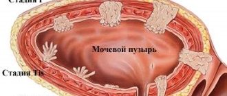
Hyperplastic changes occur in 80-90% of cases of all polypous formations in the stomach and are classified as tumor-like lesions. In 10-20% of cases, adenomatous polyps occur - benign epithelial tumors. They usually take the form of rounded formations protruding into the lumen of the stomach, located on a thin stalk or a wide base.
Precancerous polyps include glandular adenomatous polyps. Dysplasia of varying severity in adenomatous polyps is observed in 40-60% of cases, and grade III dysplasia in 5-10%. If hyperplastic polyps degenerate into cancer in 1-2% of cases, then flat adenoma - in 6-21%, and papillovirus adenoma - in 20-75% of cases;
Clinic. Single polyps can be asymptomatic and be an incidental finding. Clinically, they manifest themselves, as a rule, with concomitant chronic gastritis. If the tumor disintegrates, bleeding is possible; if a polyp is strangulated, an attack of pain in the epigastrium, nausea, and vomiting may occur.
Diagnostics. The leading role in recognizing polyps and gastric polyps belongs to x-ray and gastroscopic methods.
Endoscopy biopsy allows for cytological and morphological characteristics of polyps.
Treatment. Adenomatous polyps are treated surgically. For single polyps, endoscopic polypectomy is possible; for multiple polyps, gastrectomy or gastrectomy is performed.
Operated patients are under clinical observation. At least once every 6 months, an endoscopic examination is carried out, as well as periodically complex treatment, including a rational diet, vitamin therapy, sedatives, and spa treatment.
Pernicious anemia (Addison's disease) can be defined as the absence of intrinsic factor associated with complete atrophy of the fundic mucosa and loss of parietal cells. The disease is diagnosed when complete atrophy of the mucous membrane of the fundus of the stomach is noted, as a rule, this happens after 50 years. At the time of detection of the disease, there is no inflammation in the mucous membrane, i.e. the pathological process is atrophy without gastritis.
Some evidence points to an immunological and hereditary basis for pernicious anemia, also called type A gastritis.
With this disease, in 90% of cases, the body contains antibodies to the parietal cells of the stomach; and in 60% of cases - antibodies to the internal Castle factor (both blocking and binding it). Anemia usually develops gradually and unnoticed, progressing as vitamin B stores in the liver are depleted.
Stomach cancer with pernicious anemia occurs in 0.5-12.5% of cases.
Patients with pernicious anemia are usually observed by a general practitioner and hematologist. For patients over 50 years of age, endoscopic examination of the stomach is required once a year.
Ménétrier's disease is a lesion of the stomach of unknown etiology, manifested by very large gastric folds, large glands with slight inflammation and cystic dilatation; in this case, the submucosal layer is rarely affected; often the process is limited to the body and fundus of the stomach (the antrum is usually not affected). Sometimes the width and height of the folds reaches 3.5 cm. Characterized by increased mucus formation, hyposecretion of hydrochloric acid and pepsin, and a decrease in the content of serum proteins, leading to the appearance of hypoalbumic edema.
Clinic. Characterized by epigastric pain, weight loss, and nausea.
The course of the disease is chronic (remissions, exacerbations), sometimes spontaneous recovery occurs or transition to atrophic gastritis.
Menetrier's disease usually occurs between the ages of 30 and 60 years.
Diagnostics. X-ray and endoscopic examination reveal very large folds that never extend to the pyloric region (a biopsy of the full thickness of the fold is indicated). In 5-10% of cases, stomach cancer develops.
Treatment is conservative; in cases of severe hypoalbuminemia, gastrectomy may be necessary.
Atrophic gastritis of the resected stomach. The natural outcome of gastric resection is the development of chronic gastritis in the stump, which is accompanied by dysplasia and intestinal metaplasia of the epithelium due to a decrease in the acidity of gastric juice and the reflux of bile into the gastric stump.
Gastric stump cancer occurs in 0.4-7.8% of patients operated on for non-tumor diseases of the stomach 10, 15, 20 years after surgery.
Patients with a resected stomach require constant medical supervision throughout their lives. Once a year they should undergo a gastroscopic examination of the stomach stump.
APPOINTMENT FOR A CONSULTATION +7 (921) 951 - 7 -951
Share link:
Diagnosis of benign stomach tumor
To detect benign stomach tumors, a set of diagnostic measures is used. The leading methods are:
- laboratory tests (blood tests, serological tests to determine the presence of infection, assessment of gastric juice, etc.);
- radiological methods (radiography of the stomach, CT scan of the abdominal organs, etc.);
- Ultrasound of the gastric wall and regional lymph nodes;
- endoscopic examination of the gastrointestinal tract with targeted sampling of histological material from 6-8 suspicious areas.
Often, non-epithelial formations of the stomach are confirmed after surgery, when a morphological analysis of the removed tumor is performed.
What examinations are necessary for early detection of polyps?
The only way to early diagnose polyps is mass screening, even in the absence of any complaints. A fecal occult blood test, which is included in the list of free examinations during the annual medical examination, is the main non-invasive method to suspect the presence of villous tumors and cancer. But the “gold standard” for diagnosing polyps is an endoscopic examination of the colon, which can detect almost any formation (less than 0.5 centimeters in size).
The best prevention of the appearance of polyps in the gastrointestinal tract is regular examinations, including gastroscopy and colonoscopy.
Treatment of benign stomach tumor
Non-cancerous stomach tumors usually require surgical treatment. The most acceptable method for small epithelial formations is their endoscopic removal. Enucleation or electrocoagulation of the polyp is performed. Large and multiple polypous growths are operated on using a transperitoneal approach (part of the stomach or the entire organ is removed). In some cases, endoscopic resection of a huge tumor is performed to eliminate unwanted symptoms and reduce the risk of complications.
Surgical removal of non-epithelial benign formations is performed in various ways, depending on their characteristics. According to indications, enucleation, various types of gastric resection or gastrectomy are performed.
4.Treatment
The advisability of removing mesenchymal tumors of the stomach remains controversial today, but surgical treatment is the de facto standard. If there is such a possibility (small size of the tumor, its accessibility for manipulators), the operation is performed using endoscopic access, otherwise - open.
The prognosis is almost always favorable, the frequency of relapses is low, however, operated patients are recommended to undergo control fibrogastroscopy at least once every two years.
Forecast
For benign tumors located in the stomach, the prognosis is usually favorable. Much depends on the histological type and size of the formation. Only a doctor can assess the risk of malignancy and complications. Adenomatous polyps and most non-epithelial neoplasms are characterized by frequent malignant transformation, so they must be promptly diagnosed and removed.
After radical surgery on the stomach for benign tumors, the risk of relapse cannot be excluded, but in general there is a favorable prognosis. The patient must follow all doctor's recommendations and undergo annual follow-up examinations.
Modern methods for diagnosing polyps at the ICLINIC clinic
The only method for accurately diagnosing formations in the stomach is performing fibrogastroscopy with taking a biopsy. At the ICLINIC clinic, the procedure is performed using a video endoscope, which allows you to magnify the resulting image many times. Doctors have an excellent opportunity to study the condition of the mucous membrane in detail and diagnose all formations larger than 2 mm. If any exophytic process is detected, ICLINIC doctors immediately perform a biopsy. This painless manipulation is a mandatory component of every fibrogastroscopy in case of suspected cancer.
Important! Even in the absence of visible changes, it is necessary to conduct fibrogastroscopy every 6 months for all people over 40 years of age.
Only regular examination of the gastric mucosa by ICLINIC doctors using video gastroscopy can guarantee the absence of cancer in the upper parts of the digestive tract. If a benign tumor is detected, then you need to wait for the results of the biopsy. Histological examination will show the structure of the formation and the presence of dysplastic processes in it.
Prevention
The basis of preventive measures for benign tumors of the stomach is a systematic visit to a gastroenterologist and undergoing the examinations prescribed by him. It is necessary to diagnose and treat chronic diseases of the digestive system in a timely manner. A balanced, balanced diet plays a significant role in the prevention of stomach tumors. It is also important to adhere to the canons of a healthy lifestyle, maintain adequate functioning of the immune system, eliminate unfavorable environmental factors, etc.
Benign tumors of the stomach, as a rule, do not threaten the patient’s life, but they require a highly qualified doctor. Our clinic has various methods that allow us to provide adequate treatment even in the most difficult cases.
Book a consultation 24 hours a day
+7+7+78
Removal of a polyp from the stomach in St. Petersburg: the most important advantages of ICLINIC
Even a benign tumor in the abdomen is a dangerous painful process. Therefore, for accurate diagnosis and treatment of the disease, it is important to choose a clinic whose doctors have practical experience in successfully removing tumors. ICLINIC has only the latest Japanese video gastroscopes with high resolution and image magnification. Doctors at our clinic can easily diagnose the smallest neoplasms ranging in size from 2 mm.
If exophytic growth is detected, a biopsy is required. After receiving the results of the histological examination, ICLINIC doctors will offer surgery to remove the formation. Further healing of the mucous membrane takes place under the supervision of an experienced gastroenterologist who works in our medical institution. The operation completely preserves all the functions of the stomach, an indispensable organ for normal digestion.
Even in the absence of dyspeptic symptoms at any age, there may be problems in the upper parts of the digestive system. Therefore, it is important to regularly perform fibrogastroscopy at the ICLINIC clinic in order to prevent the development of malignant neoplasms. You can sign up online today for a convenient date.