Causes of stomach ulcers
Most often, peptic ulcer disease is associated with an imbalance between aggressive and protective factors affecting the mucous membrane of the stomach or duodenum. Aggressive factors include hydrochloric acid, which is part of the gastric juice, liver bile acids, which enter the duodenum, as well as the reflux of the contents of the duodenum into the stomach. Protective factors are mucus secreted by the gastric mucosa, normal blood circulation, timely restoration and renewal of mucosal cells.
Peptic ulcer
Ideas about the pathogenesis of peptic ulcer disease changed depending on the prevailing views in certain periods. In most of the proposed theories of pathogenesis, they tried to explain the cause of ulceration from some single point of view: inflammation, mechanical damage, vascular disorders, acid-peptic effects, impaired mucus formation, reverse diffusion of H+, duodenogastric bile reflux, etc.
Numerous clinical and experimental studies in recent years have significantly expanded our understanding of the local and neurohumoral mechanisms of ulcerogenesis. From a modern point of view, the pathogenesis of peptic ulcer disease appears to be the result of an imbalance between the factors of aggression of gastric juice and the protection of the mucous membrane of the stomach and duodenum.
Aggressive factors include hydrochloric acid, pepsin, impaired gastric emptying and duodenogastric reflux.
Acid-peptic factor. The greatest importance in the mechanisms of formation of gastroduodenal ulcers is given to the acid-peptic factor. As evidence of the role of hydrochloric acid in ulcerogenesis, the following data are provided: the virtual absence of cases of gastroduodenal ulcers with reliably established achlorhydria; the occurrence of ulcers only in areas of the digestive tract in contact with active gastric juice; good therapeutic effect of antacids and agents that block the release of hydrochloric acid; recurrence of ulcers after surgery only if gastric acid secretion persists. This position is confirmed by a “natural experiment” - Zollinger-Ellison syndrome.
Another component of the acid-peptic factor, in addition to hydrochloric acid, is the proteolytic enzyme - pepsin. The significance of the proteolytic activity of pensin in the genesis of ulcer formation is assessed ambiguously. According to some authors, ulceration is associated with peptic digestion, and hydrochloric acid plays the role of a regulator of enzymatic activity.
However, the opinion remains unrefuted that pepsin, although one of the essential factors in ulcer formation, is not endowed with a corrosive ability in itself.
Gastroduodenal motility disorders. In the mechanism of ulcer formation, in addition to the aggressiveness of the environment, a significant role is played by the duration of contact of the acidic gastric contents with the mucous membrane of one or another part of the gastroduodenal region. If there is a long-term retention of contents in the stomach, conditions are created for the development of stomach ulcers; on the contrary, with an intensive flow of acidic contents from the stomach into the duodenum or a delay in evacuation through it due to duodenostasis, ulcers form in this section.
Duodenogastric reflux, which occurs as a result of discoordination of the motor function of the duodenum and stomach against the background of pyloric weakness, is given a certain importance in the mechanisms of development of gastric ulcers. With duodenogastric reflux, bile acids and lyso-lecithin enter the stomach. Under their influence, the barrier function of the mucous membrane is disrupted, the reverse diffusion of hydrogen ions is enhanced, which leads to local tissue acidosis and tissue necrosis with the formation of an ulcerative defect.
However, a number of authors do not exclude the adaptive-compensatory nature of duodenogastric reflux as one of the mechanisms for regulating the acidity of gastric juice.
The resistance of the mucous membrane of the stomach and duodenum to the damaging effects of hydrochloric acid, pepsin and bile acids depends on a complex of interrelated protective factors: the usefulness of the mucous barrier, sufficient blood supply to the mucous membrane and some other factors.
Protective barrier of the mucous membrane. It is customary to distinguish two components of the protective mucosal barrier: a layer of visible, insoluble mucus (the “first line of defense”) and a layer of epithelial cells of the mucous membrane (the “second line of defense”).
Visible mucus (mucin) covers the entire mucous membrane of the stomach and duodenum with a thin layer of 1-1.5 mm. Mucus is tightly bound to the surface epithelium by colloidal strands. The composition of mucus is determined by two groups of substances: mucopolysaccharides and glycoproteins, which form complex high-molecular structures that form a gel. The viscosity and ability of visible mucus to resist the digestive properties of gastric juice is ensured with the participation of fucoglycoproteins and N-acetylneuraminic acid, which belongs to the group of sialomucins.
With a peptic ulcer, the overall production of mucus may decrease or its qualitative composition may change. The cause of the development of peptic ulcers is the genetically determined characteristics of fucoglycoproteins, which complicate their secretion.
The next anatomical substrate of the protective barrier is the cells of the surface epithelium of the mucous membrane. The apical cell membrane is important in the barrier function of the mucous membrane. The stability of the mucous membrane of the stomach and duodenum largely depends on its integrity and continuous renewal.
Active regeneration of the surface epithelium is considered as one of the important elements that ensures a sufficiently high resistance of the mucous membrane, and if it is damaged, rapid healing of the defect. Chronic gastritis, which is based on dieregenerative changes, in this regard can contribute to the development of peptic ulcer disease.
Gastritis associated with peptic ulcer disease is primarily Helicobacter gastritis. The formation of gastric ulcers is associated with the fact that HP can secrete protease and cytotoxins, damage the surface epithelium and, by destroying the mucous barrier, create conditions for proteolysis of the stomach wall. The involvement of HP in the development of duodenal ulcers remains unclear. Scientists propose the following “pathogenetic cascade” initiated by Helicobacter gastritis. Active inflammation of the antrum leads to increased motor function of the stomach and the discharge of acidic gastric contents into the duodenum. Hyperproduction of HC1 is associated with the urease activity of HP. Urease breaks down urea to form ammonia, ammonia stimulates G-cells that produce gastrin, which in turn leads to hypersecretion of HC1. “Acidification” of the duodenum is accompanied by the appearance of islands of gastric metaplasia in it. In the islets of metaplasia, HP can be colonized, and active inflammation develops, as in the stomach; in the duodenum, these areas are quickly destroyed and ulcers form.
In the mucous membrane of the gastroduodenal zone, physical and biochemical processes constantly occur that prevent the reverse diffusion of hydrogen ions. Normal secretion of bicarbonates and mucus allows the pH on the surface of epithelial cells to be maintained at a level of 7.1-7.4 with a wall pH of 1.4-2.0. Decreased bicarbonate secretion by the gastric mucosa may play an important role in the formation of gastric ulcers.
In the pathogenesis of duodenal ulcer, the possibility of decreased production of pancreatic bicarbonates and “acidification” of duodenal contents should be taken into account.
The role of the vascular factor. As evidence of the important role of the vascular component in the development of gastroduodenal ulcers, the following facts are given:
- changes in blood vessels in the ulcer area (sclerotic lesions of terminal arterioles and their obliteration, dilation of veins and capillaries, microcirculatory disorder);
- the incidence of peptic ulcer disease in individuals with severe stenosis of the celiac trunk and with specific changes in the vessels of the submucosal layer, characteristic of hypertension and diabetes mellitus, is many times higher than the standardized incidence rates of peptic ulcer disease;
- results of experimental studies showing that ischemia has a significant effect on the state of the protective mucosal barrier;
- good effect of hyperbaric oxygenation in the treatment of peptic ulcer.
Although the leading role of vascular lesions as one of the links in the pathogenesis of peptic ulcer disease is beyond doubt, this factor acquires independent significance only in certain types of symptomatic gastroduodenal ulcers. Thus, microcirculation disorders in the mucous membrane are the main pathogenetic link of stress ulcers, damage to regional arteries - “senile” ulcers, and specific changes in the vessels of the submucosal layer - ulcers in hypertension.
Currently, the role of prostaglandins and immune disorders in the pathogenesis of peptic ulcer is being actively studied.
Prostaglandins. Prostaglandins are biologically active substances and are derivatives of fatty acids. Clinical and experimental observations have shown that prostaglandins have an antiulcer effect and can prevent the development of gastroduodenal ulcerations when exposed to alcohol, salicylates, indomethacin, and glucocorticoid drugs.
The mechanism of the protective action of prostaglandins is not fully understood. It is believed that prostaglandins provide reliable stability of the gastroduodenal mucosa due to their ability to inhibit the production of hydrochloric acid, stimulate mucus formation and the production of the alkaline component of gastric juice, and increase the level of blood flow in the mucosa and its regenerative potency. However, the role of prostaglandins in ulcerogenesis needs further study, since it has been noted that their concentration in the blood serum and in the gastroduodenal mucosa in healthy people and patients with peptic ulcer disease may be practically the same.
Immune factors. Research in recent years has shown that various immune changes are observed in peptic ulcer disease.
The balance between the factors of aggression of gastric juice and protection of the mucous membrane of the stomach and duodenum in various phases of digestion under changing conditions of the external and internal environment of the body is maintained by the coordinated interaction of the neuroendocrine system. Violations of the integration and coordination functions of this system can play an important role in the pathogenesis of peptic ulcer disease.
Neuroendocrine factors of pathogenesis. The neuroendocrine system, which regulates various functions of the digestive organs, includes the parasympathetic link of the autonomic nervous system; sympathetic-adrenal system; system hypothalamus - pituitary gland - peripheral endocrine glands and gastrointestinal hormones. The central place in this system belongs to the hypothalamic region. The cerebral cortex exerts its influence through the hypothalamus, which, however, does not mean constant interference of the cortex in its function; cortical influence is activated only in “extreme”, extreme situations. Under normal conditions, the hypothalamus constantly monitors and corrects the activity of the endocrine glands and the autonomic nervous system. It has now been established that there is self-regulation of tissue and cellular levels brought to perfection (protein synthesis by the cell, transport of electrolytes and water, cell energy, etc.), closed systems of self-regulation, for example gastroduodenal, afferent connections of these systems with the higher parts of the central nervous system with the presence of feedback.
The discovery of gastrointestinal hormones—peptides that regulate various functions of the gastrointestinal tract—is rightfully considered one of the greatest achievements in biology in recent years. Their total number is huge, the mass significantly exceeds the mass of any endocrine gland. Endocrine cells are localized mainly around nerve endings and vessels of the mucous membrane, which suggests a neuroendocrine complex. The close relationship and mutual influence of the nervous and hormonal regulatory systems is indicated by the discovery of some peptide hormones of the gastrointestinal tract (gastrin, bombesin, vasoactive intestinal polypeptide) in the structures of the brain, and, conversely, hormones first discovered in brain cells (somatostatin, neurotensin, substance P, enkephalins), as well as “pituitary” hormones are found in endocrine cells and nerve fibers of the gastrointestinal tract.
Gastrointestinal hormones have a wide spectrum of effects on various functions of the stomach and duodenum, violations of which can be of no small importance in the pathogenesis of peptic ulcer disease. Polypeptide hormones stimulate (gastrin, bombesin) or inhibit (somatostatin, vasoactive intestinal peptide, gastric inhibitory peptide) the production of hydrochloric acid, stimulate (secretin, substance P) or suppress (somatostatin, neurotensin) the active secretion of pancreatic bicarbonates, control the motor activity of the gastrointestinal tract (motilin), increase the production of gastric mucus (somatostatin, bombesin), enhance blood flow and regenerative processes in the gastric mucosa (gastrin).
Assessment of the role of neuroendocrine disorders in the pathogenesis of peptic ulcer disease is controversial. For the occurrence of a peptic ulcer, it is necessary to sum up a number of etiological factors and include in a certain sequence a complex and multicomponent system of pathogenetic links, which ultimately leads to the formation of ulcers in the gastroduodenal zone
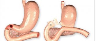
The morphological substrate of peptic ulcer disease is a chronic ulcer of the stomach or duodenum. Gastric ulcers are single in 85% of cases and multiple in 15% of cases. Duodenal ulcers are usually solitary. In 3% of cases, a combination of gastric ulcer and duodenal ulcer occurs.
A gastric ulcer is usually located on the lesser curvature in the prepyloric and pyloric sections, and much less often in the cardiac and subcardial sections; It is usually found at the border of the zones of the gastric and pyloric glands proper, where, during movements of the stomach, the greatest stretching of its muscular membrane is observed. Duodenal ulcers are most often localized in its upper (bulbar) part, less often in other parts of the intestine (extrabulbar ulcers). Usually it is located in the area of the transition of the mucous membrane of the duodenum to the mucous membrane of the stomach, i.e., in an area that is also subject to stretching during peristaltic movements of the intestine.
The ulcer has an oval or round shape (ulcus rotundum) and sizes from a few millimeters to 5-6 cm. It penetrates the wall of the stomach to varying depths, sometimes reaching the serous layer. The bottom of the ulcer is smooth, sometimes rough, the edges are raised like a roller, densely calloused (callous ulcer, from the Latin callus - callus). The edge of the ulcer facing the esophagus is somewhat undermined, and the mucous membrane hangs over the defect, and the edge facing the pylorus is gentle, looking like a terrace, the steps of which are formed by layers of the wall - mucous membrane, submucosal and muscular. This type of edges is explained by the displacement of layers during gastric peristalsis, going from the esophagus to the pylorus. On a cross-section, a chronic ulcer has the shape of a truncated pyramid, the narrow end of which faces the esophagus. The serous membrane in the area of the ulcer is thickened, often fused with adjacent organs - the liver, pancreas, omentum, transverse colon.
The microscopic picture of a chronic gastric ulcer at different periods of the course of peptic ulcer disease is different. During the period of exacerbation of peptic ulcer disease, a wide zone of fibrinoid necrosis appears in the area of the bottom and edges. On the surface of the necrotic masses there is fibrinous-purulent or purulent exudate. The necrosis zone is delimited by granulation tissue with a large number of thin-walled vessels and cells, including many eosinophils. Deeper behind the granulation tissue is coarse fibrous scar tissue. An exacerbation of the ulcer is indicated not only by exudative-necrotic changes in its bottom, but also by fibrinoid changes and inflammatory infiltration of the walls of blood vessels, often with blood clots in their lumens, as well as mucoid and fibrinoid swelling of scar tissue in the bottom of the ulcer. In the intramural nerve plexuses, ganglion cells undergo hydropic degeneration, their nuclei become pyknotic; the plexuses themselves are infiltrated with lymphocytes and histiocytes. Nerve fiber growths are detected in the area of the ulcer. In its edges, hyperplasia and dysplasia of the proliferating epithelium are noted. Due to these changes, the size of the ulcer increases, and there is a possibility of destruction of the entire wall of the stomach, which can lead to serious complications.
In cases where the exacerbation gives way to remission (healing of the ulcer), the inflammatory changes subside, the necrotic masses are rejected and in their place granulation tissue appears, which matures into coarse fibrous tissue. The surface of the ulcer is covered with regenerating flattened epithelium, which over time becomes taller and begins to secrete mucus; gastric pits and glands are formed, which, compared to a normal stomach, have a simpler structure. Regeneration of the muscular plate of the mucous membrane is also possible, but restoration of the elastic frame does not occur. The muscular membrane in the area of the ulcer is replaced by fibrous tissue. As a result of fibrinoid and inflammatory changes in the vessels, sclerosis of their walls and obliteration of the light vessels As can be seen, the submucosa and muscular layer, and not the gastric mucosa, undergo cicatricial changes (“scar after an ulcer”). After healing of duodenal ulcers, scars may not form
Thus, exacerbation of peptic ulcer disease, even in cases of a favorable outcome, leads to increased cicatricial changes in the stomach and aggravates the disturbance of the trophism of its tissues, including newly formed scar tissue, which is easily destroyed during the next exacerbation of peptic ulcer disease
During the period of remission of a peptic ulcer, based on the characteristics of the healing of the ulcer, scar tissue is found in the edges, the mucous membrane at the edges is thickened, hyperplastic. In the bottom area, the destroyed muscle layer and the scar tissue covering it are visible, and the bottom of the ulcer is covered with a layer of epithelium. Here, in the scar tissue , many vessels (arteries, veins) with thickened walls. In many vessels, the lumens are narrowed or obliterated due to proliferation of intimal cells (endovasculitis) or proliferation of connective tissue. Sometimes, at the bottom of the ulcer, among the scar tissue, a proliferation of nerve fibers is observed, similar to amputation neuromas.
The pathological anatomy of a chronic duodenal ulcer is not fundamentally different from that of a chronic gastric ulcer.
Characteristic changes in peptic ulcer disease are also observed in areas distant from the ulcer. In case of peptic ulcer with localization of the ulcer in the stomach in its antrum and in the region of the edges of the ulcer, lymphoid cell infiltration of the lamina propria increases compared to the norm, and the number of cells secreting predominantly IgG increases. Accumulations of the same cells are detected in the connective tissue of the bottom of the ulcers.
These data indicate the activity of the immune system in gastric ulcers, which is due to the antigenic properties of the decay products of the bottom of the ulcer, viruses and microorganisms, as well as the formation of autoantibodies. In case of duodenal ulcers, the gastric mucosa contains hyperplastic gastric glands proper, there is an increase in the number of main granulocytes, as well as an almost twofold increase in the number of parietal granulocytes compared to the norm. Using an electron microscopic study, it was established that along with hyperplasia of the glands, their accelerated differentiation occurs, due to which the number of mature functionally active cells producing hydrochloric acid increases. In patients with duodenal ulcers, hyperplasia of gastrin-producing cells is also noted, the number of which is almost twice as high as in gastric ulcers.
In the area of the gastric glands proper (in the area of the fundus of the stomach), changes of the type of chronic gastritis predominate; in the distal parts of the stomach, signs of atrophic gastritis with symptoms of intestinal metaplasia are often detected.
Various complications arise with peptic ulcer disease.
- ulcerative-destructive (bleeding, perforation, penetration);
- inflammatory (gastritis, duodenitis, perigastritis, periduodenitis);
- ulcerative-scar (narrowing of the inlet and outlet sections of the stomach, deformation of the stomach, narrowing of the lumen of the duodenum, deformation of its bulb);
- malignancy of an ulcer (development of cancer from an ulcer);
- combined complications.
Bleeding is one of the common and dangerous complications of peptic ulcer disease. There is no relationship between the frequency of bleeding and the location of the ulcer in the stomach; when the ulcer is localized in the duodenum, bleeding is more often caused by ulcers located in the posterior wall of the bulb. Bleeding occurs due to corrosion of the walls of blood vessels (arrosive bleeding), so it usually occurs during an exacerbation of peptic ulcer disease. Blood often remains in the stomach or intestines and is partially excreted in vomit and bowel movements. Vomit resembles coffee grounds due to changes in blood pigment under the influence of gastric juice. Feces become tarry.
Perforation (perforation) is also usually observed during an exacerbation of peptic ulcer disease. More often, pyloric gastric ulcers or ulcers of the anterior wall of the duodenal bulb perforate. Perforation of the ulcer leads to peritonitis. Initially, inflammation in the form of fibrinous deposits on the peritoneum appears only in the area of the perforation, then it becomes widespread and not fibrinous, but fibrinous-purulent. In the presence of adhesions, perforation may be accompanied by only limited peritonitis. Chronic peritonitis is rare. Then the masses of gastric contents are encapsulated, and granulomas of foreign bodies are formed on the peritoneum and in the omentum. In rare cases, when the perforation is covered by the liver, omentum, pancreas, or quickly appearing fibrin deposits, they speak of a covered perforation. Penetration of an ulcer is its penetration beyond the wall of the stomach or duodenum into neighboring organs. Ulcers of the posterior wall of the stomach and the posterior wall of the duodenal bulb usually penetrate, and more often into the lesser omentum, the head and body of the pancreas, into the hepatoduodenal ligament, and less often into the liver, transverse colon, and gall bladder. Penetration of a stomach ulcer in some cases leads to digestion of an organ, such as the pancreas.
Complications of an inflammatory nature include peri-ulcerous gastritis and duodenitis, perigastritis and periduodenitis, resulting in the formation of adhesions with neighboring organs. Rarely, a stomach ulcer is complicated by phlegmon. Severe complications of ulcers are caused by cicatricial stenosis of the pylorus. The stomach expands, food masses are retained in it, and vomiting often occurs. This can lead to dehydration, chloride depletion and the development of chlorohydropenic uremia (gastric tetany). Sometimes the scar tightens the stomach in the middle part and divides it into two halves, giving it an hourglass shape.
In the duodenum, cicatricial stenosis and deformation are most often caused by ulcers of the posterior wall of the bulb. Malignancy of chronic gastric ulcer occurs in 5-10% of cases. One of the types of ulcerated stomach cancer develops from a chronic ulcer, or ulcer-cancer.
The transition of a chronic duodenal ulcer to cancer is an extremely rare occurrence. Among the combined complications, the most common are perforation and bleeding, bleeding and penetration.
Symptoms of a stomach ulcer
The most common complaint characterizing the presence of a peptic ulcer in a patient is pain in the stomach.
Abdominal pain
The pain of a stomach ulcer is in most cases sharp, burning, aching. However, the pain can also be vague, dull or felt like a feeling of fullness in the stomach (heaviness in the abdomen), sometimes reminiscent of hunger. In people suffering from duodenal ulcers, abdominal pain occurs 1.5-3 hours after eating. Night attacks of pain often occur.
A comprehensive analysis for the diagnosis and monitoring of treatment of H. pylori-associated gastritis and gastric ulcers.
Synonyms Russian
Tests for the diagnosis of gastritis and gastric ulcers, serological examination of H. pylori, RT-PCR of H. pylori, gastrin, pepsinogens 1 and 2.
English synonyms
Workup of Gastritis and Gastric Ulcer, H. pylori serology, Gastrin, Pepsinogen I and II, H. pylori PCR.
What biomaterial can be used for research?
Cal.
General information about the study
Symptoms caused by gastritis or gastric ulcer (dyspepsia) are a common reason for patients to consult a gastroenterologist. Despite the fact that gastritis and gastric ulcers can be caused by a variety of damaging factors (NSAIDs, alcohol, tobacco, ischemia), the most common cause of these diseases is Helicobacter pylori (H. pylori) infection. H. pylori is a gram-negative spiral-shaped rod that can colonize the gastric mucosa, provoke and maintain the inflammatory process. Helicobacteriosis can be detected in almost all cases of chronic gastritis and in 50-70% of cases of gastric ulcer. Laboratory tests, including identification of H. pylori, greatly help in the diagnosis of gastritis and gastric ulcers.
Various methods and combinations of methods can be used to identify H. pylori. Serological tests can detect specific immunoglobulins in the blood directed against H. pylori. H. pylori infection is accompanied by the development of a local and systemic immune response. Following a transient increase in the titer of class M immunoglobulins (IgM), there is a prolonged and significant increase in IgG, as well as IgA antibodies in the blood serum. IgG is found in 95-100% of cases of H. pylori infection, and IgA in 68-80%. Recently, clinicians are less likely to use serological tests as the main method for diagnosing Helicobacteriosis, but prefer to combine them with new, more accurate methods.
One such method is real-time polymerase chain reaction (RT-PCR). RT-PCR is a molecular diagnostic method that allows one to detect fragments of genetic material (DNA) of the infectious agent in biological material. RT-PCR is characterized by greater sensitivity than serological tests. The combination of two methods for identifying H. pylori allows one to achieve almost 100% sensitivity. This high sensitivity means that a negative test result can exclude H. pylori as a cause of inflammatory changes. This is of fundamental importance in the differential diagnosis of NSAID-associated, stress and ischemic gastritis and ulcers.
Unlike other tests for H. pylori (eg, urease breath test or stool antigen test), the accuracy of serological tests and RT-PCR is not affected by the use of proton pump inhibitors (eg, pantoprazole). However, these tests also have some limitations. Thus, analysis of a tissue sample or aspirate with a very low bacterial load (for example, obtained while taking certain antibiotics) may be characterized by a false negative RT-PCR result. The results of serological tests, in turn, depend on the characteristics of the patient's immune response. The immune response of elderly people, patients with HIV infection and people taking cytostatic drugs (for example, glucocorticosteroids) is characterized by a reduced production of specific antibodies (any, including to H. pylori). These features should be taken into account when interpreting the analysis result.
Chronic inflammation caused by H. pylori is in some cases accompanied by impaired synthesis of specific proteins of the digestive tract (pepsinogen I, II and gastrin).
Pepsinogen is an inactive precursor to the enzyme pepsin. It has been proven that the concentration of pepsinogen in the blood reflects functional and morphological changes in the gastric mucosa, such as inflammation, atrophy, meta- and neoplasia. Pepsinogen exists in two biochemically and immunologically distinct variants – pepsinogen I and II. Secretion of pepsinogen I occurs only in the cells of the glands of the body of the stomach. Secretion of pepsinogen II occurs in the cells of the glands of the body, cardiac and pyloric parts of the stomach, as well as in the Brunner's glands of the duodenum. Normally, small amounts of pepsinogen I and II enter the blood and can be measured.
Gastrin is a hormone produced by G cells in the antrum of the stomach and cells of the islets of Langerhans in the pancreas. Gastrin is essential for the digestion process: it stimulates the production of hydrochloric acid by the lining cells of the stomach. Normally, gastrin is produced in response to a food stimulus - alkalization of the stomach environment, stimulation of the vagus nerve, stretching of the antrum of the stomach and the entry of proteins and amino acids into the stomach. The subsequent increase in gastric acidity inhibits gastrin secretion. Other functions of this hormone include increasing gastrointestinal motility and stimulating pepsin synthesis. Some gastrin is absorbed into the blood and can be measured.
In diseases of the stomach, the level of gastrin, pepsinogen I and II changes, which can be used to diagnose these diseases.
H. pylori often colonizes the antrum of the stomach, resulting in antral gastritis. With antral H. pylori-associated gastritis, the normal rhythm of gastrin secretion is disrupted and hypergastrinemia occurs. Hypergastrinemia leads to increased activity and proliferation of the main and parietal cells of the stomach, which is accompanied by increased secretion of pepsinogen I and II and hydrochloric acid. Therefore, H. pylori-associated antral gastritis is characterized by a persistent increase in the concentrations of gastrin and pepsinogen I and II, which can be used to diagnose this disease.
A minority of patients develop H. pylori-associated gastritis with predominant damage to the body of the stomach. The inflammatory reaction is accompanied by inhibition and gradual loss of the main and parietal cells of the mucous membrane, which is morphologically manifested as atrophic gastritis. Atrophy of the gastric mucosa is accompanied by a decrease in the concentration of pepsinogen I and hydrochloric acid. In the absence of negative feedback from hydrochloric acid, gastrin is produced in large quantities. Unlike H. pylori-associated antral gastritis, hypergastrinemia in atrophic gastritis is not the cause of the disease, but a compensatory response. Excess gastrin stimulates the production of pepsinogen II by the cells of the cardiac and pyloric parts of the stomach, so its concentration, unlike pepsinogen I, does not decrease, but, on the contrary, increases as a result of hypergastrinemia. Thus, atrophic gastritis of the gastric body is characterized by an increase in the concentration of gastrin and multidirectional changes in the concentrations of pepsinogen I and II. With atrophic pangastritis, which affects both the body and the antrum of the stomach, there is a decrease in all three specific proteins of the digestive tract (pepsinogen I and II and gastrin).
With effective treatment of gastritis, regardless of its type, and healing of the gastric mucosa, normalization of the levels of pepsinogen I, II and gastrin is observed. For this reason, gastrin and pepsinogen I and II studies can be used to evaluate the effectiveness of treatment.
Approximately 10-15% of patients with H. pylori-associated gastritis develop gastric ulcers. Most often, the ulcer is located in the antrum (60%) and on the lesser curvature of the stomach at the junction of the body of the stomach with the antrum (25%). Therefore, changes in gastrin, pepsinogen I and II, characteristic of antral gastritis (increased concentrations of all three proteins), are more often observed with ulcers.
It should be noted that none of the listed components of this complex analysis allows for a differential diagnosis between gastritis and gastric ulcer. Also, none of these tests can differentiate an active, current infection from a past infection. Therefore, this comprehensive analysis is not intended to evaluate the effectiveness of eradication therapy.
In most cases, if gastritis is suspected, this comprehensive laboratory analysis allows you to obtain all the necessary information. The situation is slightly different with suspected gastric ulcers, since in this case a mandatory study is a histological evaluation of the biopsy to exclude gastric cancer. In any case, the results of the study are assessed taking into account additional anamnestic, clinical and instrumental data.
What is the research used for?
- For the diagnosis and monitoring of treatment of H. pylori-associated gastritis or gastric ulcers.
When is the study scheduled?
- When examining a patient with signs of dyspepsia: discomfort or pain in the epigastric region, nausea, vomiting, rapid satiety, feeling of heaviness in the stomach, appetite disturbances.
What do the results mean?
Reference values
For each indicator included in the complex:
- [07-027] Helicobacter pylori, IgA (quantitative)
- [07-028] Helicobacter pylori, IgG (quantitative)
- [08-071] Gastrin
- [08-097] Pepsinogen I
- [08-099] Pepsinogen II
- [09-053] Helicobacter pylori, DNA [real-time PCR]
Methods for diagnosing stomach ulcers
In the diagnosis of gastric ulcers, instrumental diagnostics and laboratory methods are used. The main importance is attached to the endoscopic method - EGDS (gastroscopy).
Gastroscopy
Gastroscopy allows you to identify ulceration, clarify its location, size and depth, and determine whether bleeding is occurring. Gastroscopy also makes it possible to evaluate the relief and elasticity of the mucosa.
More information about the diagnostic method
X-ray
For stomach ulcers, a diagnostic method such as fluoroscopy can be used. Fluoroscopy is a real-time X-ray examination. The patient's stomach is filled with a contrast agent. Using X-rays, an image of the contours of the stomach is obtained. You can evaluate the dynamics of the passage of the contrast agent. In case of a gastric ulcer, examination reveals “niches” - a persistent accumulation of contrast agent on the relief of the mucous membrane or on the contour of the organ wall.
General blood analysis
With a peptic ulcer, you should expect that a general blood test will show an increase in the number of red blood cells, an increase in hemoglobin and a slowdown in ESR.
More information about the diagnostic method
Fecal occult blood test
The standard of examination for suspected gastric ulcers usually includes a stool occult blood test.
Detection of Helicobacter pylori
The cause of the inflammatory process in the stomach may be the bacterium Helicobacter pylori. In order for the treatment to be effective, it is necessary to establish its presence in the patient’s stomach. For this purpose the following may be carried out:
- taking a biopsy of the gastric mucosa during endoscopy for subsequent microscopic or cultural examination;
- urease breath test;
- stool analysis for detection of Helicobacter pylori antigen;
- serological blood test;
- PCR diagnostics.
Sign up for diagnostics To accurately diagnose the disease, make an appointment with specialists from the Family Doctor network.
Autumn exacerbation
Autumn is a time of golden leaves, hot tea and mulled wine, long walks and cozy gatherings with friends. But unfortunately, it is not always possible to enjoy the comfort of autumn due to health problems. In autumn, some chronic diseases traditionally worsen and the frequency of colds and depression increases. Let's discuss what the most common fall health problems are and how you can avoid them.
Ulcer
Ulcer of the gastric and duodenal mucosa
- a local defect of the mucous and submucosal layer (as opposed to erosions), formed under the influence of aggressive factors: acid, pepsin and bile. The ulcer heals with the formation of a scar. Duodenal ulcers are 4 times more common than gastric ulcers. Peptic ulcer disease is characterized by alternating periods of exacerbations (usually in spring or autumn) and periods of remission.
Peptic ulcer
- This is one of the most common problems of the gastrointestinal tract. According to various sources, it affects from 3 to 18% of the world's adult population. Moreover, in more developed countries this percentage is noticeably higher; This disease occurs twice as often in men as in women.
Until recently, it was believed that ulcers were caused by stress. This was explained as follows: a stressful state leads to an outflow of blood from the stomach, as a result of which the possibility of healing of the mucous membrane is reduced. Over time, this leads to the fact that the tissue located under the mucous membrane is destroyed by gastric juice and an ulcerative defect occurs.
At the end of the 20th century, Australian doctors proved that the cause of ulcers in most cases is the bacterium Helicobacter pylori. According to recent data, 95% of gastric ulcers and 85% of duodenal ulcers worldwide are associated with Helicobacter pylori infection. Since 2005, the whole world has considered peptic ulcer disease an infectious disease.
Symptoms of peptic ulcer
depend on the location of the ulcer, the duration of the disease, and the patient’s individual sensitivity to pain. The most typical are:
- pain in the epigastric region (projection of the stomach): with a stomach ulcer - during and immediately after meals, with a duodenal ulcer, on the contrary, on an empty stomach, the so-called “hunger pains”;
- belching sour;
- vomiting, reducing abdominal pain.
Diagnosis of peptic ulcer
Endoscopic examination
- the most informative method for diagnosing gastric and duodenal ulcers. It visually confirms the presence of an ulcerative defect, allows you to clarify its location, depth, shape, size, allows you to assess the condition of the bottom and edges of the ulcer, identify accompanying changes in the mucous membrane, perform a biopsy followed by a histological examination of the resulting material, which allows you to exclude the malignant nature of the ulcerative lesion. During the study, mucosal material is collected to identify Helicobacter pylori through cytological examination (microscopy). This technique is the most reliable for diagnosing this pathogen.
HELIC test
— non-invasive respiratory diagnostics of the presence of the bacterium Helicobacter pylori in the body. A quick, simple, safe, painless and fairly accurate diagnostic method that determines the presence of the urease enzyme in exhaled air as a marker of the presence of this bacterium in the stomach.
X-ray research method
using a contrast agent - the images clearly reveal a direct sign of peptic ulcer disease - a “niche” (defect) on the contour of the mucous membrane.
Also, if indicated, the doctor may prescribe additional tests: general clinical and biochemical blood tests, stool examination, etc.
How to avoid peptic ulcers
The basis for preventing the appearance of peptic ulcers, especially in people who have close relatives suffering from this disease, is a test for the presence of the bacterium Helicobacter pylori. Now this can be done simply and inexpensively. A gastroenterologist will prescribe the optimal diagnostic method and assess the risk of developing a peptic ulcer in a particular patient.
In addition, as mentioned above, an unbalanced diet, bad habits, stress, etc. contribute to the occurrence of ulcers. Accordingly, general recommendations for the prevention of not only ulcers, but also many other diseases, are advice to lead a healthy lifestyle, establish a diet, pay attention to the quality of food, excluding fast food and fatty, fried, spicy foods, do not abuse alcoholic beverages, don't smoke, be less nervous. If an ulcer has already been diagnosed, it is necessary to visit a gastroenterologist to determine the causes of its occurrence, determine the prognosis of the condition and the possibilities of individual prevention of exacerbation of this disease.
Modern methods of diagnosing and treating peptic ulcer disease make it possible to solve this problem as simply and quickly as possible.
If you notice symptoms of a peptic ulcer, do not put off visiting a gastroenterologist. Remember that an advanced ulcer, in addition to unpleasant painful sensations, can lead to other consequences - even death, which is possible if the ulcer perforates and bleeds from it.
ARVI
As soon as it gets colder, people immediately appear around them with a “cold.” What we simply call a “cold” in medicine is called ARVI or acute respiratory infections. ARI (acute respiratory disease) or ARVI
ARVI (acute respiratory viral infection) is the general name for the entire group of diseases that occur with predominant damage to the upper respiratory tract and general intoxication, which are caused by viruses
There are a large number of viruses that cause the disease, they are combined into one group - since the symptoms and treatment of the disease are similar. Only the influenza virus is included in a separate group because this disease is more severe than a regular viral infection, and the influenza virus itself spreads faster.
How to avoid ARVI
Unfortunately, there is no 100% protection against ARVI. Moreover, for people with normal immunity, the norm is to get ARVI 1-3 times a year for an adult, up to 8 times for a schoolchild and up to 10 times for a preschooler. Moreover, this is not just normal, but even useful for maintaining the immune system in good shape.
The immune response to invading respiratory viruses is a kind of “reserve troop exercise.” To fight the virus, a certain cascade of immunological reactions is launched, during which interactions between various immune cells are worked out. The next time a more formidable enemy appears, the immune system will respond faster and more effectively.
But this whole theory, of course, is little consolation at a time when a cold ruins your plans and puts you to bed with a headache, runny nose and cough. Therefore, it still makes sense to do everything possible to reduce the risk of disease. How can I do that?
There are three main causal components in the development of any cold: contact with infection, hypothermia, and stress. In each individual case of illness, all three factors are always present, but one of them predominates. In order to increase your resistance to respiratory viral infections, you must follow simple, but at the same time complex rules for many modern people:
- Maintain a healthy regimen: get enough sleep (an adult's sleep duration should be at least 7–8 hours a day), move more, and strengthen yourself.
- Do not smoke or abuse alcohol.
- Eliminate foci of chronic infection (check your teeth, tonsils, digestive tract), since parasitism and intoxication sharply reduce the body's reserve capabilities.
- Take vitamin-mineral complexes in the autumn-winter-spring period.
- During seasonal outbreaks of ARVI, add onions, garlic, ginger, and lemons to your diet.
- Maintain humidity indoors at home and at work; dry mucous membranes are much more susceptible to infection; ventilate the premises more often.
- Avoid contact with patients with respiratory diseases; during an outbreak of acute respiratory viral infections and influenza, minimize your stay in places with large crowds of people.
- During ARVI epidemics, wash your hands more often and use local saline solutions to rinse your nose.
- Use aroma lamps with eucalyptus, fir, and pine oils.
- Try to switch to a balanced diet without processed foods, with an increase in the proportion of vegetables, fruits, and fish in your diet.
- Monitor regular bowel movements: the body needs to get rid of toxins and waste products in a timely manner, otherwise it will be busy only with detoxification and will not be able to resist infection.
- If possible, take general massage courses at least 2 times a year.
Allergy
In the fall, seasonal allergies to mold appear, and cold allergies can also begin.
Mold is a fungus that grows in warm, moist environments.
Favorite places for mold to appear are basements, bathrooms, window frames, air conditioner filters, that is, those areas where there is a high level of humidity and insufficient lighting. In the fall, more mold appears indoors due to the decrease in the amount of sunlight entering the apartment. Humidifiers make the problem even worse because they increase humidity in the home and create condensation zones, creating the perfect environment for mold to grow.
In addition, mold also grows outdoors - on fallen leaves, decaying plants and organic material, and its spores fly in the air. This is a test for allergy sufferers who work in the country, where September–October is the time for harvesting, cultivating beds, and making hay.
How to avoid allergies?
It is impossible to completely protect yourself from developing a mold allergy, but you can minimize the risk of its exacerbation. To do this you need to do the following:
- eliminate areas of moisture condensation, maintain low humidity levels (up to 35–50%) in rooms where there is a tendency for mold to grow; it is necessary to eliminate any water leakage and dampening of the walls;
- ventilate the room well;
- get rid of all pieces of furniture and textiles that smell like mold or have visible signs of mold infection;
- treat affected surfaces with mold-killing agents;
- when working with fallen leaves and hay, use a mask;
- Wash fruits and vegetables thoroughly and remove their skins.
Allergy to cold is manifested by local swelling, redness and itching of the skin, the formation of blisters, and rashes. It can cause swelling of the larynx, trachea, and bronchi, which will be manifested by difficulty breathing, shortness of breath, and cough. In severe cases, it can even lead to Quincke's edema!
How can you tell if you have a cold allergy?
Apply a piece of ice to the skin for a couple of minutes - if redness, blisters, or itching appear, there is a high probability that this is an allergic reaction. In this case, we recommend consulting with an allergist - the doctor will prescribe antihistamines and advise ways to prevent allergies.
To finally understand the causes of allergies, it is necessary to undergo a special allergological examination. If your problem is allergies, remember that you need to seek medical help not only at the time of exacerbation of the disease, but also during the period of remission. It is at this time that real help can be provided - nonspecific and specific hyposensitization.
Depression
In autumn, the days become shorter, darker and colder. Bad weather, rain, wind, lack of sunlight in sensitive people with a labile psyche and prone to depression can trigger the onset or worsening of depression. There is even such a thing as “seasonal depression.” Seasonal depression is usually associated with disturbances in the metabolism of serotonin, a biologically active substance responsible for a person’s mood. In winter, the content of serotonin decreases, this is due to the fact that the production of this useful substance is regulated by melatonin. And the latter depends on the amount of light.
Depression (depressive syndrome)
- This is a completely officially recognized disease that is treated, depending on the severity of its course, on an outpatient basis or in a hospital.
Depression is an extremely common mental disorder, affecting about 10% of people over 40, of whom two thirds are women. After age 65, depression reduces the quality of life for more than 30% of people. Even children can experience depression—approximately 5% of patients under 16 suffer from the disease.
Symptoms of depression
- constant bad mood, melancholy,
- nervousness,
- sleep disorders,
- slower reaction
- decreased overall body tone,
- indigestion,
- pain of unknown origin.
How do you understand that you have depression - a condition that requires the help of a neurologist or psychiatrist, and not just a bad mood? The most obvious sign: a bad mood, unlike depression, cannot last for weeks or months. If you suspect yourself or a loved one has depression, don’t wait until it “goes away on its own,” consult a doctor, the sooner the better!
Factors that increase the risk of developing depression:
- Chronic fatigue.
- Psychotraumatic situation.
- Serious illness.
- Uncontrolled use of certain drugs that affect the central nervous system (CNS).
- Mental instability, emotional excitability.
- Some endocrine diseases, as well as conditions that arise against the background of hormonal changes in the body - pregnancy and the postpartum period, menopause, etc.
- Hereditary predisposition.
- Autumn-winter period.
Prevention
- A lack of vitamins can cause a bad mood, poor health, blues, fatigue, and loss of strength. Add more foods rich in vitamins and microelements to your diet
- Chocolate is an excellent antidepressant - the cocoa beans it contains promote the production of the “happiness hormone”. It is best to choose real dark chocolate - it has the maximum content of cocoa beans, and it is less harmful to the figure than milk.
- Bright fruits can be used not only as a source of vitamins, but also for aroma and color therapy. For example, citrus fruits are not only rich in vitamin C and bright colors, but their smell has been proven to invigorate and improve mood. The smell of chocolate, as we have already said, improves your mood, so as not to overindulge in this sweetness, you can buy shower gel, soap or even eau de toilette with the scent of chocolate.
- Essential oils of orange, rosemary, neroli, eucalyptus, incense, cardamom, nutmeg, and sage can relieve stress and relieve depression.
- A massage with aroma oils is a great way to pamper yourself with health benefits.
- Follow your daily routine.
- Play sports.
- Take every opportunity to walk during daylight hours - sunlight helps increase serotonin levels in the blood.
- The brighter the light around, the better - replace the light bulbs at home and in the office with brighter ones.
- Hang out with optimists. Our social circle greatly influences our emotional state, and if your friends are constantly unhappy with life, you willy-nilly emotionally join them and also fall into melancholy.
- Take up an interesting hobby - it will give you a boost of energy and good mood.
Remember that your health depends on you! Be attentive to the condition of your body, take care of it, lead a healthy lifestyle and consult a doctor in a timely manner. We hope that with our tips your autumn will be bright and pleasant!
Treatment methods for stomach ulcers
The course of treatment for peptic ulcer disease is selected strictly individually and includes a whole range of drugs aimed at combating Helicobacter pylori, reducing the secretion and acidity of gastric juice.
As soon as a person suspects an ulcer, he should immediately seek advice from a gastroenterologist. You can contact any of the clinics of JSC “Family Doctor”. Our doctors will prescribe the necessary examinations and treat gastric and duodenal ulcers in accordance with the latest international standards.
Make an appointment Do not self-medicate. Contact our specialists who will correctly diagnose and prescribe treatment.
Rate how useful the material was
thank you for rating
Prevention of exacerbations
Our experts recommend following some rules that prevent relapses of the disease:
- compliance with the diet and diet (spicy and salty foods, sour and rough foods are excluded, and the intake is fractional, up to 6 times a day);
- prohibition of smoking and drinking alcohol;
- elimination of physical and mental overload.
All patients with gastric and duodenal ulcers are subject to clinical observation. Gastroenterologists at the Yauza Clinical Hospital will select courses of anti-relapse treatment on an individual basis (they take place twice a year). In some cases, patients are prescribed ongoing maintenance therapy.
Treatment of peptic ulcers in our hospital is carried out in comfortable conditions, efficiently and effectively, thanks to highly qualified specialists, modern equipment and an individual approach to the treatment of each patient.