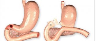
Peptic ulcer and pregnancy
Peptic ulcer
– a multifactorial, chronic, cyclical disease, with a varied clinical picture and ulceration of the mucous membrane of the stomach and duodenum during the period of exacerbation.
Peptic ulcer disease occurs in one in 4,000 pregnant women. It is possible that the data provided are underestimated, since diagnosing this disease during pregnancy is a little difficult. However, there is an opinion that the risk of developing peptic ulcers during pregnancy is significantly reduced.
There is a classification of peptic ulcer according to the form:
- spicy,
- chronic;
and by localization:
- stomach ulcer;
- duodenal ulcer.
According to modern concepts, the leading role in the development of the disease is played by the spiral-shaped bacterium Helicobacter pylori
.
80-85% of Russian residents are carriers of Helicobacter, but not everyone gets peptic ulcers. Additional factors are required for the development of the disease
- psycho-emotional stress, depression;
- family history of peptic ulcer disease (in close relatives);
- poor diet, abuse of spicy, rough foods, alcohol and smoking;
- uncontrolled and careless intake of medications (aspirin, NSAIDs, hormonal drugs).
In pregnant women
with peptic ulcer disease, exacerbations are observed more often in the 1st trimester, in the spring-autumn period, 2-3 weeks before childbirth and in the early postpartum period. The main symptom of the disease is pain in the epigastric and mesogastric areas associated with food intake. In some cases, the ulcer may be asymptomatic. The pain can be “hungry”, passing after eating and, conversely, occurring after eating, depending on the location of the ulcer. The pain syndrome may be accompanied by heartburn, nausea, belching of air or eaten food, and vomiting. You should take into account the high likelihood of developing iron deficiency anemia, bowel disorders in the form of constipation or diarrhea.
The most serious complication of peptic ulcer disease is gastrointestinal bleeding. If bleeding occurs, the risk of fetal death and complications in the mother increases sharply. As a rule, gastrointestinal bleeding is an indication for emergency surgery. Sometimes bleeding is the first manifestation of so-called “silent” or asymptomatic ulcers. Classic signs of gastrointestinal bleeding may include vomiting with dark-colored blood (“coffee grounds”), dizziness, fainting, and pale skin. Then hypotension and unformed stools of a dark, almost black color may appear. Currently, such dangerous complications of peptic ulcer disease during pregnancy are becoming less and less common, this is due to timely diagnosis (esophagogastroduodenoscopy is performed regardless of the stage of pregnancy), and to patients being well informed about their disease.
Women with gastric and duodenal ulcers during pregnancy should be observed not only by an obstetrician-gynecologist, but also by a general practitioner (gastroenterologist). In the spring-autumn period, when the course of pregnancy is complicated by the development of toxicosis, 2 weeks before birth and in the early postpartum period, it is advisable to carry out a course of preventive antiulcer therapy. When prescribing treatment, the possible effect of the drug on the fetus and uterine tone must be taken into account. Drug therapy is indicated in the absence of effect from following diet 1-1b, the inclusion of so-called “food” antacids, mineral waters Essentuki 4, 17, “Mirgorodskaya” and the threat of complications. Exacerbation of peptic ulcer disease must be confirmed clinically, as well as by laboratory and endoscopic methods. The modern arsenal of drugs includes antacids (Gaviscon, Rennie, Maalox), in the 2nd trimester it is possible to use proton pump inhibitors (Losec MAPS), antispasmodics (Drotaverine, No-shpa), etc. The criterion for the effectiveness of treatment is the regression of pain and dyspepsia. , negative results of stool occult blood test, ulcer scarring during endoscopic control. Eradication (complete destruction) of helicobacteriosis during pregnancy is not carried out.
Information prepared for you by:
Petushinova Vasilisa Mikhailovna
- gastroenterologist, therapist. Conducts receptions in the clinic building on Novoslobodskaya.
CLINICAL COURSE, DIAGNOSIS AND TREATMENT OF Peptic ULCER IN WOMEN DURING PREGNANCY
Attention is drawn to the fact that peptic ulcer may exacerbate in the first and third trimesters despite its favorable course during pregnancy. Epigastrial pain is the leading clinical symptom. In 46.5% of cases, pregnancy is complicated by early gestosis - vomitus gravidarum which may last as long as 16-20 weeks. For diagnosis of exacerbated peptic ulcer in pregnants, it is necessary to do thorough scrupulous clinical and instrumental studies involving esophagogastroduodenoscopy and echography of the stomach and duodenum. Drug therapy with inassimilable antacids and a diet are used in a exacerbation evidenced by clinical and endoscopic examinations. Particular emphasis is placed on the fact that H2-receptor antagonists and proton pump inhibitors should not be used in the treatment of peptic ulcer in pregnancy due to the fact that the effects of the agents on the fetus have been little studied.
S. G. Burkov, Dr. med. Sciences, leading researcher at the laboratory of gastroenterological research at the Research Center at the Department of Propaedeutics of Internal Diseases (head - Academician of the Russian Academy of Medical Sciences, Prof. V.T. Ivashkin) MMA named after. THEM. Sechenov. SG Burkov, MD, Leading Researcher, Laboratory of Gastroenterological Studies, Research Center, Department of Internal Propedeutics (Head - Prof. VT Ivashkin, Academician, of the Russian Academy of Medical Sciences), IM Sechenov Moscow Medical Academy.
AND
It is known that pregnancy has a beneficial effect on the course of peptic ulcer disease (PU): in most cases, remission of the disease is observed. This is facilitated by changes in the secretory and motor-evacuation function of the stomach, improved blood supply and activation of proliferation processes in the mucous membrane, caused by changes in the level of sex (estrogens, progesterone) and gastrointestinal (gastrin, VIP, bombesin, motilin, somatostatin) hormones, prostaglandins, endorphins, and others biologically active substances [1]. Some researchers tend to explain the favorable course of ulcerative disease in the gestational period by simply following a diet, eating regularly, and giving up bad habits. However, it is necessary to remember that exacerbation may occur. Complications of ulcers, such as perforation or bleeding, are extremely dangerous for the life of the mother and unborn child if not recognized in time. According to R. Dordelman [2], the frequency of surgical complications of ulcers in pregnant women is 1 - 4 per 10,000, while maternal mortality reaches 16%, and perinatal mortality - 10%. The purpose of this work was to study the clinical course, possibilities of diagnosis and treatment of ulcers in women during pregnancy. Under observation were 88 pregnant women who suffered from ulcer with localization of the process in the duodenal bulb. The duration of the disease ranged from 2 to 18 years, the average age of women was 32.7 ± 3.6 years. Clinical manifestations of ulcers were varied and were determined by the presence of a peptic ulcer, the general condition of the woman, the duration of pregnancy, and concomitant toxicosis of pregnancy. A survey of observed women showed that pain is the leading symptom in the clinical picture of the disease and occurs in 51.8% of patients in the first trimester, in 27.6% in the second, and in 42.1% in the third. Moreover, in almost all cases there was pain of multiple localization (in the epigastric region, back, left and/or right hypochondrium). The most common concern was pain in the epigastric region (up to 50% in the first trimester), which decreased as the duration of pregnancy increased (32.8% in the second trimester) and became more frequent again towards its completion (40.7%). In 11.1% of pregnant women, irradiation of pain into the interscapular space, typical for ulcer, persisted. As the duration of pregnancy increased, the nature of the pain changed: the number of patients suffering from dull, aching pain increased, with a significant decrease in acute, cramping pain. More and more women experienced a feeling of heaviness or fullness in the epigastric region (up to 64.4% in the third trimester). “Hungry” pains were the most frequent, which, however, is typical for duodenal ulcers. In most patients, the pain subsided after eating or drinking milk. Of the dyspeptic symptoms in pregnant women, the most common concerns were nausea and heartburn; vomiting, belching of air, bloating, and unstable stools were somewhat less common. However, if the frequency of nausea and vomiting decreased as pregnancy progressed, heartburn and belching occurred in more patients at the end of pregnancy than at the beginning. Throughout the entire period of pregnancy, 48 women presented certain complaints characteristic of PU, and 18 associated them with errors in diet, 16 with early toxicosis, neuro-emotional experiences, 7 with fear of the upcoming birth and its possible unfavorable completion. Palpation of the abdomen often revealed pain in the epigastric region (up to 53.7%), and at the end of pregnancy - in the right hypochondrium (40.7%), which is apparently explained by the addition of gallbladder dyskinesia. The course of ulcer was generally favorable. An exacerbation, confirmed clinically and endoscopically, was detected in 22 (25%) of 88 examined: in 13 in the first trimester, and in 8 against the background of vomiting of pregnancy, and in 9 in the third (2 - 4 weeks before the expected date of birth). In 4 cases, an ulcer of the bulb was endoscopically diagnosed, in 15 - pronounced erosive bulbitis against the background of cicatricial-ulcerative deformation of the bulb, in 3 - cicatricial-ulcerative deformation of the bulb and erosive gastroduodenitis. In 3 women, pregnancy was complicated by gastrointestinal bleeding (in one case in the first trimester, in another during childbirth, and in the third on the 1st day after birth). If outside of pregnancy the seasonal nature of exacerbations is considered typical for ulcerative disease, then in pregnant women it was not possible to identify it. All exacerbations were distributed almost evenly across the seasons: winter - 5, spring - 6, summer - 4, autumn - 7. In 46.5% of patients, pregnancy was complicated by early toxicosis - vomiting of pregnancy. 30 women had a mild form, 8 had a moderate form and 3 had a severe form. Vomiting lasted up to 16 - 20 weeks, in 8 cases it was one of the causes of exacerbation of the disease. 13 patients complained of drooling, 11 of painful nausea. In 18 cases, late toxicosis (dropsy, nephropathy) developed, but the connection with ulcer could not be traced. 23 women developed hypochromic iron deficiency anemia. The gold standard for diagnosing ulcerative disease is radiological and endoscopic methods. However, the first one is unacceptable in pregnant women. Having started to use esophagogastroduodenoscopy (EGDS) as an urgent method (together with Prof. R.M. Filimonov), we came to the conclusion that EGDS can also be used for routine examination of pregnant women, in diagnostically unclear cases, when complications are suspected. Having conducted 127 endoscopy, not only in patients with ulcerative disease, in no case did we note a negative effect on the course of pregnancy; all women tolerated the study well. As already mentioned, in 22 cases, an exacerbation of ulcer was endoscopically identified, and in 8 patients, hiatal hernia, cardia insufficiency, and reflux esophagitis were additionally identified. In 3 women, endoscopy allowed identifying the source of gastrointestinal bleeding. In diagnostically clear cases, with a benign course of ulcer, one can limit oneself to clinical observation, periodic examination of stool for hidden bleeding, determination of hemoglobin content, hematocrit, number of red blood cells in the blood, as well as ultrasound examination of the abdominal organs. Ultrasound made it possible to detect hypersecretion of the stomach on an empty stomach in 86.8% of cases; in 18 patients, palpation under ultrasound control revealed pain in the area of the duodenal bulb. Moreover, in 13 patients with an ulcer or erosive lesion of the bulb, the pain was more pronounced than in patients who did not have such disorders. This symptom disappeared when the patient’s well-being returned to normal after the ulcer had scarred. Motor disturbances of the stomach were detected by ultrasound in almost all patients suffering from ulcerative gastric ulcer. Unfortunately, in no case were we able to visualize the ulcerative defect. Ultrasound was the main method that allowed a differentiated diagnosis of ulcer with exacerbation of chronic cholecystitis and cholelithiasis. When carrying out differential diagnosis with vomiting of pregnant women, it should be remembered that dyspeptic syndrome caused by ulcer is always accompanied by abdominal pain, vomiting in most cases brings relief, and is not always preceded by nausea. Vomiting during pregnancy is characterized by painful, almost constant nausea, aggravated by exposure to various odors, and drooling; vomiting does not depend on food intake, often occurs in the morning, and pain, as a rule, is absent. It should be remembered that a stenosing gastric outlet ulcer can simulate excessive vomiting of pregnancy. Treatment of exacerbation of ulcer in pregnant women should be comprehensive, strictly individual and based on the principles we propose [3]. Drug therapy is carried out only during an exacerbation of the disease, confirmed clinically and by laboratory and instrumental methods (except x-ray). It is also indicated in the absence of effect from following a diet, diet, or the inclusion of “food” antacids; with the development of complications. It is necessary to take into account the possible harmful effects of drugs on the condition of the fetus and myometrial tone. All observed women were prescribed split meals (at least 5 times a day) and adherence to a table type diet No. 1. In the practice of gastroenterology, drugs from the group of H2-receptor antagonists (cimetidine, ranitidine, famotidine), proton pump inhibitors (omeprazole, pantoprazole) have been widely used for the treatment of ulcers; however, they should not be prescribed to pregnant women due to the lack of knowledge of their effect on the fetus. Pregnant women should not take atropine and bismuth-containing drugs (roter, de-nol), traditionally used to treat ulcer. As recommended by H. Kümerle and K. Brendel [4], “if drug therapy is necessary, drugs that have been widely used during pregnancy for many years should be used, preferring them to newer drugs.” That is why we chose non-absorbable (insoluble) antacids, which realize their effect through two main mechanisms - neutralization and adsorption of hydrochloric acid produced by the stomach. Due to the lack of absorption, these drugs are most suitable for pregnant women. As our research has shown, the most justified is the use of Maalox, which has proven itself well during clinical trials conducted in an obstetric clinic. Maalox is a well-balanced mixture of magnesium hydroxide and aluminum hydroxide, which provide high acid-neutralizing ability of the drug. It is distinguished by high efficiency, good taste, and absence of constipation during prolonged use (which is important for pregnant women). Maalox was prescribed 15 ml 3 times a day 60 minutes before meals and 4 times at night for 14 days. In addition, 6 women with severe pain were administered metacin (0.5 ml of a 0.1% solution, subcutaneously) for 5 days, once a day. 8 patients with exacerbation of ulcerative ulcer and vomiting of pregnancy were additionally prescribed cerucal (10 mg 2 times a day). The complex treatment provided was effective in all cases. It was possible to achieve a lasting improvement in well-being: the disappearance of pain (in 6 pregnant women on the 2nd day, in 12 on the 3rd - 5th day and in 4 - on the 6th - 7th day), heartburn, and reduction in nausea. During a control ultrasound of the stomach and duodenum, there was no pain on palpation in the area of the bulb or it was insignificant. 26 women with hypochromic iron deficiency anemia were additionally treated with ferroplex (2 tablets 3 times a day) or ferrogradument (1 tablet a day). The treatment turned out to be ineffective, anemia was difficult to treat, and in 11 pregnant women it persisted until the end of pregnancy. For flatulence and symptoms of intestinal dyspepsia, it was additionally recommended to take enzyme preparations (Creon, Digestal, 1 - 2 capsules with meals). In addition, multivitamins (gendevit, oligovit, 1 - 2 tablets per day), and alkaline mineral waters were prescribed. However, it should be remembered that water is not used in the second half of pregnancy when symptoms of late toxicosis develop, when it is necessary to limit fluid intake. Peptic ulcer had no effect on the outcome of pregnancy; all women gave birth to live, full-term children with normal weight and height parameters. Thus, in most cases, a benign course of ulcer during pregnancy is observed, although in 25% of cases an exacerbation may develop. Pain is the main symptom in the clinical picture of exacerbation of the disease. Drug treatment should be carried out in case of exacerbation of ulcer, confirmed clinically and endoscopically, and the effect of drugs on the condition of the fetus must be taken into account.
Literature:
1. Shekhtman M.M., Korotko G.F., Burkov S.G. Physiology and pathology of the digestive organs in pregnant women. Tashkent: “Medicine” UzSSR, 1989;160. 2. Durdelmann P. Chirurgische Komplicationen der Gravidat. GynKk Prax 1983;7(2):201-9. 3. Burkov S.G., Polozhenkova L.A. Diseases of the digestive system and pregnancy / Guide to gastroenterology / Ed. F.I. Komarova, A.L. Grebeneva. M.: Medicine, 1996;3:720. 4. Clinical pharmacology during pregnancy/Ed. H.P. Kümerle, K. Brendel. In 2 volumes. T. 1. M.: Medicine, 1987;328.
Colpitis (vaginitis) - symptoms and treatment
The goals of treatment are to eliminate inflammation and restore vaginal microflora.
Treatment regimen for vaginitis
Treatment always consists of two stages. The first stage is the fight against inflammatory agents. This stage sometimes begins with a slight acidification of the vaginal environment (only if indicated). The second stage is the restoration of microflora in the vagina and intestines, followed by transition to preventive measures to reduce the risk of relapse.
Medicines
Treatment of specific and nonspecific vaginitis. Depending on the causative agent of the disease, systemic therapy with antibacterial drugs (amoxicillin, jazzomycin, clindamycin, ornidazole, metronidazole, tinidazole, etc.) may be required. Suppositories, capsules or vaginal tablets are prescribed locally, most often containing combination drugs (Poliginax, Macmiror complex, Terzhinan, Neo-penotran, etc.).
Treatment of candidal vaginitis (thrush). For thrush, antimycotic (antifungal) drugs of local and systemic action are prescribed.
Treatment of atrophic vaginitis. For atrophic vaginitis, the use of vaginal creams, tablets or rings with estrogen is indicated [13].
Lifestyle and aids
During treatment, sexual abstinence is recommended. After the main course of therapy, it is necessary to carry out a course of restoring the microflora in the vagina with preparations containing lactobacilli.
Physiotherapeutic procedures
In case of a chronic and often recurrent process, treatment should be comprehensive and include physiotherapeutic procedures (ultrasonic sanitation with the stage of restoration of vaginal biocinosis). It is important not only to eliminate inflammation, but also to restore damaged microflora, immune defense and remove the influence of the causative factor (sanitize foci of chronic infection, change personal hygiene products or contraception, compensate for diabetes with insulin).
It is also worth noting that there is an additional method of treating and restoring the vaginal microflora - this is low-frequency ultrasound sanitation with the Gineton-MM apparatus. Advantages of this treatment method:
- direct bactericidal effect of ultrasonic vibrations with a frequency of 22-44 kHz;
- effective hydrodynamic rehabilitation;
- increasing the concentration of the drug at the site of inflammation;
- vibration and hydromassage in sonified tissues, stimulation of microcirculation, improvement of trophism and tissue metabolism.
This method is used as an additional method to the main treatment [2][4][6][9][11].
Surgical operations
Surgery is not required to treat vaginitis.
Diet for vaginitis
Nutrition does not have a significant effect on the course of vaginitis. When taking antibiotics, alcohol should be avoided.
Restoring and improving quality of life
If you follow the doctor's prescriptions, a complete cure and restoration of quality of life is possible.
Treatment of vaginitis during pregnancy
During pregnancy, careful monitoring of the microflora is necessary. This is due to the likelihood of infection spreading to the fetus and membranes, the threat of miscarriage and premature birth, miscarriage and pregnancy loss. The drugs are prescribed by the doctor individually depending on the test results and the timing of pregnancy.
How to treat vaginitis without potent drugs
Vaginitis caused by a bacterial infection cannot be cured without the use of antibiotics.
Is douching used for vaginitis?
Douching is not required to treat vaginitis.
How is a partner treated for vaginitis?
For specific vaginitis, the woman’s sexual partner is treated with antibacterial agents. For nonspecific vaginitis, the partner is not treated.
Traditional methods of treating vaginitis
The use of traditional medicine often not only does not lead to a cure, but also aggravates the situation.


