Perforated ulcer of the stomach and duodenum
Perforation, or perforation, of stomach and duodenal ulcers is a breakthrough of the ulcer into the free abdominal cavity with the entry of gastroduodenal contents into it. In 75% of cases, a perforated ulcer is located in the duodenum, most often observed in men aged 20–40 years with a short history of ulcers (up to 3 years). Sometimes perforation of an ulcer can occur in people who have never previously complained of epigastric pain and were not aware of the presence of an ulcer. In young people, perforation of duodenal ulcers predominates, and in middle-aged and elderly people, perforation of gastric ulcers predominates. Perforation of ulcers is more often observed in autumn and spring.
Classification. By localization: a) stomach ulcers: lesser curvature (cardiac, antral, prepyloric, pyloric), anterior wall (antral, prepyloric, pyloric), posterior wall (antral, prepyloric, pyloric); b) duodenal ulcers: anterior wall, posterior wall. Along the flow: a) perforation into the free abdominal cavity, b) covered perforation, c) atypical perforation. There are 3 phases of the clinical course of a perforated ulcer: 1) the shock phase, 2) the phase of “imaginary well-being,” and 3) the phase of widespread peritonitis.
Etiology and pathogenesis. The main factor leading to the development of perforation is the exacerbation of peptic ulcer disease, when the processes of inflammation and destruction in the ulcer intensify, it deepens until the formation of a hole in the wall of the organ. Through this opening, the contents of the stomach and duodenum - gastric juice, air (gas bubble of the stomach), eaten food - enter the abdominal cavity. Hydrochloric acid from gastric juice suddenly entering the abdominal cavity causes a chemical burn of the peritoneum of the upper abdominal cavity (chemical peritonitis). In response, the peritoneum begins to produce fluid - exudate, which, diluting the acid, reduces its concentration and the strength of its irritating effect. At the same time, a large amount of biologically active substances are released into the blood, which determine the first clinical phase of the disease - the shock phase. The second phase - “imaginary well-being” - is due to the fact that gastric contents cease to flow into the abdominal cavity (most often due to clogging of the perforation with a lump of food). Acid diluted with exudate irritates the peritoneum less, and the pain receptors of the burned peritoneum become less sensitive. Subsequently, pathogenic microorganisms that enter the abdominal cavity from the stomach and infect the peritoneum begin to multiply, release toxins and cause the development of the third phase of the disease - widespread peritonitis.
Complaints. The main complaint when an ulcer perforates is pain in the upper abdomen. In most patients, perforation of stomach and duodenal ulcers begins suddenly, accompanied by sharp pain in the abdomen. The pain can be so severe that patients compare it to a “dagger blow.” They are permanent in nature, localized first in the epigastric region or in the right hypochondrium, and then spread relatively quickly throughout the abdomen, often along the right lateral canal. In 30-40% of patients, pain radiates to the shoulder, scapula or supraclavicular region: on the right - with perforation of pyloroduodenal ulcers, on the left - with gastric ulcers. When an ulcer perforates, general symptoms are also observed: dry mouth, thirst, nausea. In 30-40% of patients, reflex vomiting occurs, which becomes more frequent as peritonitis progresses.
Anamnesis. Before ulcer perforation, 80-90% of patients have a typical ulcer history or vague gastric complaints, against which perforation occurs. In 10-15% of patients, “without anamnesis” or “silent” perforated ulcers occur, when perforation is, as it were, the first symptom of a peptic ulcer. In 50-60% of patients, prodromal symptoms of perforation or exacerbation of peptic ulcer disease are observed (increased pain, general weakness, low-grade fever, nausea, vomiting).
Examination of the patient. The condition of the patients is serious. Pallor, cold extremities, and cold sweat on the face are noted. Breathing is frequent, shallow, the patient cannot take a deep breath. The pulse in the first hours after perforation is slow or normal, and with the development of peritonitis it becomes more frequent. Body temperature is initially normal or subfebrile, and later rises to 38 degrees. and more. There is also retention of stool and gas. The appearance of the patients is characteristic: they take a forced position on their back or side with their knees brought to their stomach, and avoid changing it. The facial expression is frightened, suffering.
Characteristic symptoms of perforation are revealed by objective examination. The abdomen is often scaphoidally retracted or flat and does not participate in the act of breathing. Tension of the muscles of the anterior abdominal wall is a very characteristic and constant symptom of a perforated ulcer. In this case, most patients experience a board-like tension in the abdominal muscles. It can cover the entire abdomen or its upper section. However, in elderly patients, sometimes muscle tension may not be pronounced. On palpation, in addition to muscle tension, sharp pain is noted, more in the upper abdomen, the Shchetkin-Blumberg symptom. Percussion often reveals a very important sign - “disappearance of hepatic dullness” or a decrease in its size as a result of the entry of free gas from the lumen of the stomach through the perforation into the abdominal cavity. In addition, a high-pitched tympanic sound (Spizharny's symptom) and dullness in the lateral parts of the abdomen may be detected in the epigastric region - due to the accumulation of liquid stomach contents there, poured out through the perforation, and exudate produced by the peritoneum in response to its sharp irritation with acidic gastric juice. Auscultation may reveal the absence of intestinal motility, listening to heart sounds up to the level of the navel (Gusten's symptom). Digital rectal examination may reveal sharp pain in the pouch of Douglas (Kulenkampf's sign).
The shock phase (up to 6 hours) is characterized by daggering, excruciating pain in the abdomen. The condition of the patients is serious, they are excited, pale, covered in cold sweat, and show fear and suffering. Breathing is frequent and shallow. The pain is localized in the epigastric region or right hypochondrium, and can radiate to the right shoulder and collarbone. Characteristic is “board-shaped” tension of the muscles of the anterior abdominal wall in the epigastrium. Percussion often detects the symptom of “disappearance of hepatic dullness.” Phase of “imaginary well-being” (6 – 12 hours). In this phase, the patient's condition improves. Abdominal pain and muscle tension in the anterior abdominal wall are reduced. Breathing becomes equal. The Shchetkin-Blumberg sign is positive in the epigastrium, the right half of the abdomen. Diagnostic errors most often occur in this phase. The phase of widespread peritonitis (more than 12 hours). The condition of the patients again deteriorates significantly. Bacterial purulent peritonitis develops. As a result of intoxication, the general condition worsens, body temperature rises to 38° or more, pulse quickens, blood pressure decreases, and bloating appears. Facial features become sharper, tongue is dry. The clinical picture of a perforated ulcer during this period does not differ from that of widespread peritonitis of another etiology.
Diagnostics. General blood analysis. Leukocytosis and a neutrophil shift in the leukocyte count to the left are observed.
Plain radiography of the abdomen. Free gas is detected in the abdominal cavity (pneumoperitoneum). On photographs with the patient in an upright position, it is revealed as a crescent-shaped clearing under the right, less often under the left, or both domes of the diaphragm. The most characteristic is a crescent-shaped clearing between the liver and the right dome of the diaphragm, that is, on the right. Pneumoperitoneum with perforation of an ulcer is found in 60-80% of patients and is a direct symptom of perforation, but its absence does not exclude a perforated ulcer.
Pneumogastrography. In the absence of pneumoperitoneum on a plain radiograph of the abdomen, 500–700 ml of air is introduced through a probe into the stomach after it has emptied, which partially passes through the perforation into the free abdominal cavity and is found under the diaphragm. Fibroesophagogastroduodenoscopy (FEGDS). During the procedure, you can detect a perforated ulcer, and after the procedure, you can detect free gas in the abdominal cavity.
Diagnostic laparoscopy. It is possible to detect the presence of exudate in the abdominal cavity, signs of inflammation of the peritoneum, and the perforation of the stomach or duodenum itself. Laparocentesis. During an abdominal puncture performed below the navel, the trocar stylet is directed to the right hypochondrium. After this, a 30-centimeter vinyl chloride tube is inserted, from which the exudate is aspirated. If there is any doubt about the nature of the exudate, the Neimark diagnostic test can be used. To perform this test, add 4-5 drops of 10% iodine tincture to 2-3 ml of exudate found in the abdominal cavity. If the liquid contains an admixture of gastric contents, then under the influence of iodine tincture it acquires a dark, dirty-blue color (due to starch residues). Diagnosis of “covered” perforation often presents significant difficulties. Free gas in the abdominal cavity in such patients is detected less frequently than with open perforation. In the diagnosis of this form of perforation, an ulcer history in the past, an acute onset of the disease, and two phases in the clinical course - pronounced perforation syndrome and extinction of clinical symptoms - are important.
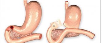
Treatment. Patients with a perforated gastric or duodenal ulcer are subject to immediate hospitalization in the surgical department and emergency surgery. Possible options for operations: Suturing the ulcer with a single-row suture in the transverse direction with application of a greater omentum. Suturing the ulcer according to Opel-Polikarpov (with tamponade of the perforated hole with a strand of the greater omentum). In this case, a strand of the greater omentum is inserted into the perforated hole, and then, using interrupted sutures in the transverse direction, the edges of the perforation hole and the strand of the greater omentum inserted and stitched into it are tightly brought together. Gastric resection is performed rarely, according to strict indications and taking into account contraindications. Operations for perforated ulcers are necessarily accompanied by thorough sanitation of the abdominal cavity, removal of exudate and spilled gastric contents from the abdominal cavity, drying and draining it. Suturing an ulcer when it perforates can be performed either openly or using laparoscopic techniques.