- Causes of peritoneal carcinomatosis
- What diseases does it develop in?
- Diagnosis of peritoneal carcinomatosis
- Stages of abdominal carcinomatosis
- Symptoms of abdominal carcinomatosis
- How is peritoneal carcinomatosis treated?
- Which therapy methods give the best results?
- Prognosis for carcinomatous lesions
- Read also:
Peritoneal carcinomatosis is a tumor lesion of the layers of the mucous membrane covering the organs and the inner wall of the abdomen. Mainly caused by the growth of cancer metastases in the abdominal cavity, sometimes it is possible to develop a primary malignant process - mesothelioma in the peritoneum itself.
It is more correct to call metastatic lesions “carcinomatosis”, since carcinoma is a synonym for cancer. Similarly, metastases of sarcoma to the peritoneum are called “sarcomatosis.”
A frequent, but not obligatory, manifestation of peritoneal carcinomatosis is the production of ascitic fluid. With or without ascites, damage to the peritoneum by a malignant process always threatens the patient’s life and requires very difficult treatment.
Causes of peritoneal carcinomatosis
Not every cell that breaks away from the mother’s cancerous tumor is capable of becoming a metastasis; the lion’s share of circulating malignant cells die in the bloodstream. To gain the ability to become a metastasis, a cancer cell must change internally - learn to produce substances that allow it to live independently and invade another place, suppressing normal cells.
Cells detached from the node migrate over long distances, pushing apart normal cells, are implanted into the abdominal mucosa, and can even penetrate inside other cells. After establishing itself in the area, reproduction and the formation of an entire cell colony begin.
In addition to the transfer of metastatic cells through the blood and lymph, the spread also occurs inside the abdominal cavity - transcoelomically. It is not entirely clear why malignant cells linger in the peritoneum; a beneficial effect of the microclimate is assumed. Most metastases are found in places with a calmer environment and weak peristalsis of organs, or where intra-abdominal fluid is actively absorbed.
Cells are often “scattered” during surgery and during laparoscopic surgery the likelihood of contamination is half as high as during classical surgery. During surgery, it is necessary to prevent cancer dissemination through repeated treatment with special solutions, but the most effective way to cleanse dissemination is intracavitary chemotherapy with hyperthermia (HIPEC).
Book a consultation 24 hours a day
+7+7+78
Development of pathology
Oncological carcinomatosis occurs in several stages.
- The first stage is the emergence of cells and their gradual spread throughout the body. Moreover, it should be noted that the development of pathology can be triggered not only by altered cells, but also by certain interventions in the body, for example, surgery or injury to internal organs. Once inside the peritoneum, the cells begin to move as the organs contract. This is why the disease often manifests itself in the lower abdominal cavity.
- Second phase. At this moment, the changed cells begin to grow to the internal organs, becoming fixed in the serous tissues. Especially often, cells that have undergone mutation accumulate in the rectum, in the omentum, and in the cecum. At the same time, they begin to actively reproduce. The process is accelerated due to contact of the peritoneum and affected organs with blood vessels.
What diseases does it develop in?
Peritoneal carcinomatosis is diagnosed in every third patient with a neoplasm of the gastrointestinal tract. Metastases to the peritoneum are characteristic of carcinomas of the stomach and pancreas; up to 40% of patients are affected. In intestinal cancer, carcinomatosis is detected only in a tenth of patients. The highest percentage is due to malignant processes of the ovaries - at the time of diagnosis of the disease, two out of three patients already have tumor nodes on the peritoneum.
The likelihood of carcinomatosis depends on the degree of aggressiveness of cancer cells and the size of the primary tumor, so with total infiltrative gastric cancer it is detected more often than with a local process that has not destroyed the outer serous membrane of the organ.
However, in none of the malignant processes of any localization, be it breast or prostate cancer, lung or nasopharynx, intraperitoneal metastasis is excluded. Posthumously, carcinomatous changes are detected in every third person who died from the progression of the disease.
For sarcomas, such localization of metastases is atypical; peritoneal sarcomatosis is detected in hardly three out of a hundred patients. In the rarest cases, mucinous adenoma of the appendix and ovarian cystadenoma, completely benign in histology, can also lead to contamination of the peritoneum with the production of a gel-like secretion.
One in a million, and much more often it will be a woman, is diagnosed with a mucinous adenoma of the appendix or a mucinous cystadenoma of the ovaries, which subsequently often leads to colonization of the peritoneum. The spread of adenomucinous cells in the abdominal cavity with the production of a gel-like secretion is already called “pseudomyxoma”; often at this stage of the disease it is not possible to determine the original source of the tumor.
Lifespan
Peritoneal carcinomatosis and ascites, the prognosis of which depends on many factors, may affect a small part of the peritoneum. If the operation is performed in a timely manner, the prognosis is relatively favorable. However, for this, the patient must strictly adhere to the recommendations of the oncologist.
If the cancer has spread to a large area of the serous membrane, death may occur within a few months. However, high-quality palliative therapy can improve the prognosis and alleviate the patient’s condition.
To begin treatment of carcinomatosis and improve the prognosis, contact the oncology clinic of the Yusupov Hospital. Experienced specialists in the field of cancer treatment will conduct a comprehensive examination and, together with other specialists, will develop an effective treatment program. You can make an appointment with an oncologist at the Yusupov Hospital using the feedback form on the website or by phone.
Diagnosis of peritoneal carcinomatosis
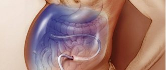
It is not difficult to identify tumor lesions of the peritoneum with ascites; in the absence of the production of pathological secretions, diagnosis is based on visualization - ultrasound and CT with contrast.
With ultrasound , on the inner layer adjacent to the muscles of the abdominal wall, which is normally very thin and invisible, you can see layers several centimeters thick, only small nodules are practically not visible.
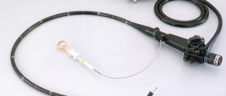
Contrast-enhanced CT is much more informative than ultrasound and can detect centimeter-sized formations. The most accurate diagnostic method is laparoscopy. This examination is mandatory for gastric carcinoma; for ovarian cancer, surgery is preferable - diagnosis and treatment at the same time.
During laparoscopy or puncture, ascitic fluid is obtained to examine and determine the primary source of the malignant process. A precipitate is isolated from the exudate, which is examined under a microscope and specific reactions are carried out - PCR and IHC.
PET scan at the stage of primary diagnosis is not always informative, since not all malignant cells of the lung, liver, and kidneys are capable of accumulating isotopes.
Without a doubt, the most optimal diagnostic method is to obtain a piece of tumor tissue for examination. Biopsy is not appropriate if the source of metastases is known and after recent treatment of the primary cancer.
Stages of abdominal carcinomatosis
Staging of peritoneal carcinomatosis cannot be called accurate; all classifications are approximate in determining the volume of damage and do not specify the location of the nodes. Often, staging gives a general idea of the prognosis for the effectiveness of treatment measures, rather than informing about the actual condition inside the abdominal cavity. The gradation of tumor spread into three degrees, developed by Japanese specialists, takes into account the total volume of the lesion, without the number and size of foci:
- P1 - limited;
- P2 - lesions separated by normal tissue;
- P3 is a set of nodes.
During surgery, surgeons determine the peritoneal carcinomatosis index (PCI), measuring nodules in 13 regions of the cavity; the total score affects treatment tactics, primarily the possibility of removing the peritoneum - peritonectomy and the advisability of intracavitary chemotherapy. In some malignant processes, complex formulas for calculating PCI are used. The best idea of the size of the cancerous lesion is given by staging by degree:
- 0 - the cavity is clean,
- I - nodules up to 5 mm in one anatomical zone,
- II - multiple nodules up to 5 mm,
- III - local lesion 0.5–2 cm,
- IV - 2 cm nodules.
The course of carcinomatosis is determined not so much by the size of the metastatic node as by the cellular potency for progression and production of ascitic fluid, the total area of tumor transformation and clinical manifestations.
Stages of the disease
Inside the human abdominal cavity there are many important organs. However, they do not have clear boundaries. That is why it is difficult even for an experienced specialist to diagnose and determine the stage of carcinomatosis. An important role in this situation is played by the primary disease, due to which carcinomatosis subsequently develops.
Most often, the doctor can determine the extent of damage to the cavity by assessing the condition of the serous membrane.
Now experts note three stages in determining the diagnosis of caceromatosis:
- the tumor contour has one affected area of serous tissue;
- several areas are affected, between which there is a healthy membrane;
- strong changes are observed in the cavity, a large area of tissue is affected. This mainly happens at stage 4 of oncology.
Symptoms of abdominal carcinomatosis
Peritoneal carcinomatosis of small extent may not manifest symptoms, especially in the absence of ascitic fluid production. On the other hand, fluid can be produced in the absence of visible metastases. As a rule, the symptoms are nonspecific, and a different set may include:
- painful sensations that change localization, and more often - incomprehensible discomfort in the abdominal cavity;
- increasing weakness to the point of loss of ability to work;
- weight loss with a stable diet regimen;
- progressive loss of appetite;
- functional disorders of the gastrointestinal tract.
Further growth of cancerous damage is accompanied by tumor intoxication, compression of the stomach by tumor nodes is complicated by nausea and vomiting, intestines - constipation and diarrhea with worsening partial obstruction. The disintegration of large nodes can cause pain and fever.
Ascites disrupts the breathing process and causes heart failure with constant edema, and frequent evacuation of pathological fluid leads to protein deficiency.
How is peritoneal carcinomatosis treated?
None of the modern methods of treating carcinomatosis guarantee radical removal of the tumor or can cure, but can improve the condition and significantly prolong life.
Surgical treatment of carcinomatosis is technically difficult for the operating team and difficult to tolerate by the patient, since it involves removal of the primary cancer, enlarged lymph nodes, omental bursae and all visible tumor formations along with the peritoneum.
Peritonectomy is a multi-stage intervention, including the removal of several organs and parts of the abdominal cavity. As a result of the operation, the patient may be left without a spleen, gallbladder, part of the intestines, uterus and appendages.
The standard treatment for carcinomatosis is systemic and local chemotherapy - intraperitoneal after removal of ascites or through a laparoport installed during surgery.
The effectiveness of drug therapy is low, with the exception of cases of primary ovarian cancer. Targeted and immuno-oncological drugs are only being studied in clinical trials.
Treatment
Carcinomatosis of the abdominal cavity, the prognosis of which largely depends on the adequacy of therapy, seriously affects the patient’s health. The first stage of treatment for a malignant tumor is to identify the primary lesion, its location and stage. Therapy for pathology is prescribed only after the specialist receives the necessary data.
Surgical methods of therapy are applicable when the stage and location of the cancer tumor allows it. Stage 4 abdominal carcinomatosis, for which the prognosis is unfavorable, does not require surgical intervention. Peritoneal carcinomatosis and ascites require treatment with chemotherapy.
For this disease, symptomatic therapy is carried out, which involves pain relief, removal of accumulated fluid, prevention of intoxication, removal of excess fluid and improvement of digestion.
Which therapy methods give the best results?
The highest effect is demonstrated by a combination of three cancer treatment methods:
- An operation with the maximum possible removal of malignant tumors is cytoreduction.
- Local intraperitoneal hyperthermia.
- Intracavitary administration of chemotherapy drugs.
The use of intraperitoneal hyperthermic chemotherapy (IHCT or HIPEC) during surgery makes it possible to maintain a very high concentration of the cytostatic directly in the affected area for as long as possible and enhance the drug effect by heating the tissue. With very modest historical results for surgical intervention of cytotoxic-resistant pseudomyxoma, only HIPEC offers patients the prospect of a long life.
The IHCT technology is as follows: for an hour and a half, a heated chemotherapy drug is delivered under pressure into the abdominal cavity in a dose significantly exceeding the maximum allowed for intravenous administration. Due to local use, the spectrum of toxic reactions changes, life-threatening damage to hematopoiesis is excluded, but abdominal pain and temporary disruption of the functioning of the gastrointestinal tract are possible.
Intraoperative photodynamic therapy (PDT), when tumor foci identified using a photosensitizer are treated with a laser, is inferior to HIPEC in terms of effectiveness, since it is impossible for the laser to penetrate into all the “nooks and crannies” of the abdominal cavity. However, it is advisable to use photodynamic therapy for large and few cancerous nodes.
Signs and symptoms
Peritoneal carcinomatosis, the treatment of which requires the patient to stay in the hospital and be monitored around the clock, is a secondary disease. The clinical picture for this diagnosis is determined by the signs of a primary malignancy. Peritoneal carcinomatosis in cancer is characterized by the formation of ascites - the accumulation of free fluid in the abdominal cavity.
The main signs of peritoneal carcinomatosis:
- body weight decreases quickly and the belly increases
- indigestion
- profuse sweating;
- dull, severe pain in the abdominal area
- Characterize carcinomatosis of the peritoneum; pain under the breast
- the patient shows signs of severe intoxication
- loose stools that may contain blood.
As the oncological process progresses, the patient may experience loss of consciousness and a state of delirium. Peritoneal carcinomatosis due to cancer requires immediate treatment, so the Yusupov Hospital accepts patients in serious condition every day, 24 hours a day.
Carcinomatosis and ascites can threaten the patient’s life, so people at risk should know the symptoms of the disease in order to promptly consult an oncologist. Specialists from the Yusupov Hospital answer questions from patients: abdominal carcinomatosis - what it is, what treatment methods exist and what is life expectancy.
Prognosis for carcinomatous lesions
The course of the process is influenced by the volume of the lesion at the time of initiation of therapy, the degree of malignancy of the tumor, which in turn determines the sensitivity to chemotherapy. The talent and experience of the surgeon, and undoubtedly the correct choice of treatment tactics, have a fundamental influence.
Only HIPEC showed clearly revolutionary results in clinical trials. After intraoperative hyperthermic chemotherapy, the five-year survival rate for gastric cancer carcinomatosis increased to a maximum of 20%; all other methods excluded such a long survival. With colon cancer with metastases to the peritoneum, every third patient lived more than 5 years, with carcinoma of the cecum and appendix - six out of ten entered the second five years of life.
Read also:
- Hyperthermic intraperitoneal chemotherapy
- Peritoneal carcinomatosis
- HIPEC
- Treatment of ascites
- Ascites
- Pleural carcinomatosis
Book a consultation 24 hours a day
+7+7+78
Bibliography
- Davydov M.I., Ter-Ovanesov M.D., Buydenok Yu.V. et al. / Hyperthermic intraoperative intraperitoneal chemotherapy for gastric cancer: is there a real opportunity to change the prognosis? // Bulletin of the Russian Cancer Research Center named after. N. N. Blokhin RAMS; 2010 T. 21; No. 1
- Stepanov I.V., Paderov Yu.M., Afanasyev S.G./ Peritoneal carcinomatosis // Siberian Journal of Oncology; 2014; No. 5
- Akiyama H., Yamaoka H., Tanaka K., et al./ Continuous hyperthermic peritoneal perfusion for peritoneal dissemination of gastric cancer // Hepatogastroenterology; 1998.
- Bozzetti F., Bonfanti G., Morabito A., et al/ A multifactorial approach for the prognosis of patients with carcinoma of the stomach after curative resection // Surg. Gynecol. Obstet.; 1986.
- Cotte E., Passot G., Gilly FN, Glehen O. /Selection of patients and staging of peritoneal surface malignancies // World J. Gastrointest. Oncol.; 2010; Vol. 2.
- Chua TC, Moran BJ, Sugarbaker PH, et al. /Early- and longterm outcome data of patients with pseudomyxoma peritonei from appendiceal origin treated by a strategy of cytoreductive surgery and hyperthermic intraperitoneal chemotherapy// J. Clin Oncol 2012.
- Deraco M., Santoro N., Carraro O., Inglese MG, et al./ Peritoneal carcinomatosis: feature of dissemination. A review // Tumori; 1999.
- Lansom J., Alzahrani N., Liauw W., Morris DL /Cytoreductive Surgery and Hyperthermic Intraperitoneal Chemotherapy for Pseudomyxoma Peritonei and Appendix Tumors // Indian J Surg Oncol. 2016 Jun.
- Sugarbaker PH/ Overview of peritoneal carcinomatosis // Cancerologia; 2008.