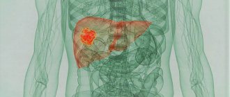
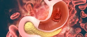
- What are cancer metastases to the liver?
- Where do cancer cells metastasize to the liver?
- Why do many tumors metastasize to the liver?
- How do cancer metastases to the liver manifest? What symptoms should you see a doctor for?
- How are cancer metastases to the liver diagnosed?
- How are liver metastases treated?
- Liver recovery after removal of metastasis
- What complications may arise? When is emergency action needed?
- What is the prognosis for cancer metastases to the liver?
- Where to treat liver metastases in Russia?
What are cancer metastases to the liver?
Metastases are secondary lesions that occur when cancer cells from the main, “maternal” tumor break off and migrate with the blood or lymph flow to different parts of the body. Metastases can occur in different organs. They are often found in the liver.
If the tumor initially develops from liver tissue, primary liver cancer occurs. Metastatic cancer is called secondary - it always originates from other organs. Most malignant liver tumors are secondary cancers.
I was diagnosed with metastases in the liver with a primary focus in the intestine. I really hoped that we would have time to treat the intestines, but we didn’t have time - I delayed for a long time with the paperwork at another clinic. So, when they were discovered, I naturally gave up, and I already mentally imagined the remaining weeks. Fortunately, I believe in modern pain management, but I didn’t believe that you could survive with lesions in the liver. This would have been the case if I had not ended up with Dr. Pylev. He found a way out. And that saved me.
Show in full »
I don’t remember the exact name of the operation - it is very long, with some indices, but, in fact, as Andrei Lvovich explained to me on his fingers - fortunately for me, three lesions formed in one lobe of the liver. Unfortunately for me - in the one that is larger, and therefore more important. You can't just take it and cut it off. The remaining piece will not withstand the load. The solution consisted of two operations. The first is that it somehow redirected blood flow to the liver so that the smaller part of the liver received more blood than the larger part. Due to this, a small share began to grow! And that means she began to take on more and more load. A little less than a month later it was already of a size that is enough for almost normal functioning of the liver as an organ. With a second operation, he removed the infected lobe. And the little one now works alone, but adapts to the loads. I didn’t think, of course, that in my life I would have to learn about all this, much less experience it myself. But, as the doctor said, until recently such operations were not performed at all. Six months have already passed since the initial diagnosis. I think I wouldn’t have had them if it weren’t for my luck in getting to these doctors. Are there even enough words to express the gratitude from me and the children? Don't think. But anyway, thanks to them.
Stenting
If a tumor in the head of the pancreas blocks the flow of bile, stenting is used. A stent is installed in the bile duct - a hollow cylindrical frame with a mesh wall. It expands the lumen of the duct and helps restore the outflow of bile.
Typically, the stent is placed using an endoscope inserted through the mouth into the duodenum. If this fails, percutaneous transhepatic cholangiography and a stent placed during this.
Where do cancer cells metastasize to the liver?
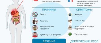
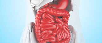
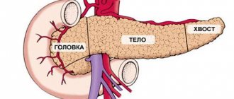
Metastatic liver cancer often occurs from the lungs, stomach, colon and rectum, mammary glands, esophagus, and pancreas.
In lung, stomach and colorectal cancer, liver metastases are found in 50% of cases, in breast cancer, melanoma - in 30% of cases.
Rarely, malignant tumors of the uterus and ovaries, pharynx, oral cavity, bladder, and kidneys metastasize to the liver. Metastases in the liver in brain cancer practically do not occur.
Classification
Depending on the number, metastases are:
- sole, or solitary;
- single (up to 10);
- multiple (more than 10).
The sizes are small (up to 1 cm in diameter), medium (1-3 cm) and large (more than 3 cm). The speed of their appearance depends on the degree of aggressiveness of the primary tumor.
Metastatic tumors are not classified according to the TNM system. The presence of metastases in the liver itself indicates stage 4 of the primary tumor.
Why do many tumors metastasize to the liver?
The liver is one of the largest organs. It performs important functions: cleanses the blood of toxins, produces bile, produces various proteins, enzymes, stores glycogen reserves, which is a source of energy.
A huge amount of blood passes through the liver - approximately 1.5 liters per minute. About 30–35% of the blood enters through the arteries, the remaining 70–75% through the portal vein from the intestines. Inside the liver there are special sinusoidal capillaries (sinusoids), in which the blood flow slows down, arterial blood mixes with venous blood, and together they return to the heart through the inferior vena cava.
This special blood supply to the liver promotes the spread of cancer cells.
Before establishing its own “colony”—a metastatic tumor—in the liver, a cancer cell has to travel a long way. It must break away from the mother tumor, penetrate a blood or lymphatic vessel, travel throughout the body and settle in the liver tissue. It can die (and many cancer cells do) at any stage.
Up to a certain point, the maternal tumor and immunity inhibit the growth of metastases. The migrated cancer cells are either inactive or multiply very slowly. Then their rapid growth begins. Scientists do not fully know why this happens. As the number of cancer cells in the metastasis increases, they begin to produce growth factors that stimulate the growth of new blood vessels that feed the tumor.
Targeted therapy
In recent decades, molecular genetics has been actively developing. Many mutations have been studied that lead to the development and progression of cancer, the products of defective genes are proteins that help cancer cells multiply uncontrollably, survive, and “hide” from attacks by the immune system. Thanks to this knowledge, a new method of treating liver cancer and other oncological diseases has emerged - targeted therapy.
Targeted drugs block molecules vital for cancer cells. Three targeted drugs are effective for liver cancer:
- Sorafenib (Nexavar)
blocks kinase enzymes that are involved in the formation of new blood vessels, programmed cell death. The drug is used for inoperable liver cancer. - Regorafenib (Stivarga)
also inhibits kinases, thereby inhibiting the proliferation of cancer cells and the growth of new blood vessels in tumor tissue. It is recommended for use in cases where sorafenib is ineffective. - Cabozantinib (Kometrik)
is an inhibitor of the enzyme c-Met tyrosine kinase and vascular endothelial growth factor 2 (VEGFR2). Studies have shown that this drug suppresses the progression of liver cancer and reduces the risk of patient death.
How do cancer metastases to the liver manifest? What symptoms should you see a doctor for?
In the early stages, as with many malignant tumors, cancer metastases in the liver do not manifest themselves in any way. Over time, the lesions increase in size and begin to interfere with blood flow and bile outflow. Liver function is disrupted and various symptoms occur:
- Weakness, fatigue, decreased performance.
- Weight loss to the point of extreme exhaustion - cachexia.
- Deterioration of appetite up to anorexia.
- Nausea, vomiting.
- Sallow skin color or jaundice.
- Dull pain under the right rib. Feeling of heaviness, fullness, pressure.
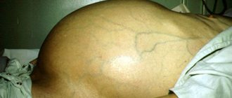
- Enlarged abdomen due to dropsy (ascites).
- Dilated veins under the skin of the abdomen (often the picture is very characteristic: the veins diverge in all directions from the navel and resemble the “head of a jellyfish”).
- Spider veins on the skin.
- Increased heart rate.
- Temperature increase.
- Skin itching.
- Intestinal dysfunction, bloating.
- Bleeding in the esophagus.
- Gynecomastia (enlargement and engorgement of the mammary glands in men).
Such disorders occur not only in liver cancer. Of course, there is no reason to panic if from this list you are only worried about weakness, fever and bloating.
The most serious symptoms that should prompt an immediate visit to the doctor: persistent vomiting: more than 1 day, more than 2 times a day, vomiting blood, rapid unexplained weight loss, black stools, severe abdominal enlargement, jaundice.
Metastases in any organ, including the liver, can cause constant excruciating pain.
Get a doctor's consultation
Efficiency forecast
Timely therapy gives positive results in most cases. Conservative treatment may be complicated by the sensitivity of the organ to the drug. The administered antitumor drugs destroy actively dividing cells and slow down tumor growth. Therapy allows you to reduce the size of the tumor for subsequent surgery. Single cancer metastases are removed surgically. Proper nutrition, lifestyle, daily routine are components of complex therapy.
The prognosis for the effectiveness of treatment largely depends on the phenotype, the nature of the lesion, the number of metastatic nodes and the location of the primary tumor. Metastases are dangerous due to the risk of internal bleeding, compression of blood vessels, a toxic increase in bilirubin levels, and impaired bile outflow.
The presence of extrahepatic metastases and multiple advanced liver lesions, unfortunately, give an unfavorable prognosis.
How are cancer metastases to the liver diagnosed?
The examination may include various studies and analyses:
- Liver ultrasound is a simple and accessible diagnostic method and is often used for screening. But it does not always help to find metastases and obtain the necessary information about them.
- Imaging methods: multislice CT, MRI, PET, angiography (a study during which a contrast agent is injected into the vessels). They help not only to detect metastases in the liver, but also to assess their size, quantity, location, growth pattern, detect suppuration and decay, spread to neighboring tissues and organs.
- Often, in order to prescribe effective treatment, the doctor needs to know what structure the tumor tissue has at the microscopic level, how much cancer cells differ from normal ones. To do this, a biopsy is performed: a fragment of tumor tissue is obtained using a needle (fine-needle aspiration biopsy) or a special instrument - a trephine (core biopsy, trephine biopsy). The procedure is carried out under ultrasound control.
- Blood tests, in particular the level of liver enzymes, help to understand how severely the liver is functioning.
“During a biopsy, a needle is inserted into the tumor. Could this cause cancer cells to break off and metastasize?” It is a myth. A biopsy does not increase the risk of metastasis.
Often, during the examination, metastases in the liver are first detected, and then they begin to look for the primary tumor. The task is made easier by a biopsy: knowing what cancer cells look like under a microscope, the doctor can understand which organ they come from.
Chemoembolization
First, an entrance is made into the vessels supplying the malignant neoplasm with blood. Chemotherapy drugs are injected into them using microinjections. The blood supply to the tumor is blocked and a prolonged effect of intra-arterial chemotherapy is ensured. To better bind the chemotherapy drug to tumor cells, lipiodol is added to it. The technique is highly effective in the treatment of liver metastases.
How are liver metastases treated?
Treatment tactics will depend on several factors:
- Number of metastases: whether they are single or multiple.
- Type of cancer.
- The severity of dysfunction of the liver and other organs.
The main methods of treatment are the same as for other cancers. Single metastases (or several small ones) can be removed surgically. Lobar, segmental, and atypical resection (removal of part of an organ) is performed.
Courses of chemotherapy and radiation therapy are prescribed.
Euroonco doctors in Moscow use a modern method of treating metastatic liver cancer, which is often practiced in foreign clinics - percutaneous transhepatic radiofrequency ablation (RFA).
Treatment results
| 3A - cancer metastases in the liver 3B - tumor reduction after chemoembolization 3C - reduction of metastases after radiofrequency ablation (RFA) 3D - the result of therapy after 6 months |
Book a consultation 24 hours a day
+7+7+78
During the procedure, a special needle-electrode is inserted into the metastasis and radio waves are applied through it, which destroy cancer cells. As a result, controlled aseptic necrosis of the tumor occurs without damage to surrounding healthy tissue. This can significantly improve survival and reduce the risk of relapse.
RFA is unique in that it can be reused if new metastases are detected in the liver. The technique is successfully used in primary liver cancer, when there is simultaneously cirrhosis and a high risk of liver failure.
For example, we were able to achieve stable remission and, possibly, complete recovery in one patient who was diagnosed with breast cancer with single metastases in the liver. Doctors at the Euroonko clinic in Moscow performed a radical mastectomy and lobar resection of the liver, which were supplemented with a course of chemotherapy.
Also, a good result was achieved in a patient with colon cancer and five small metastases in different lobes of the liver. We performed a resection (removed part of the intestine), administered a course of chemotherapy and radiofrequency ablation of the liver.
Treatment of cancer metastases to the liver has some difficulties. For example, metastatic cancer often does not respond to drugs that worked against the primary tumor. We have to select the optimal therapy and combine different types of treatment. Liver metastases also respond poorly to systemic chemotherapy. The best effect is obtained by administering drugs into the hepatic artery.
Chemotherapy drugs help slow down the growth of metastases, reduce their number, prolong the patient’s life and relieve painful symptoms. In the early stages, a course of chemotherapy reduces the risk of metastasis. In cases where this is necessary, our doctors use implantable venous and arterial port systems and regional intra-arterial infusion of chemotherapy drugs.
Radiation therapy for cancer metastases to the liver helps relieve pain, but does not increase life expectancy.
Targeted therapy involves the use of drugs that have a specific “target” - a specific molecule necessary for the growth and survival of cancer cells. For metastatic liver cancer, the only targeted drug with proven effectiveness is sorafenib. It is registered in more than 60 countries for the treatment of primary and metastatic liver cancer.
Embolization is a promising method for the treatment of liver metastases and other malignant tumors, which is used in the department of interventional oncology and endovascular surgery. The essence of the method is that a special drug is injected into the vessel feeding the tumor, which disrupts the blood flow.
Chemoembolization is most effective when microspheres are introduced into the vessel, which release the chemotherapy drug. Chemoembolization is currently the “gold standard” of treatment in cases where surgical tumor removal or transplantation cannot be performed.
During chemoembolization, a double effect is achieved. The microspheres block the blood flow, depriving the tumor of necessary substances, and the released chemotherapy attacks the tumor cells.
All existing drugs for chemoembolization are available at Euroonco.
About the possibilities of modern high-tech medicine in the treatment of liver tumors in the Health Kitchen program on the Dozhd TV channel.
Get a treatment program
New treatments
Radiofrequency ablation
Modern surgery allows this method to be used for metastatic liver disease. A source (electrode) of high-frequency radiation is brought through the skin and muscles to the tumor node. The accuracy of the electrode location is controlled by electroanatomical mapping. Radiation directly affects the metastasis and destroys tumor cells. At the same time, the impact on surrounding tissues and the risk of bleeding are minimal. The procedure can also be performed under local anesthesia.
Chemoembolization of tumor
Used when ablation is contraindicated. Having provided access to the vessels feeding the tumor, a chemotherapy drug is injected into them through a needle. This substance “solders” the walls of the arteries, blood stops flowing through the vessels into the metastasis, and without nutrition the tumor node dies. Aseptic necrosis and death occurs.
Targeted therapy with biologics
Specific drugs obtained as a result of high-tech synthesis (Sorafenib, in particular) selectively act specifically on cancer cells. Substances toxic to tumor cells accumulate in them and prevent further growth of cancerous foci. The method is used in complex therapy, as an addition to other treatment methods, in the presence of contraindications to radical treatment.
Liver recovery after removal of metastasis
If, after surgical treatment for metastases in the liver at stage 4 cancer, cancer cells are no longer found in the organ, the doctor will recommend taking pictures (ultrasound, CT or MRI) and taking tests (for the level of alpha-fetoprotein, substances that characterize liver function) once every 3–6 months for the first two years, then every 6–12 months. This helps to detect relapse or possible side effects of treatment in time.
A healthy diet and physical activity in accordance with the doctor’s recommendations help speed up the recovery of the liver and the entire body.
Intra-arterial chemotherapy
The entrance to the artery leading to the liver is made through the inguinal artery using angiography. The drug is injected directly into the organ once every 2-3 weeks. The possibility of placing a stationary port catheter in the inguinal artery avoids the need for constant angiography. This simplifies the intra-arterial chemotherapy session.
What complications may arise? When is emergency action needed?
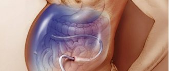
A tumor in the liver can compress the portal vein, inferior vena cava, and bile ducts. In the latter case, the outflow of bile is disrupted. The toxic breakdown product of hemoglobin contained in it - bilirubin - begins to enter the blood. The skin, sclera and mucous membranes turn yellow - obstructive jaundice occurs. This condition is dangerous because bilirubin is toxic to the brain and other organs; a strong increase in its level can lead to death. In addition, due to obstructive jaundice, it is impossible to perform surgery and conduct a course of chemotherapy.
The outflow of bile is restored surgically, under the control of ultrasound (puncture cholangiography) or X-ray television. There are two types of drainage:
- External - bile is removed outside.
- External-internal: part of the bile is excreted outside, part into the intestinal lumen.
If the tumor compresses several bile ducts in different parts of the liver, several drains are installed. Euroonco doctors use a modern method of one-stage stenting. In this case, external drainage is left for only 1–2 days, or you can do without it altogether.
Total irradiation
Total radiation therapy is used for multiple metastases. With this procedure, the entire volume of this organ is irradiated. As a rule, the total dosage is 24 Grays for 10-12 fractions or 30 Grays for 12-15 fractions.
Not every radiologist will undertake such a procedure; exceeding these standards can lead to radiation hepatitis. When the hepatic gate is compressed by enlarged lymph nodes, local irradiation can be applied. Thanks to such radiation, you can get a better result, with less risk of side effects, than with chemotherapy.
What is the prognosis for cancer metastases to the liver?
Patients who come to Euroonko clinics with this diagnosis are primarily concerned with the question: “is it possible to cure liver metastases in stage 4 cancer?” The effectiveness of treatment will depend on the type of cancer, its molecular genetic characteristics, location and grade of malignancy of the tumor. Most patients, after they are first diagnosed with liver metastases, live for 6–18 months. For colon and rectal cancer, after major cytoreductive operations, the prognosis is more favorable.
If metastases are present not only in the liver, but also in other parts of the body, for example, metastases in the liver and bones, the prognosis worsens. However, surgical treatment is also possible.
Euroonco doctors have extensive experience in the combined treatment of liver metastases. Thanks to this, we can significantly prolong the lives of patients. The best results are obtained when colon cancer metastasizes to the liver. We have developed clear criteria, guided by which, sometimes we can refrain from surgical treatment if lesions are found in both lobes of the liver. In such cases, treatment begins with a course of chemotherapy.
If metastases in the liver arise from a tumor of the lung, pancreas, stomach, etc., removal of part of the liver as an independent treatment method is ineffective, but it can work well in combination with chemotherapy.
If there are simultaneously metastases in the liver and lymph nodes, this significantly worsens the prognosis. Five-year survival rate is approximately halved. But even in this case, treatment is still possible.
Where to treat liver metastases in Russia?
Many Russian patients, faced with this terrible diagnosis - “cancer with metastases”, believe that this is a death sentence and nothing can be done, or that effective help can be obtained, but only abroad. In fact, all modern technologies and drugs are available in Russia. Modern qualified assistance can be obtained at Euroonco clinics.
| Read more about treatment at Euroonko | |
| Emergency oncology care | from 12,100 rub. |
| Palliative care in Moscow | from 44,300 rubles per day |
| Oncologist consultation | from 5,100 rub. |
| Chemotherapy appointment | RUB 6,900 |
We believe that you can always help, and therefore we take on the treatment of patients with cancer at any stage. Our doctors have extensive experience and carry out complex invasive procedures and surgical interventions, chemotherapy according to international protocols. We know how to help.
Read also:
- Hepatocellular carcinoma
- Diagnosis of liver cancer
- Liver cancer treatment
- Metastases in the brain
- Metastases in the lungs
- Bone metastases
- Metastases in the spine
- Metastases in the peritoneum
- Metastases in breast cancer
- Metastases in sarcoma
- Metastases in lymph nodes
- Metastases in prostate cancer
- Metastases in pancreatic cancer
- Krukenberg metastasis
- Effective methods of treating cancer with metastases
- Treatment of metastases in uterine cancer
- Skin and subcutaneous metastases in cancer
Book a consultation 24 hours a day
+7+7+78
Bibliography:
[1] The prognostic value of tumor cells blood circulation after liver surgery for cancer lesions - Patiutko IuI, Tupitsyn NN, Sagaĭdak IV, Podluzhnyĭ DV, Pylev AL, Zabezhinskiĭ DA. — Khirurgiia (Mosk). 2011;(6): 22–6.
[2] Surgical and combined treatment of multiple and bilobar metastatic affection of the liver. — Patiutko IuI, Sagaĭdak IV, Pylev AL, Podluzhnyĭ DV. — Khirurgiia (Mosk). 2005;(6): 15–9.
[3] Modern approaches to the treatment of colorectal cancer metastases in the liver - A. L. Pylev, I. V. Sagaidak, A. G. Kotelnikov, D. V. Podluzhny, A. N. Polyakov, Patyutko Yu.I.,. - Bulletin of Surgical Gastroenterology - Number: 4 Year: 2008 Pages: 14–28.
[4] Ten-year survival rate of patients with malignant liver tumors after surgical treatment - Yu. I. Patyutko, A. L. Pylev, I. V. Sagaidak, A. G. Kotelnikov, D. V. Podluzhny, M. G. Agafonova. - Annals of Surgical Hepatology 2010.
[5] Surgical and combined treatment of patients with metastatic liver and lymph nodes invasion by colorectal cancer - Patiutko IuI, Pylev AL, Sagaĭdak IV, Poliakov AN, Chuchuev ES, Abgarian MG, Shishkina NA. — Khirurgiia (Mosk). 2010;(7): 49–54.
Symptoms
Often, the first symptoms of stomach cancer appear in the later stages, when metastasis has already occurred. Symptoms depend on which organ the cancer cells have spread to:
- In the peritoneum : abdominal pain, abdominal enlargement due to the accumulation of fluid inside ( ascites ), loss of appetite, causeless severe weight loss.
- In the liver: loss of appetite and weight loss, dark urine, enlarged abdomen, jaundice, pain in the upper abdomen on the right (under the right rib), nausea, vomiting, increased sweating.
- In the lungs: chest pain, persistent chronic cough, blood in the sputum, wheezing, shortness of breath, weight loss.
- In the bones: pain, pathological (from slight effort) fractures.
- In the brain: headaches, nausea, vomiting, weakness, numbness in the arms and legs, impaired coordination of movements, personality and behavior disorders, speech, swallowing, urinary and stool incontinence.
All of these symptoms can be caused by other diseases.