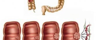
Surgery is one of the main methods of treating rectal cancer. Radiation or chemoradiotherapy is sometimes needed before surgery to make it easier to remove the tumor. It also reduces the likelihood of the disease returning.
The Asaf HaRofe Clinic treats colorectal cancer in the early stages and in advanced states, when metastases have affected surrounding organs and systems. Among our advantages:
- Professional doctors with extensive experience in the treatment of cancer of the rectum, colon, and gastrointestinal tract.
- Innovative therapeutic regimens combining traditional techniques with original ones.
- Latest generation medicinal formulas developed at the hospital’s research sites.
We provide all types of therapeutic interventions at affordable prices, regulated by the Israeli Ministry of Health.
Before surgery for rectal cancer
If the patient smokes, it is necessary to quit or reduce smoking. This will help reduce the risk of complications (respiratory tract infections) and help the wound heal after surgery.
Surgeons collect tests to find out whether the patient can undergo surgery.
These may include blood tests, blood pressure checks, heart function tests (ECGs), and several others.
A member of the surgical team meets with the patient to discuss the operation. If an ostomy is to be created, the nurse will provide all the necessary information on this issue.
Surgeries for colorectal cancer at Asaf HaRofeh Hospital
There are different techniques and types of operations that can be used for this disease. The choice of surgery is determined by factors such as the stage of the cancer, the location of the tumor, and your overall health.
After the operation, the tissues that are removed by the surgeon are sent to a pathologist for examination. He checks the edges of the surgical area for the presence of abnormal cells. If they are found, it is possible that the cancer was not completely removed. When abnormal cells are found, repeat surgery or radiotherapy may be suggested.
Local resection
For a small tumor at stage 1, the cancer can be removed using local resection. An endoscope is used, a long and flexible tube with a small camera at the end. The operation is called transanal endoscopic microsurgery.
If the tumor is located very low in the rectum, near the anus, the surgeon may not use an endoscope. The malignant tumor will be removed using surgical instruments inserted through the anus. This type of surgery is called transanal rectal resection.
Total mesorectumectomy
This is a commonly used surgery for colorectal cancer. The surgeon removes the part of the organ that contains the tumor, as well as sections of healthy tissue on either side. In addition, the fatty tissue (mesorectum) around the rectum, including the blood vessels and lymph nodes, is resected. Its removal reduces the risk of relapse.
Types of mesorectumectomy
There are several types of mesorectumectomy. The choice of surgery is influenced by the location of the tumor in the rectum, its size, and the distance of the tumor from the anus.
Anterior resection
This type of surgery is usually used when the cancer is located in the upper and middle part of the rectum (near the large intestine).
After the part of the intestine containing the tumor is removed, the surgeon repairs the connection by bringing the two ends together. Some patients have a temporary stoma (ileostomy) created after this. The operation to close it is performed after a few months.
Proctectomy with coloanal anastomosis
This surgery is performed when the cancer is located low in the rectum.
The surgeon removes the entire rectum, connecting the large intestine to the anus.
Sometimes doctors create a bag (reservoir) from the colon (coloplasty) instead of a straight one to store feces.
After a proctectomy, a temporary stoma (ileostomy) may be created to allow time for the intestines to heal.
The stoma is closed after a few months.
Abdominoperineal extirpation of the rectum
This type of surgery (BPEP) is performed when the tumor is located very low in the rectum, near the anus. To remove all the malignancy, the surgeon performs a resection of the rectum and anus. In this case, a stoma is required and created - a colostomy. One incision is made in the peritoneum, the other in the perineum, near the anus. Through the final incision, the surgeon removes the anus and surrounding tissue.
Open or laparoscopic surgery for rectal cancer
Colorectal surgery is performed using an abdominal or laparoscopic approach.
Abdominal surgery is performed through a large incision running approximately from the breastbone (breastbone) to the belly button. In some patients, the incision is made across rather than along the abdomen.
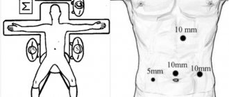
During laparoscopic surgery, the surgeon makes 4 or 5 small incisions in the peritoneal cavity. A laparoscope equipped with a light and a camera is inserted through one of them, and surgical instruments are inserted through the others to remove the tumor.
Recovery from laparoscopic surgery is generally faster compared to open surgery. The surgeon will tell the patient what type of surgery is appropriate in this case.
Stoma (colostomy/ileostomy)
After surgery to remove colorectal cancer, some patients have an ostomy created to remove bowel movements from the body. This is an artificial hole in the wall of the abdomen, to which a bag is attached to collect feces.
A stoma is created from an open part of the intestine. If it is done using the large intestine, then this operation is called colostomy, when - through the small intestine (ileum) - ileostomy.
This artificial hole is used for a certain period of time or can be permanent. This is temporary to allow the intestines to heal after rectal surgery. Colostomy can be loop or end. To create a loop, the surgeon pulls a small loop of intestine out through an incision in the abdomen. Then he makes a hole in the loop and sews it to the skin. A loop stoma is called a double-trunk stoma, since two branches are removed.
To create an end stoma, the surgeon removes one end of the intestine through an incision and sutures it to the skin. The hole is placed on the left side of the abdomen. Often this type of colostomy is permanent.
If the hole is created temporarily, a second operation will be required to close it several months later to reconnect the bowel, twelve weeks after the initial surgery.
When the tumor is located very low in the rectum, near the anus, a permanent stoma will most likely be required. The surgeon will give information for each specific case whether the stoma will be permanent or temporary.
Hartmann's operation
For the treatment of cancer of the sigmoid colon, rectosigmoid colon, as well as tumor lesions of the upper ampullary part of the rectum (if it is impossible to form an anastomosis - suturing two sections of the intestine), Hartmann's operation is used: resection of the area of the colon affected by the tumor and the imposition of a single-barrel colostomy with the possibility of subsequent delayed restoration of the intestine .
Each of these interventions can be definitive (usually for stage 4 cancer) or temporary, performed to prepare the patient for subsequent stages. These surgical interventions are aimed at eliminating an immediate threat to the patient’s life. Subsequent stages are performed within 4-6 months after the initial operation, during which time adjuvant chemotherapy is performed and the patient is under medical supervision. [1]
Surgeries for late stage rectal cancer
Pelvic exenteration
If the cancer has affected other nearby organs, extensive surgery is sometimes required to remove it. For example, pelvic exenteration.
Pelvic exenteration in men
This surgery is used to treat malignant diseases in the pelvic area.
It involves removal of the bladder, rectum and prostate gland. This surgery is only done if there are no other signs of cancer anywhere else in the body.
Only specially trained and experienced surgeons are required to perform pelvic exenteration.
Before making a decision, the doctor tells the patient about the benefits and risks of this surgical procedure. This is a serious and major operation, but it can cure cancer in some patients.
Pelvic exenteration is recommended for recurrent rectal cancer. During this procedure, the surgeon removes the bladder, rectum, anus and prostate gland. And it creates two new holes - for draining urine (urostomy) and for removing feces from the body - colostomy.
Pelvic exenteration for women
This type of surgery is used for recurrent cervical cancer, to treat uterine cancer, vaginal cancer and vulvar cancer. During the operation, the surgeon removes the bladder, part of the intestines, ovaries, uterus, cervix and vagina. Surgery is serious, but sometimes it can cure cancer. Surgery is only performed if there are no symptoms of cancer anywhere else in the body.
There are several types of pelvic exenteration:
- Anterior exenteration – removal of the bladder and internal genital organs.
- Posterior exenteration – resection of the rectum and internal reproductive organs.
- Total exenteration – removal of the bladder, rectum and organs of the reproductive system.
The type of surgery is determined based on the type of cancer and your individual situation.
Lung resection
The main treatment for cases that have spread to the lungs is chemotherapy. But in some cases, surgery to remove the affected part of the lung is recommended. Only when the cancer is located in one part of the organ and nowhere else in the body.
Liver resection
If secondary lesions appear in the liver, they most often turn to cytostatic agents. The goal of treatment is to shrink the tumor and control it for as long as possible.
But some patients may have surgery to remove the diseased part of the liver. Sometimes liver resection can cure the patient.
This is a major surgical procedure that lasts 3-7 hours.
It is performed only in specialized hospitals in Israel, by doctors with experience in liver surgery. Quite rarely, such treatment is considered as an option for liver metastases.
Surgery to remove colorectal cancer and liver resection are performed simultaneously or as separate surgeries.
Chemotherapy usually precedes liver resection.
Tumor removal during colonoscopy or sigmoidoscopy
In some cases (with preinvasive and microinvasive or intramucosal cancer), endoscopic intraluminal removal of tumors is possible during colonoscopy, which can be combined with electro- and argon plasma coagulation.
Endoscopic treatment is also used in patients in serious condition (advanced age with multiple organ failure, severe chronic concomitant diseases), when traditional surgical treatment is impossible. In particular, when intestinal obstruction develops, Euroonko clinics install a stent in the colon. This treatment is called palliative and is aimed at prolonging life and improving its quality in patients with a tumor in advanced stages. [1,2]
Treating a Blocked Bowel
Sometimes cancer blocks the intestines, causing symptoms like pain and vomiting. As a rule, urgent medical attention is required. This condition is treated in two ways.
Stenting
During the procedure, the surgeon uses a colonoscope to place a stent in the blocked area. The stent then expands to keep the bowel open.
The cancer causes a blockage that can be removed through surgery at a later time.
Surgery
Sometimes intestinal obstruction is corrected through surgery by removing the blocked section of the rectum. After this surgery, most patients have a temporary or permanent stoma. The surgeon sometimes combines this type of surgery with tumor removal.
Rehabilitation after surgery for rectal cancer
After surgery, the patient will be encouraged to start moving as soon as possible, which will prevent complications such as infections and blood clots from developing. The physiotherapist will provide information on leg and breathing exercises.
By the evening after surgery or the next day, the patient will be assisted to get out of bed or sit up for a short time.
Pain
After surgery, the patient experiences pain and discomfort, which is controlled with analgesics. Any pain or discomfort should be reported to nurses.
They will provide medicine to relieve the condition. You may need to change the dosage or change the analgesic.
Spinal anesthesia is also provided. This is an injection of a long-lasting pain reliever into the fluid around the spinal cord. They relieve pain for up to 24 hours. Another alternative is to administer a continuous dose of painkiller into the cerebrospinal fluid using a pump - an epidural.
Painkillers are given through an IV placed in a vein in the hand or forearm. The drip is connected to a pump or pump (patient-controlled analgesia). By pressing a button, you can receive an additional dose of analgesic. Effective equipment programming prevents overdose.
Droppers and drainages
A dropper is installed that supplies fluid to the body into a vein in the hand or forearm - intravenous infusion. Once the patient is able to take food and fluids independently, it will be removed.
During the operation, a catheter is placed in the bladder to drain urine.
Some patients have a nasogastric tube installed, a tube that goes through the nose into the stomach. It is used to remove fluid from the stomach until the intestines begin to function.
Drains may be placed in the surgical wound area to drain excess fluid. They are removed after a few days.
Eating and drinking
Soon after surgery, the patient can eat and drink independently. To speed up the recovery process, the patient is provided with additional drinks for several days.
Stoma
If a stoma has been created, it will be swollen at first, but will shrink to its normal size within a few weeks. A loop colostomy uses a rod for support while healing occurs. The rod is removed after a few days.
The nurse will teach you how to care for your stoma. For most patients, 3-4 days are enough to learn and cope with this situation.
Extract
Depending on the type of surgery performed, the period of hospital stay is 3-7 days. An appointment is made for a postoperative examination, where the doctor will advise you on further treatment - radiation therapy or chemotherapy.
If sutures, clips or staples are placed on the wound, they are removed 7-10 days after surgery.
Sex life after surgery
The doctor will provide information on how long it will take for rehabilitation after surgery for rectal cancer and when you can resume sexual life. For most people this takes at least 6 weeks, and often longer.
For patients with an ostomy, it takes longer to adjust.
Laparoscopy in colorectal surgery
The possibility of performing the operation using laparoscopic technology can be determined already at the initial appointment. Modern technologies allow resection of the tumor-affected area of the intestine through small incisions on the skin (5 - 12 mm) in full compliance with the rules of ablastics - oncological operations to protect healthy tissues from infection by cancer cells of the tumor being removed.
In addition to pronounced aesthetic advantages, laparoscopic surgery allows you to shorten the postoperative period, speed up healing and reduce pain, the number of postoperative, incl. infectious complications, including interintestinal adhesions.
Restoration of normal intestinal activity occurs in a short time and allows patients not to interrupt their usual life for a long time. [4,5]
A distinctive feature of laparoscopic interventions for low cancers
The Euroonko clinic in Moscow has all the capabilities to conduct operations that are unique in their structure using laparoscopic technologies. So, if the tumor is located low, we can perform operations without bringing the intestine to the anterior abdominal wall. An operation is performed to bring down the overlying sections of the colon either into the anus or into the perineal wound; to protect this type of surgical aid, a temporary colostomy is sometimes performed as in other cases. [3]
When removing the anal canal (extirpation of the rectum), it is possible to lower the sigmoid colon into the perineal wound - essentially the same colostomy, only in a familiar place; with certain care, a relatively comfortable life and preservation of working capacity are possible. This surgical intervention requires separate discussion and decision together with the patient.
Bowel function after surgery for rectal cancer
Most people experience changes in their bowel habits after rectal surgery.
If local resection is performed, patients recover quickly. After a total mesorectumectomy, more time is needed - several months - to restore bowel function.
When there was radiation therapy or chemoradiotherapy before surgery, this will also affect the functioning of the organ. This means that it will take longer for bowel function to return to normal.
After rectal surgery, the following changes in the functioning of the organ are possible:
- Diarrhea or constipation.
- Frequent stools.
- Fecal incontinence.
- Bloating.
These disturbances pass over time. The doctor will give recommendations on how to normalize the condition, prescribe medications, and may refer you to another specialist.
Diet after surgery for rectal cancer
Regular eating will help restore organ function. If you have problems with your appetite, it may be easier to eat small meals several times a day. You should drink at least 1-2 liters of fluid per day, especially if you have diarrhea.
High protein foods - fish, meat, eggs - will help the body heal after surgery.
It is important that your diet includes a wide range of foods for a healthy, balanced diet. However, some types of food cause problems. Keeping a food diary of what foods a person eats and how they affect them can help.
If you have diarrhea, you should give preference to foods low in fiber - white bread and pasta instead of wholemeal pasta. One should eat green leafy vegetables, cook vegetables and eat fruits after peeling them.
After intestinal function returns to normal, it is worth gradually introducing foods that cause problems. A person may find that they no longer affect the functioning of the organ. If your diet after colorectal cancer surgery is still limited, you should definitely consult a nutritionist.
Some tips on how to eliminate bloating:
- You need to eat slowly and chew your food thoroughly.
- It is important to remember that beans, beer, chewing gum, soda and onions cause bloating.
- Capsules with mint, dill, and mint tea can help.
Medicines
The doctor will recommend taking antidiarrheal medications. The most commonly used drug is loperamide (also known as Imodium or Diareze®), which slows down bowel movements.
Taking loperamide regularly, half an hour before meals, helps in some cases. This drug is also available in syrup form and the dose can be adjusted as needed. It may take time to find the optimal dosage. It is recommended to start with a small amount and increase the amount until the drug has the desired effect.
It is safe to take loperamide long-term as needed, but discuss this with your doctor.
Stress management
Emotions can affect bowel function. Anxiety and stressful situations contribute to diarrhea. Loss of control over the functioning of an organ is itself stress.
Learning to relax will benefit both the intestines and the entire body. Some support groups offer relaxation courses.
Exercises for the pelvic floor muscles
There are exercises you can do to strengthen your bowel muscles: the sphincter muscles (in the anal area) and the pelvic floor muscles (also important for bladder control and sexual function).
These exercises are useful for stool incontinence. The doctor can teach you the technique for performing them. It will take at least 12 weeks to regain muscle strength, doing the exercises three times a day.
Maintaining a Healthy Weight
Excess weight puts pressure on the pelvic floor muscles. Therefore, it is especially important to maintain a healthy body weight if you have problems with bowel control. The doctor will give recommendations on this issue.
results
Results of the first stage of the study. Before treatment, the pressure readings at rest in women averaged 26.2±6.2 mmHg, and with volitional contraction - 91.2±30.0 mmHg. (with the norm being 41-63 and 110-178 mm Hg, respectively); the average score on the Wexner scale is 12.8±3.9 points. In men, the same parameters were 23.5±7.0 and 103.9±46.6 mmHg. (with the norm being 43-61 and 121-227 mmHg) and 13.0±3.5 points, respectively. After treatment in women, the positive dynamics of indicators averaged 15.3% (p = 0.12) and 17.3% (p = 0.24): the average resting pressure was 30.2 ± 7.9 mm Hg ., pressure during volitional contraction - 107.0±36.3 mm Hg.
In men, the positive increase in pressure indicators at rest and during volitional contraction was 14.9% (p = 0.23) and 18.1% (p = 0.11) and, respectively, 27.0 ± 8.5 and 122.7 ± 49.9 mmHg When analyzing pressure indicators, significant differences between the parameters of volitional contraction before and after treatment were noted without taking into account gender differences - in total for men and women. Thus, the dynamics of indicators during contraction was 17.7% (p = 0.047) (initial 98.7 ± 40.7 mm Hg versus 116.2 ± 44.8 mm Hg after treatment).
Clinically, after treatment, complaints of incontinence of stool components were subjectively assessed by patients as 11.0±4.6 points in women (a decrease of 14.1%, p=0.42) and 10.5±4.7 points (a decrease of 19 .2%, p=0.155) in men, no statistically significant improvement was found after treatment.
Results of the second stage of the study. According to sphincterometry, pressure indicators at rest and during volitional contraction before/after treatment in women were 29.3±3.8/36.0±6.4 and 104.4±22.0/131.4±25.6 mmHg, which indicated a dynamic improvement in the tone and contractility of the sphincter apparatus by 22.9% (p=0.01) and 25.9% (p=0.025), respectively.
In men, similar values were 29.3±6.4/35.2±4.1 and 162.3±28.0/189.0±50.4 mmHg. respectively. The increase in values was 20.1% (p=0.069) and 16.3% (p=0.18) and was not statistically significant. It should be noted that further research is needed for changes in resting blood pressure, as the lack of statistical significance at the 5% level (p approaching, although not reaching, the 0.05 threshold) may be due to the small sample size.
According to a study of the reservoir function of the reduced intestine, the threshold of constant sensitivity to filling before treatment was determined only in 9 (52.9%) of 17 patients, while after treatment it was determined in all patients. The feeling of an urge to defecate, which before treatment occurred in only 11 (64.7%) of 17 patients, was observed in all patients after treatment and was noted already with a volume of more than 72.6 ml. The most significant seems to be the appearance of a constant intense urge to defecate: before treatment, it was noted in only 6 (35.3%) out of 17 with a volume of 80 ml; after treatment - in 14 (82.4%) patients with 120 ml or more (Table 1) . There were no adverse events or complications during the study.
Table 1. Dynamics of reservoir function parameters in patients with SNPR ( n = 17)
| Parameter Parameter | Volume, ml of air (norm) Volume, ml air (normal) | Before treatment Before treatment | After treatment After treatment | ||
| volume, ml of air volume, ml air | number of patients patients' number | volume, ml of air volume, ml air | number of patients patients' number | ||
| First sensitivity threshold Rectum first level of sensitivity to fill | 36,7±19,7 | 65,4±26, | 16 | 35,4±15,8 | 16 |
| Constant sensitivity threshold Constant level of rectum sensitivity to fill | 66,7±24,2 | <42,5 | 9 | <42,5 | 16 |
| Feeling the urge to defecate Feeling the urge to defecate | 110,0±37,4 | <72,6 | 11 | >72,6 | 16 |
| Constant urge to defecate Constant urge to defecate | 150,0±51,0 | <99 | 6 | >99 | 14 |
On the SNPR scale before treatment, patients assessed their condition at 37.6 ± 6.3 points (pronounced SNPR), after treatment in all patients the average score decreased by 46.5% - to 20.1 ± 5.7 points (weakly expressed SNPR). Upon closer examination, the dynamics of subjective indicators in women was 44.1%, in men - 49.4% (Table 2).
Table 2. Dynamics of indicators on the SNPR scale ( n = 17)
| Patients | Before treatment, points Before treatment, scores | After treatment, points After treatment, scores | Dynamics of indicators, % Dynamics of parameters, % | p |
| Women | 39,5±2,8 | 22,1±22,1 | 44,1 | 0,02 |
| Men | 36,0±8,2 | 18,2±7,4 | 49,4 | 0,069 |
| All patients/Total cohort | 37,9±6,4 | 19,8±5,8 | 47,8 | 0,002 |
Results of the third stage of the study. 3-6 months after the course of conservative rehabilitation, a telephone survey was conducted with filling out the SNPR scale in 11 (64.7%) of 17 patients (the survey was not conducted in 6 patients with shorter periods after completion of the course). Based on the data obtained, in 4 (36.4%) of 11 patients the positive dynamics of the indicator on the SNPR scale remained, in 6 (54.5%) patients the results slightly worsened, in 1 (9.1%) patient they returned to the original values (Table 3).
Table 3. Dynamics of indicators on the SNPR scale in 3-6 months after completion of the course ( n = 11)
| Positive effect of treatment Positive treatment effect | Number of patients, abs. (%) Patients' number, abs. (%) | Dynamics of indicators | p | |
| at the end of the rehabilitation course, points At the end of the rehabilitation course, scores | 3-6 months after the rehabilitation course (telephone survey), points 3—6 months after the rehabilitation course (telephone survey), scores | |||
| Saved Remains | 4 (36,4) | 19,5±6,7 | 19.5±6.7 (without negative dynamics) (no negative dynamics) | 0,13 |
| Decreased Reduced | 6 (54,5) | 20,0±4,6 | 24,7±2,7 (unstable effect) | 0,148 |
| Completely lost Completely lost | 1 (9,1) | 23 | 41 (return to original state) (return to initial condition) | 0,317 |
Based on the clinical data obtained by the staff of the laboratory of clinical pathophysiology of the Federal State Budgetary Institution “National Medical Research Center of Coloproctology named after. A.N. Ryzhikh" of the Ministry of Health of Russia with consultation by specialists of the neurosurgical department of the Center for the provision of surgical care to patients with degenerative diseases and acute spinal trauma of the State Budgetary Institution "City Clinical Hospital No. 67 named after. L.A. Vorokhobova" DZM developed a complex of exercise therapy for training the pelvic floor muscles and obturator apparatus and treating anal incontinence. In order to strengthen the rehabilitation course, this complex can be recommended for use by patients both in a medical institution and at home to maintain the achieved positive effect.
Stenting
In some cases and with inoperable tumors in patients in serious condition with partial intestinal obstruction, decompression of the gastrointestinal tract can be achieved by endoscopic installation of a stent in the colon.
In almost 55% of patients with colorectal cancer, due to the characteristics of the venous outflow from the colon, metastases to the liver are detected. Often, in addition to tumor intoxication and pain, they aggravate the situation, causing compression of the biliary tract and creating conditions for the development of obstructive jaundice. In our clinic, metastases are removed surgically during liver resection; in addition, radiofrequency ablation, chemoembolization of liver vessels, feeding the metastatic focus can be used. [1]
