- home
- Coloproctology
- Rectal cancer
- Stages of colorectal cancer and their symptoms
Stages of rectal cancer
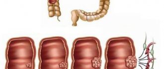
Rectal cancer is an oncological disease in which a tumor localized in the distal part of the large intestine occurs against the background of pathological changes in the mucous membrane. There are different forms of cancer: the focus of pathology can grow into the lumen of the intestine, over time blocking it, or spread deep into the intestinal wall. Infiltration of the intestinal wall is possible, and the tissue loses its elasticity, which affects its functionality. Gradually, cancer cells penetrate into nearby tissues, and if left untreated, metastases affect other organs, most often the lungs, kidneys, liver, ovaries, etc.
Rectal cancer accounts for 5% of all malignant tumors of the digestive system. For the most part, the disease affects people 40-60 years old, although in recent years, oncologist patients are increasingly becoming younger people. The risk group for morbidity includes patients suffering from ulcerative colitis, proctitis, diffuse polyposis, having fistulas, ulcers, anorectal fissures, rectal polyps - in this case, the risk of malignancy is especially high. Large polyps are dangerous; if the size exceeds 3 cm, then the risk of degeneration into cancer reaches 40-85%. The process of malignancy of a polyp can last several years, but after degeneration the tumor grows rapidly.
How many stages of colorectal cancer are there?
In domestic clinics, the disease is usually divided into stages; rectal cancer has four stages. The classification is based on the size of the tumor, the existing invasion of the intestinal wall and the degree of spread of malignant cells to other tissues and organs.
| Stage | Tumor size and spread | Main symptoms |
| 0 | small tumor within the mucosa; | there are no signs of disease; |
| I | the formation does not exceed 2 cm, mobile, within the intestinal wall, only the mucous and submucosal layer is affected; | asymptomatic; |
| II | the tumor is no more than 5 cm, spreads beyond the intestinal wall, metastases are either absent or present in regional lymph nodes; | indigestion, feeling of incomplete bowel movement, false urge to defecate, rectal discharge; |
| III | the tumor size exceeds 5 cm, the tumor grows into the intestinal wall, there are multiple metastases in the lymph nodes; | persistent constipation, bloating, rumbling in the abdomen, when the intestinal lumen is blocked by a tumor, intestinal obstruction occurs; |
| IV | the tumor is large, immobile, metastasizing to distant organs; | rectal bleeding, pain, stool retention, fever, weakness, vomiting, cachexia. |
Staging is important for choosing the optimal treatment tactics and prognosis of the disease. If at stage 0 or I of rectal cancer it is possible to get by with polypectomy during colonoscopy or intestinal resection, then in advanced cases extensive intervention is assumed. To determine the stage, it is necessary to undergo an examination. In our clinic, patients have access to the most effective examination methods.
written consultation, in order to determine the indications for surgery, as well as choose the correct surgical treatment tactics, you can send me a full description of the colonoscopy, histology data, if possible, MSCT data of the abdominal cavity with contrast, indicate your age and main complaints. Then I will be able to give a more accurate answer to your situation.
Circumferential resection margin / circular resection margin / lateral resection margin
In the illustration on the left:
- T2 tumor limited to the bowel wall
- T3 tumor with wide circumferential resection margin or T3 CRM-
- T3 tumor involving the lateral resection margin or T3 CRM+ (red arrow)
- T4 tumor with invasion into seminal vesicles and prostate
If there are visible lymph nodes and tumor screenings within 2 mm of the mesorectal fascia, this should always be reflected in the description as they may reflect involvement of the lateral resection margin (blue arrow).
An MRI scan should determine the following:
- Tumor location
- Level (lower, middle or upper ampullary rectum), size, growth circumference
- T stage: T1, T2, T3 or T4
- Distance from the tumor to the mesorectal fascia. Involved or not?
- Tumor or lymph node growth within 1 mm of the resection margin?
- N-stage: Are there lymph nodes within or outside the perirectal tissue?
Clinical picture
At the initial stage of the disease, there are no unpleasant symptoms, but as the tumor grows, a number of signs appear. Symptoms can be divided into two groups: characteristic of a given pathology and general, occurring in many diseases.
| Symptoms characteristic of cancer | General signs |
|
|
The nature of the discharge is of great importance. For example, with a tumor located in the upper, middle or ampullary section, particles of scarlet blood are present in the stool; with a low tumor location, the discharge resembles fresh blood in appearance. The presence of pus in the stool indicates the development of an infection. The presence of a tumor may be indicated by the appearance of ribbon-shaped feces.
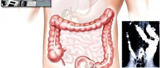
Diagnostics
The simplest methods for diagnosing a rectal tumor are finger examination and examination with a rectal speculum. The proctologist detects pathological formations on the mucous membrane, but the presence of a malignant process can be confirmed or refuted only with the help of laboratory and instrumental methods. During the examination, a biopsy of the pathological tissue is performed.
- Sigmoidoscopy. The examination is carried out by pumping air into the rectum to straighten the folds and carefully examine the mucous membrane using a rectoscope.
- Irrigoscopy. The rectum is filled with a contrast solution and examined using an X-ray machine.
- Ultrasound of the pelvic organs. The study allows you to detect damage to neighboring organs and lymph nodes.
- CT scan of the pelvic organs. The study is prescribed to clarify the X-ray and ultrasound data.
- Blood test for tumor markers.
- Histological analysis of a biopsy is necessary to establish malignancy and identify the type of cancer cells.
- Cytological analysis of biopsy material, mucus and pus taken from the rectum allows us to study in detail the structure of the cancer cell.
Attention!
You can receive free medical care at JSC “Medicine” (clinic of Academician Roitberg) under the program of State guarantees of compulsory medical insurance (Compulsory health insurance) and high-tech medical care.
To find out more, please call +7, or you can read more details here...
Treatment
The only effective treatment for rectal cancer is radical removal of the tumor. There are a number of techniques, the choice of the appropriate one depends on the stage.
| Stage | Treatment |
| 0 | polypectomy during colonoscopy or transanal excision of the tumor |
| I | resection, less often abdominoperineal extirpation of the rectum |
| II | resection or extirpation, possibly with chemotherapy after surgery |
| III | resection in combination with pre- and postoperative chemotherapy and radiation |
| IV | resection with chemotherapy, removal of metastases, pelvic evisceration |
Along with surgical treatment for rectal cancer, radiation and chemotherapy are used. Treatment tactics are selected for each patient individually. In some cases, preoperative radiation is recommended, the purpose of which is to destroy cancer cells and reduce the risk of recurrence. For some types of tumors (for example, squamous cell carcinoma), only chemoradiotherapy is effective. Chemotherapy can be prescribed both before and after surgery.
T4
T4 - the tumor infiltrates surrounding organs (vagina, prostate, seminal vesicles or bladder) and tissues. Patients with this stage require a long course of chemoradiotherapy and extensive surgery. For invasion of surrounding organs, all diagnostic methods show similar sensitivity: 70% for TRUS, 72% for CT and 74% for MR imaging. On the left is a T4 tumor with prostate invasion.
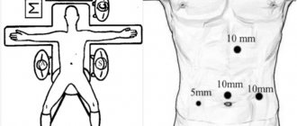
Surgery for rectal cancer
Oncological disease is treated by laparoscopic surgery
The goal of surgical treatment is to ensure the unimpeded passage of intestinal contents; for this purpose, the tumor is removed with part of the adductor and efferent intestines, if possible, with the creation of a primary anastomosis. Regional lymph nodes are also subject to removal.
One of the effective techniques is total mesorectal excision (TME), described in 1982 by the English surgeon Richard John Heald. It is based on the isolation of the rectum under visual control in the avascular zone - in the so-called correct layer - between the intestinal fascia proper (mesorectum) and the parietal fascia of the pelvis. The tissue located in the mesorectum contains lymph nodes, which, as a rule, are affected by metastases. When the rectum is isolated in the traditional way, part of the fiber is separated and remains in the small pelvis, which in 25% leads to the appearance of satellite metastases. TME excludes such a development of events. Based on this technique, intestinal resection is performed: anterior, low, abdominal-anal, abdominal-perineal extirpation.
TME is used today by many specialists, but the disadvantage of the method is the high rates of local relapses. I was able to reduce the recurrence rate from 45% to 5% by performing TME through a laparoscopic approach.
A. Primary Research
The radiological report must contain the following items:
- The location of the tumor is in the lower, middle or upper ampullary part of the rectum.
- Distance from the anorectal angle to the lower pole of the tumor.
- Tumor length.
- Circular or semi-circular. The position of the tumor is lateral, anterior or posterior wall.
- T-stages: T1/T2: tumor limited to the intestinal wall
- T3: perirectal tissue invasion, indicating the extent of invasion in mm.
- T4: Invasion of surrounding organs and tissues, indicating the organs affected.
- The shortest distance (in mm) between the tumor and the mesorectal fascia, indicating the location.
Advantages of the laparoscopic technique for total mesorectumectomy
- During the operation, there is a need to intersect nerve fibers, which can lead to urogenital disorders. To prevent such disorders, I use a nerve-sparing technique.
- In the presence of a widespread tumor or small-sized carcinoma, the operation is performed in several stages.
- To form the interintestinal anastomosis, I use modern staplers made in the USA, which helps to minimize the risk of developing postoperative complications, such as stricture or failure of the anastomotic suture.
- Using the LigaSure electrothermal tissue ligation device, ultrasonic scissors, and the Force Triad energy platform, mobilization of the colon is carried out quickly and bloodlessly, there is no need to use surgical clips and threads, and it is possible to preserve nerve fibers.
Considering the psychological state of patients when removing part of the intestine to the anterior abdominal wall, I strive, if possible, to perform organ-sparing operations while maintaining the continuity of the intestinal tube and rectal sphincter.
I have personally performed more than 300 laparoscopic interventions for diseases of the large intestine. The results of the operations are summarized in the monograph “Minimally invasive surgery of the colon.” Information about the surgical treatment methods I use can also be found in numerous peer-reviewed scientific publications.
MR protocol
T2 FSE images only, no contrast enhancement.
Only T2 FSE sequences are required. The use of gadolinium drugs does not improve diagnostic accuracy and, therefore, they are not included in the standard protocol for this pathology. Images are obtained in the sagittal, coronal and axial planes. It is necessary to start with the sagittal series; axial images are built along it, perpendicular to the walls of the rectum at the level of the tumor (blue lines). Coronal images are aligned perpendicular to the axial series (yellow line) through the distal portions of the tumor and parallel to the anal canal. This approach avoids partial volume artifact and allows for accurate assessment of the depth of tumor invasion, as well as assessment of tumor ingrowth into the anal canal. In addition, the surgeon’s direction to study the tumor level helps to correctly plan the MR sequence. The upper limit of the field of view (FOV) is L5, the caudal limit is below the level of the anal canal. A superficial coil is recommended; an endorectal coil is not necessary. There is no consensus on whether to use 1.5T or 3.0T systems. Diffusion-weighted images can be used to re-estimate the stage of a process. A high signal level on B1000 images indicates an incomplete response.
Angles
Axial images should be at the correct angle, perpendicular to the tumor axis, to avoid volume averaging. In the example on the left, the first planning of the axial images was performed at an incorrect angle, which gave the false impression of involvement of the mesorectal fascia (MRF, highlighted in red circle), when correctly changing the angle perpendicular to the axis of the tumor, it is clear that the mesorectal fascia (MRF) is not involved (yellow circle).
Do not use fat suppression programs or rectal contrast.
Fat signal suppression does not improve visualization of tumor boundaries. Patients do not require bowel preparation. The use of rectal contrast is not recommended because distension of the bowel wall may lead to false-positive results in the absence of invasion of the mesorectal fascia. In addition, it makes the assessment of distal mesorectal lymph nodes difficult. Description of the study
Prognosis for rectal cancer
If the tumor grows within the intestine itself for a long time, then in most patients, with timely treatment, the chances of a complete recovery are very high. The stage of the disease also affects survival in colorectal cancer:
- with 0-I, the five-year survival rate reaches 90%;
- in II – about 60-85% of patients recover;
- III - cure is possible in 25-60% of patients, the effect largely depends on properly prescribed treatment;
- Stage IV - has the most unfavorable prognosis, survival rate is only 7%.
Despite the availability of effective diagnostic methods, most cancer cases are diagnosed late due to a person's reluctance to see a doctor when the first symptoms appear, which reduces the chances of a full recovery.