The appearance of fatty acids in the stool indicates the development of a disease called steatorrhea. When the patient defecates, there are clearly visible parts of fat. The patient experiences frequent urges to go to the toilet. The stool is greyish in color and quite abundant.
The patient often suffers from diarrhea, less often constipation. Whatever the consistency of the feces, an oily residue remains on the surface of the toilet after bowel movements, which is quite difficult to wash off. Fatty acids in stool are the main symptom of steatorrhea.
General stool analysis - information about the study
A coprogram is a laboratory diagnostic method that includes the study of the physical and chemical properties of feces, as well as its microscopic examination. The results of a general stool analysis allow the doctor to obtain detailed information about the health of the gastrointestinal tract (GIT) and the functioning of digestion. The coprogram is used to diagnose diseases of the intestines, stomach, pancreas, and liver. For the study, stool collected after a morning bowel movement is used. A general stool analysis is carried out in the laboratory within 6 working days.
Basic therapy
In order for the doctor to prescribe the correct treatment, it is necessary to find out which disease caused the disturbances in the digestive process. In most cases, children are diagnosed with pancreatic steatorrhea, which occurs as a result of a lack of lipase, so the basis of treatment in this case will be drugs containing digestive enzymes, for example:
- "Creon";
- "Festal";
- "Pancreatin";
- "Mezim."
Steatorrhea can be cured only after the root cause of the disease has been completely eliminated. The predominant method of treatment is medication. The doctor prescribes lipase drugs in high concentrations. They will help restore fat absorption and improve the functioning of the intestinal tract. Enzyme preparations are prescribed to restore good digestion. After diagnosing steatorrhea, doctors often suggest the use of antacids. In particularly difficult cases, these drugs are also accompanied by: adrenocorticotropic hormone, vitamin complexes, hydrochloric acid.
Table. Antacids for complex therapy of steatorrhea.
| Drug name | How to use | At what age should it be given? |
| 1-2 tablets up to 3 times a day (tablets should be dissolved until completely dissolved). | From 12 years old | |
| 1-2 packets 1.5 hours after meals. The maximum duration of treatment is 3 months. | From 15 years old | |
| 10-20 ml after meals and before bed. | From 12 years old | |
| The dosage regimen is selected individually. | From 10 years of age (can be used by children under 10 years of age, but strictly as prescribed by a doctor and under his supervision) | |
| 1-2 tablets 2 to 6 times a day (the exact dose depends on the age of the child). | From 6 years old |
Auxiliary therapy may include vitamin preparations and hormonal medications, but they should only be prescribed by a specialist after a thorough study of the clinical picture of the disease.
Indications for analysis
A coprogram can be prescribed by different specialists: therapist, pediatrician, gastroenterologist, allergist, infectious disease specialist, surgeon, neonatologist.
A general stool analysis is indicated in the following cases:
- Suspicion of any diseases of the gastrointestinal tract. The coprogram is prescribed to identify the causes of prolonged diarrhea, blood in the stool, increased gas formation, nausea and vomiting, loss of appetite, frequent bloating, cramps and abdominal pain.
- Monitoring of already diagnosed diseases of the gastrointestinal tract and evaluation of the results of therapy.
- Screening for colon cancer by testing for occult blood.
- Suspicion of parasitic diseases. The coprogram allows you to find eggs of helminths (pinworms, roundworms) or parasites directly (giardia) in the stool.
- Intestinal infections. Using a coprogram, you can identify the causative agent of infection (bacteria, virus), which in some cases is necessary for the doctor to select the correct medications.
Different specialists can prescribe a coprogram to clarify the diagnosis and monitor treatment. Photo: lenblr / freepik.com
Complications and consequences
Complications develop only if treatment is not started in a timely manner. There is a disruption in the absorption of nutrients in the intestinal tract. Against this background, hypo- and avitaminosis, protein deficiency and exhaustion of the body develop. Pathology of water and electrolyte balance is manifested by a constant feeling of thirst, edema, dehydration, and convulsive attacks.
The specialist diagnoses the appearance of oxaluria (excessive pathological excretion of oxalic acid salts from the body along with urine) and the formation of urinary stones of oxalate origin. This pathological condition occurs because, under normal conditions, oxalates do not enter the blood from the intestinal tract, since their combination with calcium makes them insoluble. With the development of steatorrhea, calcium is excreted in large quantities from the body along with feces. This leads to a significant release of oxalates from the intestines into the blood.
Preparing for analysis
Before collecting material for a coprogram, preliminary preparation is recommended. 7-10 days before donating stool, it is necessary to stop taking medications, especially medications that affect the digestive system (enzymes, laxatives, antidiarrheals, antacids, anthelmintics, bismuth and iron preparations, castor and petroleum jelly, antibiotics, anti-inflammatory drugs).
For two days before collecting stool, you should not use rectal suppositories or perform enemas, or conduct an X-ray examination of the gastrointestinal tract. Feces should not be collected for analysis during menstruation, or if the patient has bleeding hemorrhoids or anal fissures.
Diet before taking the test
Depending on the purpose of the analysis and the patient’s state of health, the following diet may be recommended 3-4 days before the coprogram:
- Gentle diet (Schmidt diet). The daily diet of average calorie content (1700-2500 kcal) is divided into 5 meals. It includes milk, boiled chicken eggs, white bread, minced meat, mashed potatoes and a slimy broth.
- Nutrition with maximum food load (Pevzner diet). The diet represents the normal diet of healthy people. It includes white and black bread, fried meat, butter, sugar, fried potatoes, cereals, and fresh fruit. This diet makes it possible to identify even minor disturbances in the digestive and evacuation capacity of the gastrointestinal tract.
If it is necessary to test stool for occult blood, foods that may cause a false positive reaction for blood are excluded from the diet. These include meat, fish, all types of green vegetables, tomatoes, chicken eggs.
Treatment
Help before diagnosis
The first step in treating a patient with creatorrhoea is changing the diet. The goal of a therapeutic diet is to reduce the amount of protein products, which reduces the functional load of the digestive glands. The degree of dietary restrictions is determined by the severity of symptoms. The daily diet is selected in such a way as to provide the patient with signs of creatorrhoea with all the necessary vitamins and microelements.
Conservative therapy
To improve digestion processes and eliminate creatorrhea, enzyme preparations are selected that contain basic pancreatic enzymes. Medicines increase the digestion of protein and other types of food, normalize the frequency and consistency of stools. For low acidity, gastric juice and acidin-pepsin are prescribed. In addition to replacement therapy for creatorrhoea, etiopathogenetic and symptomatic treatment is carried out, which includes the following drugs:
- Gastroprotectors
. The effect of drugs for creatorrhoea is aimed at increasing the amount of parietal mucus and protective factors of the stomach. They are used for chronic gastritis to reduce the effect of negative external factors. - Painkillers
. Analgesics are indicated for exacerbation of chronic pancreatitis, organic pancreatic pathology, accompanied by creatorrhea. To enhance the analgesic effect, they are combined with antispasmodics. - Antidiarrheals
. The drugs are taken for a combination of creatorrhea and chronic diarrhea caused by short bowel syndrome or inflammatory bowel diseases. They normalize gastrointestinal motility, slow down the passage of food, thereby improving digestion.
During the period of remission for diseases associated with creative rheumatoid arthritis, physiotherapeutic treatment is actively used. Ozocerite and paraffin applications to the area of the anterior abdominal wall, inductothermy, and electrophoresis with medicinal substances are effective. To improve digestion processes and eliminate creatorrhoea, drinking specially selected mineral waters is effective. For general health purposes, sanatorium-resort treatment is recommended.
How to properly collect stool for research?
To carry out a coprogram, feces are collected only after spontaneous bowel movement (not after an enema or suppository).
It is recommended to urinate before collecting material to prevent urine from getting into the stool. Then you need to toilet the external genitalia and anal area. It is advisable to wash yourself with warm boiled water; for the anus area, you can use mild soap, which then needs to be rinsed thoroughly.
Defecation is carried out in a clean and dry container (not in the toilet). To collect feces, you will need a special container with a spoon built into the lid, which is used to remove feces from the container. For most tests, 10-15 g of stool (about 1 teaspoon by volume) is needed. The collected material is placed in a container and tightly closed with a lid.
Container for collecting feces. Photo: jora_abramov / freepik.com
Research standards
In adults and older children, the norms for general stool analysis are the same.
Table 1. Reference values of coprogram in adults and older children
| Index | Normal value |
| Macroscopic examination | |
| Consistency | dense |
| Form | decorated |
| Color | brown |
| Smell | fecal, not sharp |
| Slime | absent |
| Blood | absent |
| Leftover undigested food | none |
| Chemical research | |
| Acidity (pH) | 6,8—7,6 |
| Reaction to occult blood | negative |
| Reaction to protein | negative |
| Reaction to stercobilin | positive |
| Reaction to bilirubin | negative |
| Microscopic examination | |
| Detritus | insignificant amount |
| Muscle fibers with striations (undigested) | none |
| Muscle fibers without striations (digested) | units in the preparation |
| Connective tissue fibers | none |
| Fat neutral | absent |
| Fatty acid | absent |
| Salts of fatty acids (soaps) | insignificant amount |
| Intracellular starch | absent |
| Extracellular starch | absent |
| Iodophilic flora is normal | units in the preparation |
| Pathological iodophilic flora | absent |
| Crystals | none |
| Slime | absent |
| Columnar epithelium | absent |
| Epithelium is flat | absent |
| Leukocytes | none |
| Red blood cells | none |
| Protozoa | none |
| Worm eggs | none |
| Yeast mushrooms | none |
In young children, normal stool test values differ depending on the type of feeding.
Table 2. Reference values of coprogram in young children
| Index | Normal value in breastfed children | Normal value in formula-fed children |
| Macroscopic examination | ||
| Consistency | sticky, viscous (mushy) | putty-like consistency |
| Color | yellow, golden yellow, yellow green | yellow-brown |
| Smell | sourish | putrefactive |
| Slime | absent or in small quantities | absent |
| Blood | absent | absent |
| Leftover undigested food | none | none |
| Chemical research | ||
| Acidity (pH) | 4,8-5,8 | 6,8-7,5 |
| Reaction to occult blood | negative | negative |
| Reaction to protein | negative | negative |
| Reaction to stercobilin | positive | positive |
| Reaction to bilirubin | positive, negative after 9 months | positive, negative after 9 months |
| Microscopic examination | ||
| Detritus | insignificant amount | insignificant amount |
| Muscle fibers | small quantity or absent | small quantity or absent |
| Connective tissue fibers | none | none |
| Fat neutral | drops | a small amount of |
| Fatty acid | crystals in small quantities | crystals in small quantities |
| Salts of fatty acids (soaps) | in small quantities | in small quantities |
| Starch | absent | absent |
| Pathological iodophilic flora | absent | absent |
| Columnar epithelium | absent | absent |
| Epithelium is flat | absent | absent |
| Leukocytes | single in the preparation | single in the preparation |
| Red blood cells | none | absent |
| Protozoa | none | absent |
| Worm eggs | none | absent |
| Yeast mushrooms | none | absent |
References
- Azer, S., Sankararaman, S. Steatorrhea. Treasure Island (FL): StatPearls Publishing, 2022.
- Bijoor, A., Geetha, S., Venkatesh, T. Faecal fat content in healthy adults by the 'acid steatocrit method'. Indian J Clin Biochem., 2004. - Vol. 19(2). - P. 20-2.
- Kamath, M., Pai, C., Kamath, A. et al. Comparing acid steatocrit and faecal elastase estimations for use in M-ANNHEIM staging for pancreatitis. World J Gastroenterol. — 2022. — Vol. 23(12). - P. 2217-2222.
Decoding indicators
Indicators are macroscopic, microscopic and chemical. Let's look at them separately.
Macroscopic
The consistency, color, shape of feces, as well as the presence of impurities are studied.
Consistency and shape of stool
Normal stool contains about 75% water and has a dense consistency and cylindrical shape. Such feces are called formed⁴.
Changes in the shape of feces can occur as a result of eating different foods or be a consequence of diseases of the digestive system. There are the following types of violation of the consistency of stool:
1. Hard consistency of stool. The water content in solid feces is 50-60%. This form occurs due to the slow movement of the bolus of food through the colon, as a result of which too much water is absorbed. If spasms of the colon are added to slow peristalsis, the stool becomes fragmented and looks like dense balls. Such feces occur with constipation, stenosis and spasms of the colon.
2. Pasty consistency of stool. This consistency may be the result of large consumption of plant foods, which enhance intestinal motility. Also, some intestinal diseases - colitis and fermentative dyspepsia - lead to mushy feces.
Important!
How soon should the material be taken to the laboratory?
It is recommended to donate feces as soon as possible after collection.
If it is not possible to immediately take the material to the laboratory, it can be stored in a tightly closed container in the refrigerator for no more than 8 hours³. If a coprogram is performed to identify helminths and protozoa, stool should never be stored; it must be delivered to the laboratory warm immediately after defecation. 3. Fatty or ointment-like consistency of stool - steatorrhea. It occurs due to an increased content of undigested fat in the feces, which occurs as a result of impaired breakdown and digestion of fats. Most often, steatorrhea indicates a malfunction of the pancreas (pancreatitis). It can also be a consequence of diseases of the liver and biliary tract (cholecystitis, cholelithiasis) or malabsorption in the intestine.
4. Ribbon-shaped stool. Such feces occur with prolonged spasms and stenoses of the sigmoid and rectum.
5. Diarrhea. This is liquid, unformed feces with a high water content. There are several types of diarrhea:
- Osmotic. It occurs as a result of impaired absorption of osmotically active substances (proteins, fats, carbohydrates), due to which water is also retained in the intestinal lumen. Such diarrhea appears in diseases accompanied by impaired digestion and absorption (pancreatitis, Crohn's disease, etc.).
- Secretory. The cause of secretory diarrhea is the increased secretion of inflammatory exudate or mucus by the intestinal mucosa. Characteristic of inflammatory diseases of the gastrointestinal tract: enteritis and colitis.
- Motor. Occurs when increased intestinal motility, when a bolus of food quickly moves through the gastrointestinal tract and water absorption is impaired.
- Mixed. Occurs as a result of a combination of the above reasons.
Examination of stool in the laboratory for the presence of parasites. Photo: p.thongdumhyu / Depositphotos
Stool color
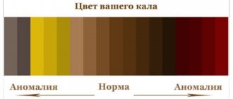
The normal color of stool is brown. The color of stool is due to the content of stercobilin. This is a pigment formed in feces from bilirubin bile.
The color of stool can normally change depending on the nature of the diet and when taking certain medications. The more plant foods you eat, the lighter brown your stool will be. A large amount of meat in the diet leads to black-brown stools. The color of feces when taking bismuth preparations becomes black, iron - black with a greenish tint.
Changes in stool color become important in the diagnosis of certain diseases:
- Grayish-white (acholic). White stool means it does not contain stercobilin. This may indicate both a violation of bile metabolism and a violation of its flow into the intestines. Such stools occur with hepatitis, acute pancreatitis, cholelithiasis (obstructive jaundice). Acholic stool in children in the first months of life may indicate malformations of the biliary tract.
- Red. Feces become red due to the admixture of blood during bleeding from the colon and rectum, hemorrhoids, and anal fissures. Blood may also be seen as speckles in the stool.
- Black stool with a runny consistency is called melena, or tarry stool. It appears when there is bleeding from the esophagus, stomach and upper small intestine. The black color is due to the fact that the blood reacts with the hydrochloric acid of the stomach, resulting in the formation of a black pigment - hematin hydrochloride. Most often this is caused by peptic ulcer of the stomach and duodenum.
Stool color in children of the first year of life
The first stool of a newborn baby is meconium.
It has a characteristic greenish-black color and sticky texture. This is how the child defecates for the first 1-2 days of life. On the third to fifth day after birth, yellow streaks appear in the stool. By the end of the first week, the stool becomes completely yellow. Breastfed babies' stool color ranges from light yellow to brownish yellow. A formula-fed baby's stool may be tan, tan, or green-brown.
In some infants, stool may normally be greenish in color in the first 3 months of life. This happens when the child’s digestive system has not yet converted bile bilirubin and stercobilin. Normally, by the fourth month of life, healthy microflora appears in the intestines, which completely converts bilirubin into stercobilin, so from this age the feces should not be green².
The color of a baby's stool may change at 6 months due to the start of complementary feeding. The less breast milk or formula your baby consumes, the darker the color of the stool. Provided that complementary foods are introduced in a timely manner and a varied diet, by the age of one year the stool character approaches that of an adult and the color of the stool becomes brown.
Smell
A normal, non-pungent fecal odor is formed due to the products of bacterial breakdown of proteins. With a large amount of meat in the diet, the smell of stool may increase; with a vegetarian diet, it may weaken or disappear.
A sharp, foul smell of stool indicates a violation of food digestion and rotting proteins in the intestines. Putrefactive dyspepsia leads to this. Stool may also have a sour odor, which is the result of increased fatty acid content in fermentative dyspepsia.
Impurities
Normally, stool should not contain food debris, visible impurities of blood, mucus, or pus.
- Lumps of undigested food may be present in the stool due to insufficiency of the enzymatic function of the pancreas or accelerated movement of the food bolus through the intestines.
- Mucus in the stool is a symptom of inflammation in the intestines. Mucus can be either mixed with feces or separate from them. Mucus in the stool occurs in the following diseases: intestinal infections, irritable bowel syndrome (IBS), malabsorption syndrome, lactase deficiency, celiac disease.
Breastfed infants may have small amounts of mucus in their stool due to the immaturity of the digestive system and the inability to digest all the fats in breast milk¹.
- Visible blood in the stool is a sign of bleeding in the gastrointestinal tract.
- An admixture of pus in the stool indicates an extremely severe inflammatory process in the gastrointestinal tract (ulcerative colitis, dysentery).
A scatological examination of stool in children under 1 year of age may detect bilirubin. This is the norm. Photo: anna_grant / freepik.com
Chemical research
A chemical study determines acidity and also reveals the presence of proteins, pigments and enzymes in the feces.
Acidity (pH)
Determining the acidity of stool is important for diagnosing diseases. The stool reaction is caused by the activity of bacteria inhabiting the gastrointestinal tract. Normally it is neutral or slightly alkaline (pH 6.8-7.6)¹.
Changes in stool reaction can be of four types:
- Strongly acidic reaction (pH less than 5.5). This fecal reaction is characteristic of fermentative dyspepsia. With this disease, the intestinal microflora is overly activated, which leads to increased formation of carbon dioxide and organic acids (fermentation) in the lumen of the gastrointestinal tract.
- Acidic reaction (pH 5.5-6.7). This stool pH occurs due to malabsorption of fatty acids.
- Alkaline reaction (pH 8.0-8.5). An alkaline reaction of feces is observed when food proteins that are not digested in the stomach and small intestine rot. Occurs when pancreatic secretion is impaired.
- Sharply alkaline reaction (pH more than 8.5). Characteristic of putrefactive dyspepsia (colitis).
Protein determination
Normally, there is no protein in the stool. A positive reaction indicates the presence of inflammatory exudate, mucus, bleeding, and undigested protein food. Protein in stool can be a sign of diseases:
- stomach (gastritis, gastric ulcer, cancer);
- duodenum (duodenitis, duodenal ulcer);
- small intestine (enteritis, celiac disease);
- colon (colitis, polyposis, cancer, increased secretory function of the colon);
- rectum (hemorrhoids, anal fissure, proctitis, rectal cancer).
Blood determination
Normally, blood in the stool should not be detected. A positive test indicates bleeding from any part of the digestive system. Blood in the stool appears due to inflammatory processes in the mucous membrane of the gastrointestinal tract, malignant neoplasms, varicose veins of the esophagus and rectum.
hidden blood
The reaction to occult blood in the stool is often a false positive. This may be due to poor preparation for the study and non-compliance with a vegetarian diet, taking iron supplements, or injury to the gums when brushing your teeth. In some cases, for an accurate diagnosis it is necessary to perform the study several times⁴.
Determination of stercobilin
Stercobilin is a pigment that should normally be present in feces. Its normal value is considered to be 40-350 mg per 100 g of feces. The absence or decrease in the amount of stercobilin in the feces leads to light-colored stools. This happens with blockage of the biliary tract, cholangitis, hepatitis, and acute pancreatitis. An increase in stercobilin content in stool indicates hemolytic anemia.
Determination of bilirubin
In a healthy adult, bilirubin is not detectable in the stool. Bilirubin can be present in the stool in children up to 9 months of age¹; this is normal.
Microscopic examination
Microscopic examination of stool allows you to determine the remains of undigested food, cellular elements of the blood, cells of the gastrointestinal mucosa (epithelium, tumor cells), as well as parts of parasites living in the intestines.
Microscopic examination of stool. Photo: Dr. Dwight F. Miller/NASA Spinoff
Normally, stool microscopy reveals:
- detritus - small particles of various sizes that are considered unrecognizable particles of food, intestinal epithelial cells and bacteria;
- digested muscle fibers;
- connective tissue fibers: plant fiber, remains of cartilage and tendons from food;
- elements of digested plant fiber.
Pathological findings on stool microscopy are:
1. Undigested muscle fibers. These elements indicate digestive disorders, which may be the result of insufficiency of the secretory function of the pancreas (pancreatitis) or accelerated intestinal motility (for example, with enteritis).
2. Starch. Starch grains are most often found in stool during diarrhea.
3. Fat and its breakdown products (fatty acids, soaps, neutral fat) are detected in the following cases:
- insufficiency of the secretory function of the pancreas (pancreatitis);
- insufficient flow of bile into the intestines (hepatitis, cholecystitis, cholangitis, cholelithiasis);
- impaired absorption of fatty acids in the intestine.
4. Cellular elements. Microscopically you can detect:
- Epithelial cells of the gastrointestinal tract. Single epithelial cells can be found in the coprogram as a normal variant, but a large number of them indicates inflammation of the intestinal mucosa (enteritis, colitis).
- Leukocytes. Leukocytes in feces are a sign of inflammatory diseases of the gastrointestinal tract: colitis, enteritis, proctitis, paraproctitis, dysentery, helminthic infestation, nonspecific ulcerative colitis.
- Red blood cells. Red blood cells in the stool indicate bleeding from the colon.
- Malignant tumor cells. This is a very rare finding in general stool analysis, even in cases of severe clinical tumors.
5. Causative agents of intestinal diseases: helminth eggs (pinworms, roundworms), protozoa, yeast fungi, pathological bacteria. Normally, these microorganisms are not found in stool.
Is it possible to recognize steatorrhea on your own?
The clinical symptoms of the pathology are quite specific, so you can suspect an increased content of neutral fat in the feces on your own. The main signs are associated with a change in the consistency of feces - they become sticky, oily, shiny, and can stain underwear. Such feces are difficult to wash off from the surface, sometimes light yellow spots remain on the laundry after washing. The color of the stool may remain the same, but in most cases the stool will lighten and become greyish. There is no strong odor with steatorrhea - on the contrary, such feces usually smell weakly or not at all.
Severe dyspeptic symptoms with steatorrhea are usually absent, but sometimes patients complain of bloating, a feeling of heaviness and fullness in the abdomen, dry mucous membranes of the oral cavity, and appetite disorders. If it lasts for a long time, the child may experience other signs not related to the functioning of the digestive tract. It can be:
- frequent headaches that turn into dizziness (in school-age children this is manifested by a decrease in academic performance and difficulties in mastering the material);
- joint pain (mainly in the back and lower back);
- bleeding gums;
- weakness and drowsiness.
In children of the first year of life, steatorrhea is manifested by increased excitability, frequent bowel movements, refusal of the breast or bottle, and poor weight gain. Feces are oily, difficult to remove from diapers and nappies, and have a stronger odor compared to children of the older age group.
One of the manifestations of pancreatic or hepatic steatorrhea may be a cough, which externally manifests itself as infrequent coughing. The cough is usually dry, not painful, goes away easily after drinking liquid, but can last up to 2-3 months. If a child suddenly begins to cough, but there is no wheezing in the lungs, and overall health remains satisfactory, a stool test should be taken for a coprogram to check the functioning of the digestive system.


