Stagnation of bile (cholestasis) is a condition in which the flow of bile into the duodenum is impaired. In this case, pronounced disturbances in the breakdown and absorption of fats develop, which leads to an increase in their concentration in the bloodstream. This entails changes in the transformation of glucose into glycogen, as well as an increase in the amount of cholesterol in the blood. Thus, stagnation of bile, left untreated, can lead to the development of diabetes mellitus, atherosclerosis and their complications. Cholestasis can develop in the bile ducts (intrahepatic stasis) and in the gallbladder (extrahepatic), and can also be acute or chronic.
Causes of bile stagnation
Cholestasis can develop for many reasons, the most common of which are:
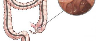
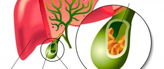
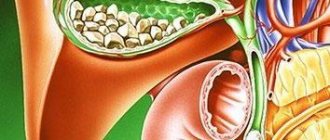
- obstruction of the gallbladder or bile ducts due to the presence of stones in them, as well as when they are compressed by a cyst or tumors;
- abnormalities in the structure of the gallbladder or ducts, when they are bent;
- inflammation of the gallbladder or ducts;
- dysfunction of the valves responsible for the free movement of bile;
- endocrine and enzymatic disorders, in which thickening of bile and changes in its chemical properties are observed.
Diagnosis
Stagnation of bile is characterized by changes in the color of the sclera and skin (yellowing), as well as complaints of itching, unstable stools (alternating constipation and diarrhea), discolored feces and dark urine. If there are even several of these symptoms, the doctor has reason to prescribe a biochemical blood test (the level of bilirubin, bile acids, liver enzymes, etc. is examined), a urine test for urobilin, ultrasound of the liver and gallbladder, fibrogastroduodenoscopy, etc. When diagnosing cholestasis, it is important to distinguish between these condition from viral, parasitic and other liver diseases that have a similar clinical picture.
Cholestasis syndrome
In the peripheral blood, target-like red blood cells, anemia, and neutrophilic leukocytosis are detected. Within 3 weeks, the content of conjugated bilirubin in the blood serum increases. Biochemical markers of cholestasis are alkaline phosphatase and uglutamyl transpeptidase, leucine aminopeptidase and 5-nucleotidase. In chronic cholestasis, the level of cholesterol lipids, phospholipids, triglycerides, and lipoproteins increases, mainly due to the low-density lipoprotein fraction. At the same time, the concentration of high-density lipoproteins is reduced. The serum contents of chenodeoxycholic, lithocholic and deoxycholic bile acids are increased. The level of albumin and globulin does not change in acute cholestasis. The activity of AST and ALT increases slightly. Bile pigments and urobilin are found in the urine.
Morphologically, the liver with cholestasis is enlarged in size, greenish in color, with a rounded edge. In later stages, nodes are visible on its surface. Light microscopy reveals 6-irubinostasis in hepatocytes, sinusoid cells, and tubules of the third zone of the lobule. “Cirrus” degeneration of the hepatocyte, foam cells surrounded by mononuclear cells, are revealed. Necrosis of hepatocytes, regeneration and nodular hyperplasia in the initial stages of cholestasis are minimally expressed. In the portal tracts (first zone), proliferation of ductules and the presence of bile thrombi are observed; hepatocytes turn into bile duct cells and form the asal membrane. Obstruction of the bile ducts is promoted by the development of fibrosis. Mallory bodies may form in cholestasis. The microvasculature of the liver and its cellular elements undergo reactive changes. Swelling of the cells lining the sinusoids, their dystrophic changes, and the presence of vacuoles containing bile components or their metabolites are observed. By electron microscopy, changes in bile canaliculi are nonspecific and include dilatation, edema, thickening and tortuosity, loss of microvilli, vacuolization of the Golgi apparatus, and hypertrophy of the endoplasmic reticulum. In the liver (hepatocytes, Kupffer cells, bile duct epithelium) there is excessive deposition of copper and metalloproteins, lipofuscin, cholesterol, and other lipids. Changes in liver biopsy in the early stages of cholestasis may be absent.
In the early stages of cholestasis, the liver is not microscopically changed, in the later stages it increases in size and has a greenish color. Microscopic signs of cholestasis in the liver are clumps of bilirubin in the cytoplasm of hepatocytes and lumps of bile (bile thrombi) in the lumens of dilated bile canaliculi. Rupture of the bile canaliculi leads to the release of bile into the intercellular space with the formation of “bile lakes”. Morphological signs of cholestasis are usually more pronounced in the central zones of the hepatic lobule. With long-term disorders of biliary excretion, these changes are visible in the intermediary and then periportal zones. As already noted, there are three forms of cholestasis: intracellular, intratubular and mixed. In the early stages, one or another form of cholestasis is rarely expressed. Intracellular cholestasis is observed with drug (aminosine ) damage, intratubular - with subhepatic jaundice, mixed - with viral liver damage. Coagulation of bile in the interlobular bile ducts is detected only in sectional preparations.
Hydropic and acidophilic dystrophy in the liver is observed already on the 7th day. In rare cases, the cytoplasm of hepatocytes, located around thrombosed bile canaliculi that do not accept dyes well, appears reticulated and contains pigment granules - “pinnate” degeneration of hepatocytes. Progressive dystrophy leads to necrotic changes in the parenchyma.
The following types of necrosis in cholestasis are distinguished:
- focal necrosis of hepatocytes (sensitivity to staining is reduced, the nucleus disappears, hepatocytes are replaced by leukocytes);
- necrobiosis of a group of hepatocytes in a state of “pinnate” degeneration ends in biliary or reticular (mesh) necrosis;
- centrilobular zonal necrosis of hepatocytes (usually in sectional preparations).
Alteration of the parenchyma is caused by the toxic effects of bile components, as well as mechanical pressure from dilated thrombosed bile canaliculi. Stagnation of bile and necrobiosis of hepatocytes are accompanied by inflammatory mesenchymal cell reactions (they occur no earlier than the 10th day of stagnation), then hyperplasia of reticulin fibers in the lobule and proliferation of connective tissue in the portal field occur - the beginning of the formation of biliary cirrhosis. Stagnation of bile is also accompanied by proliferation of cicholangioles. In the liver tissue, the content of glycogen and RNA is reduced, the amount of lipids is increased, there is a positive PAS action of glycoproteins, protein and its active groups, the activity of oxidoreductases is reduced and CP and ALP are increased. The lumen of the tubules is widened from 1 to 8 μm; there are no villi at the biliary pole of the hepatocyte, or they are shortened and take the form of a balloon or bubble. The ectoplasm of the pretubular zone of the hepatocyte is expanded, the Golgi apparatus is increased in size, and hyperplasia of the smooth ER is noted. The number of lysosomes is increased, they are randomly located in hepatocytes (not only in the peribiliary zone, but also at the vascular pole), and also extend into the space of Disse. Mitochondria show signs of dystrophic changes. The junction of cells in the area of the bile canaliculi appears intact. The ultrastructure of the altered liver is identical to intrahepatic and extrahepatic cholestasis. The existing differences are quantitative: with extrahepatic cholestasis they are more pronounced.
Bile thrombi consist of granular components (bile itself) and ring-plate formations of free bilirubin and have a coarse-grained structure localized in mesosomes. Bound bilirubin, having the form of small grains, is located in the vesicles of the EPS, and sometimes lies freely in the cytoplasm.
There are differences in the nature of damage to the intrahepatic bile ducts in various diseases. VH is characterized by the formation of catarrhal and obstructive cholangitis, PBC is characterized by destructive cholangitis, and subhepatic jaundice is characterized by pericholangitis.
Stagnation of bile in the liver is naturally accompanied by proliferation of cholangioles (ductular proliferation). Proliferating bile ducts may be no different from ordinary bile ducts. Sometimes proliferating bile ducts do not have a clear lumen and are formed by two rows of oval cells with an elongated nucleus and basophilic cytoplasm. A significant number of ducts in the portal field indicates their proliferation.
Proliferation of the bile ducts has an adaptive-compensatory significance and is aimed at correcting bile secretion. When the cause of bile stagnation is eliminated, the ductular reaction is reduced, and the portal triad is completely restored.
The results of clinical and biochemical studies do not always make it possible to distinguish between intrahepatic and extrahepatic cholestasis. The diagnostic examination algorithm is of great importance. Extrahepatic mechanical obstruction with the development of biliary hypertension is supported by pain in the abdominal cavity (observed when stones are localized in ducts, tumors), palpable gallbladder. Fever and chills may be symptoms of cholangitis. The density and tuberosity of the liver upon palpation reflect advanced changes or tumor damage to the liver. The diagnostic examination algorithm involves, first of all, performing an ultrasound of the abdominal organs, which makes it possible to identify a characteristic sign of a mechanical blockade of the bile ducts - suprastenotic dilatation of the bile ducts (the diameter of the common bile duct is more than 6 mm). When dilatation of the ducts is detected, it is advisable to conduct cholangiography. The procedure of choice is endoscopic retrograde cholangiography (ERCH). If retrograde filling of the bile ducts is impossible, percutaneous transhepatic cholangiography (PTCHG) is used. If there are no signs of extrahepatic obstruction of the bile ducts, a liver biopsy is performed. This procedure can be performed only after excluding obstructive extrahepatic cholestasis (to avoid the development of biliary peritonitis). Cholescintigraphy with technetium-labeled iminodiacetic acid also helps to identify the level of damage (intrahepatic or extrahepatic). The use of magnetic resonance cholangiography is promising.
Treatment
Treatment tactics are formed based on the examination results, but always involve eliminating the cause of this condition. If bile stagnation is caused by obstruction of the gallbladder or bile ducts, surgical intervention is indicated to restore the patency of the ducts. In other cases, treatment is carried out using a complex of drugs with choleretic, choleretic, hepatoprotective or other effects. The course of treatment is formed individually for each patient.
Prevention and prognosis
Among the preventive measures, the following should be highlighted:
- compliance with the dietary regime. Normalizing a nutritious diet allows you to restore bile flow and prevent stagnation;
- giving up alcohol;
- strict control of the dose and duration of taking hepatotoxic drugs;
- timely consultation with a doctor if dyspeptic disorders and distension in the liver area appear.
You also need to be careful when working with contaminated medical instruments. This applies to healthcare workers to avoid infection with hepatitis B and C viruses.
With timely diagnosis and treatment, it is possible to slow down the progression of pathology and normalize the functioning of the hepatobiliary tract. It is important to remember that diet therapy and preventive examinations make it possible not only to identify cholestasis at the initial stage, but also to support the functioning of the entire digestive system.
Medicines
Photo: womanjour.ru
The following groups of hepatoprotectors are distinguished:
- essential phospholipids (Essentiale Forte, Antraliv, Phosphogliv), which contain phosphatidylcholine and unsaturated fatty acids. The action of these drugs is to restore the cell wall of hepatocytes, which are largely composed of phospholipids. In addition, essential phospholipids enhance the detoxification function of the liver, prevent the appearance of connective tissue in the liver parenchyma, and also have antioxidant properties. To achieve the desired effect, these products are used for at least six months;
- amino acids (Heptral, Hepa-merz). They participate in the synthesis of phospholipids, which are a structural component of the cell wall of hepatocytes, and have a regenerating and detoxifying effect. It is recommended to use drugs in this group via intravenous administration, since when taken orally, amino acids are metabolized and reach the liver in small quantities;
- vitamins (B1 (thiamine), B6 (pyridoxine), B12 (cyanocobalamin), E (tocopherol)). Facilitate metabolic processes in the liver, thereby promoting the restoration of its cells. As a rule, they are not used as monotherapy, but complement other hepatoprotectors;
- inhibitors of lipid peroxidation (Tiogamma, Berlition). The action of the drugs is aimed at accelerating the removal of lactic acid from liver cells, which has a toxic effect. The formation of lactic acid occurs as a result of the combination of nitrogen with oxygen. These drugs can be prescribed either in the form of injections or in tablet form;
- ursodeoxycholic acid (Ursofalk). Stimulates the removal of bile from the liver, thereby achieving a hepatoprotective effect;
- combination drugs that contain components belonging to several groups of hepatoprotectors (Essel Forte, Phosphonciale, Rezalut Pro, Hepatrin, Sirepar).
In the treatment of cholestasis, preference is given to ursodeoxycholic acid drugs. Essential phospholipids are not used, since their long-term use can lead to stagnation of bile.
When a clinical picture of hypo- or avitaminosis develops, vitamin or vitamin-mineral complexes are prescribed, containing the amount of vitamins and minerals necessary for the normal functioning of the body. Currently, there are a large number of different names of these drugs on the pharmacological market. We offer to your attention the following:
- Revit – contains vitamins A, B1, B2 and C. Suitable for use by both adults and children;
- Duovit is a fairly common representative of vitamin and mineral complexes. Each package of the drug contains red capsules containing vitamins (A, E, C, D3, B1, B2, B5, B6, B12 and folic acid) and blue capsules containing microelements (calcium, phosphorus, magnesium, iron, copper , zinc, manganese, molybdenum). This division of capsules into colors greatly facilitates their use. The fact is that it is recommended to separate the intake of the vitamin and mineral complex on different days, so the created form in the form of blue and red capsules allows you not to get confused in the alternation;
- Teravit. Contains vitamins A, C, D3, B2, B1, E, PP, B5, B6, B12, as well as a number of microelements: calcium, iron, phosphorus, iodine, magnesium, zinc, selenium, copper, manganese, chromium, molybdenum and potassium . Prescribed for adults and children over 12 years of age.
- Supradin. It contains vitamins A, E, C, D2, B1, B2, B3, B5, B6, B9 and B12. The preparation contains the following microelements: calcium, magnesium, phosphorus, copper, iron, manganese, zinc, molybdenum.
As a rule, the duration of taking vitamin or vitamin-mineral complexes is 1 month; if necessary, the course can be extended.
Cholestyramine enhances the excretion of bile acids from the body and also reduces the absorption of cholesterol in the intestine. The drug is available in the form of a powder, which, according to the instructions, is diluted with water. One powder accounts for ½ cup of boiled water. It is important to note that no more than 10 minutes should pass after preparing the solution and before using it. The duration of the course of taking cholestyramine is at least 1 month.
Folk remedies
Photo: Ponchikov.net
As is known, in some cases surgical intervention is required to eliminate the cause of cholestasis. That is why it is strongly recommended not to self-medicate at home, but to immediately consult a specialist at the first symptoms of cholestasis. It is important to understand that the use of traditional medicine in this case will not only not provide any help, but can also cause harm. However, there are folk remedies that can be used for preventive purposes. We offer to your attention the following:
- take 20 g of pre-prepared rose hips and 10 g of crushed nettle leaves. Mix the listed ingredients and pour two glasses of boiling water over them, then place in a water bath and boil for 15 minutes. After which the resulting decoction should be infused for 1 hour, having previously wrapped the container containing the decoction. The drink becomes ready for use after careful straining. It is recommended to consume 50 ml throughout the day;
- Dry the birch leaves and chop them finely. Take 1 tablespoon of plant material and pour one glass of boiling water, then heat over low heat in a water bath for 30 minutes. Then the resulting decoction must be allowed to brew for half an hour, after which it should be strained. It is recommended to consume 1/3 glass 3 times a day 30 minutes before meals;
- mix chopped chicory root and dried peppermint leaves in equal proportions. To prepare the infusion, you will need 10 g of the resulting plant collection. Pour 10 g of the collection into glasses of boiling water and let it brew for 1 hour. After this, the resulting infusion should be filtered through a strainer. One glass of infusion is drunk during the day. The course of treatment varies from 1 week to 1 month;
- Grind 1 glass of flax seeds and pour them with three glasses of milk. Then place on low heat until approximately 1/3 of the original volume of liquid remains in the container. After this, the solution is carefully filtered through a strainer. It is recommended to drink one glass per day. The duration of use of this recipe is 10 days, after which a break is taken;
- take 3 drops of peppermint oil and one teaspoon of honey. Mix the listed components thoroughly. The resulting mass should be consumed 3 times a day. The duration of the course is 1 month;
- mix red beet juice with black radish juice in equal proportions. It is recommended to consume 1 glass per day, preferably in the morning. The duration of the course is 1 month.
The information is for reference only and is not a guide to action. Do not self-medicate. At the first symptoms of the disease, consult a doctor.
Description
Cholestasis is a condition characterized by a slowdown or complete cessation of bile secretion, which is accompanied by an increase in the content of substances secreted along with bile in the blood.
According to statistics, approximately 10 out of 100 thousand people experience cholestasis. It can also be noted that cholestasis is diagnosed in men somewhat more often than in women. In most cases, this pathology is observed in people over 40 years of age.
The most common causes of cholestasis include:
- damage to the liver parenchyma as a result of prolonged consumption of alcoholic beverages, taking medications that have a hepatotoxic effect;
- liver infection;
- congenital metabolic disorders, for example, galactosemia, cystic fibrosis and others;
- inflammatory damage to the bile ducts;
- cholelithiasis;
- acquired or congenital obstruction of the bile ducts;
- sarcoidosis;
- oncological disease of the hepatocellular system;
- hormonal changes in the body, for example, during pregnancy.
Cholestasis is usually divided into the following types:
- intrahepatic - occurs as a result of a decrease in the production of bile and its release into the bile capillaries;
- extrahepatic - in most cases indicates obstruction of the bile ducts.
According to the mechanism of development, the following types of cholestasis occur:
- partial – characterized by a decrease in the amount of bile produced;
- total - bile does not enter the duodenum;
- dissociative – characterized by a decrease in the body’s amount of substances necessary for the production of bile.
As a rule, the prognosis is quite favorable. In most cases, with cholestasis it is possible to achieve complete recovery or long-term remission. This is achieved through timely diagnosis and specialized treatment. In some cases, complications develop, in particular, liver failure may develop, which threatens to disrupt the normal functioning of other organ systems. It is also important to follow preventive measures, which include excluding spicy, fried foods and alcoholic beverages from the diet. In addition, it is necessary to promptly treat the pathology that causes the development of cholestasis.
Diet
Diet 5th table
- Efficacy: therapeutic effect after 14 days
- Duration: from 3 months or more
- Cost of products: 1200 - 1350 rubles per week
Nutrition plays a very important role in the treatment of choliatic syndrome. For this disease Diet Table No. 5 , which involves avoiding fried, fatty foods high in animal fats. It is very important to completely avoid caffeinated drinks and alcohol.
Meals should be light and include fruits, vegetables, and fresh juices. The number of calories consumed per day should be reduced.
The following choleretic products should be included in the menu:
- Low-fat milk.
- Vegetable oils.
- Salads from beets, radishes, radishes, tomatoes with vegetable oil.
- Cabbage – stewed and pickled.
- Oatmeal, corn porridge.
- Whole grain cereals and breads.
- Greens – spinach, dill, parsley, celery, lettuce, etc.
- Carrots, tomatoes.
- Berries and fruits.
- Water (up to 2 liters per day) with lemon juice.
- Rosehip decoction.
Proper nutrition will help prevent bile reflux and biliary reflux esophagitis .
General information
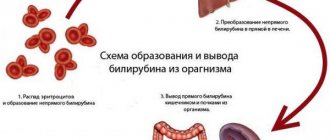
Cholestasis is a condition when the movement of bile from the liver slows down or stops completely. This condition is also defined as cholestatic syndrome . This occurs due to a violation of the formation of bile, its excretion or excretion, which is associated with a variety of pathological processes. The latter can be localized in any of the areas from the sinusoidal membranes of hepatocytes to the papilla of Vater. When the outflow of bile is disrupted, bilirubin , formed in the chord of breakdown of erythrocytes , enters the blood and gradually accumulates in it. This disorder leads to pain, discomfort, and other unpleasant symptoms and requires immediate treatment. Why this happens, what the symptoms of cholestasis are and how to properly treat it will be discussed in this article.