General information
Duodeno-gastric reflux (syn. reflux gastritis , biliary reflux , alkaline gastritis , biliary gastritis ) is a pathological process of retrograde flow of bile contents of the duodenal intestine into the stomach, which can be accompanied by clinical symptoms, histological signs and endoscopic changes of reactive (chemical) gastritis.
The term "refluxus" means "reverse flow". Under physiological conditions, bile from the duodenum should not enter the anatomically overlying parts of the digestive canal, therefore the bile reflex is considered a pathological phenomenon. DGR occurs as a result of excessive flow of bile from the duodenum due to insufficiency of the pylorus, which acts as a barrier to the retrograde flow of bile or due to a violation (reduction) of anterograde peristalsis of the stomach and duodenum. Modern scientific research indicates an increase in the number of diseases caused by the presence of pathological duodenogastric reflux in patients. At the same time, the high prevalence of GHD (in 46-52% of cases) and the frequent combination with chronic gastritis , functional dyspepsia , peptic , GERD , stomach cancer , Barrett's esophagus , sphincter of Oddi dysfunction , duodenostasis , postcholecystectomy syndrome , etc. complicates their course and therapy. GHD is quite common after surgery (in 16% of cases after cholecystectomy and in almost 55% of cases after surgery for duodenal ulcers).
In addition, duodenal reflux can cause the development of metaplasia of the esophageal epithelium, severe esophagitis and squamous cell carcinoma of the esophagus against the background of metaplasia. It should be noted that gastroduodenal reflux in its “pure” form (positioned as an isolated diagnosis) is relatively rare (10-15%) and is mainly diagnosed against the background of other diseases. That is, in most cases, GHD is a syndrome accompanying a number of diseases of the upper gastrointestinal tract.
GHD occurs due to excessive flow of bile from the duodenum due to insufficiency of the pylorus, which acts as a barrier to the retrograde flow of bile or due to a violation (reduction) of anterograde peristalsis of the stomach and duodenum.
Thus, duodenal pathological reflux complicates the course of various organic/functional diseases of the gastrointestinal tract, which necessitates its timely diagnosis, correct clinical interpretation and adequate drug correction.
Advantages of treatment of duodeno-gastric reflux at the Swiss University Hospital
- Our Center is equipped with modern equipment produced by leading European and American companies, which allows us to carry out the most complex diagnostic and therapeutic procedures with high accuracy and efficiency.
- The clinic employs specialists of the highest and first categories who have extensive experience; each of them is fluent in all techniques in their specialization.
- Our specialists have performed more than 600 surgical interventions related to duodenal obstruction.
- Patients who have concomitant diseases of the abdominal cavity and pelvis and require surgical treatment can undergo simultaneous surgery in our clinic. During one intervention, you can get rid of 3-4 pathologies (for example, kidney cysts, ovarian cysts, nephroptosis, fibroids and a number of other diseases).
Pathogenesis
The pathogenetic mechanisms of development of GDR are based on:
- failure of the sphincter apparatus, which allows the contents of the duodenal intestine to freely reach the stomach through the pyloric/lower esophageal sphincters;
- antroduodenal dysmotility (disorder of coordination between the pyloric/antral parts of the stomach and duodenal intestine), which leads to disruption of control over the direction of flow of duodenal contents;
- elimination of the antireflux barrier after surgery (partial gastrectomy ).
With the development of pathological GHD due to dysfunction of the sphincters, bile retrogradely, as part of the refluxate, enters from the duodenum into the higher located stomach. Components of duodenal contents, represented by bile acids, lysolecithin and trypsin , have an aggressive damaging effect on the gastric mucosa. Taurine conjugated bile acids and lysolecithin have the most pronounced effect, especially at acidic pH, which determines their synergy with hydrochloric acid in the development of gastritis . Trypsin and non-conjugated bile acids have a pronounced toxic effect at slightly alkaline and neutral pH, while the toxicity of non-conjugated bile acids is provided predominantly by ionized forms that can easily penetrate the coolant.
Long-term exposure of the coolant to bile acids contained in bile causes necrobiotic and dystrophic changes in the surface epithelium and leads to the condition of reflux gastritis (gastritis C).
In the presence of Helicobacter pylori, the damaging effect of the refluxant on the coolant is enhanced. The formation of DGR contributes to disruption of the motility of various parts of the gastrointestinal tract and the function of the sphincters, which leads to disruption of the digestive conveyor, has a negative effect on membrane/cavity digestion and absorption of food ingredients, and changes the water balance. The aggressive influence initially manifests itself in the form of increasing atrophy, dysplasia and metaplasia of the coolant, which form the risk of developing gastrocarcinogenesis. The gradual aggressive effect of bile with pancreatic juice contributes to the fact that superficial gastritis progresses and mucosal erosions transform into erosive and ulcerative lesions of the coolant.
Duodenum, 12 duodenum: treatment
The duodenum is an important organ of the digestive system and digestive behavior. The intestinal cavity is a reservoir into which digestive juices flow from the pancreas, liver and glands of the wall of the small intestine. Here, cavity digestion begins to take place, preparing the food bolus for the final breakdown of nutrients in the brush border of the cells of the duodenal mucosa. Between the microvilli, abundantly covered with enzymes, the bulk of the nutrients entering the human body are digested and absorbed at high speed. This digestion in the brush border is called parietal. The duodenum is richly innervated, especially in the initial section and in the area of confluence of the ducts from the gallbladder and pancreas. In the muscular lining of the intestine there are sensors for the rhythm of contractions of the entire small intestine.
The duodenum is also a hormonal organ. In the intestinal wall, under the influence of gastric contents, enterogastron is produced, which suppresses the secretion of gastric juice and relaxes the muscles of the stomach wall. Enterogastron prevents the digestive hormone gastrin from stimulating the activity of the gastric glands. Gastrin is produced by the pyloric zone of the stomach, which is close in origin and structure to the duodenum. The duodenum secretes pancreozymin, cholecystokinin, secretin and other hormones that regulate the activity of the pancreas and gallbladder, while simultaneously stopping gastric secretion. Digestive hormones from the duodenum stimulate the intestinal glands, which actively begin to secrete juice and stimulate intestinal motility. In the duodenum, hormones of general action are found that affect the body’s metabolism, endocrine, nervous, and cardiovascular systems.
Causes
The main reasons for the formation of DGR include:
- Congenital/acquired functional deficiency (weakening of the closing function) of the pyloric sphincter.
- Hyperkinetic type (with increased motility) of duodenal peristalsis.
- Inconsistency (discoordination) of the physiological cycles of relaxation/contraction of both the stomach and the DP-gut (migrating motor complex).
- Duodenal hypertension (increased pressure in the lumen of the duodenum), caused by splanchnoptosis (prolapse of internal organs), lumbar lordosis , hernias /malignant neoplasms.
- Long-term inflammation of the duodenum (chronic duodenitis , duodenal ulcer , gastroduodenitis ).
- Lack/absence of hormones ( gastrin ).
- Helminthic infestation (giardiasis).
- Anomalies in the development of the duodenal intestine.
Risk factors for developing duodenogastric reflux include:
- irregular food intake and poor quality nutrition (overeating, dry food, fatty and spicy foods that cause hypersecretion of bile);
- smoking and alcohol abuse;
- old age (after 60 years);
- long-term use of antispasmodics and NSAIDs;
- operations for resection of part of the stomach, cholecystectomy (removal of the gallbladder), anastomosis of the intestine and stomach;
- biliary dyskinesia , cholecystitis ;
- pancreatitis;
- diabetes mellitus , obesity .
Folk remedies
Treatment of GHD with folk remedies often gives the same positive effect as medication. In addition, the frequency of side effects during its implementation is significantly lower.
The most well-known folk remedies for the treatment of this disease are the following:
- Brew St. John's wort, chamomile and yarrow, taken in any proportion, with boiling water and add to tea. You need to drink the decoction 2 times a day. It will relieve heartburn, alleviate the symptoms of gastritis, minimize duodenogastric reflux, and eliminate dysbacteriosis;
- 1 tbsp. l. flaxseed, pour 100 ml of cool water, infuse until the seeds secrete mucus. Consume on an empty stomach;
- 2 tbsp. l. smoke herbs per 500 ml of boiling water. Leave for an hour, take 50 ml every 2 hours. Infusion of 2 tbsp. l. marshmallow roots in 500 ml of water, steeped for 5–6 hours, taken in small portions throughout the day. Using these folk remedies, you can prevent bilious vomiting;
- Rue leaves are effective folk remedies for improving intestinal motility. They need to be chewed after meals, 1–2 leaves;
- Mix 50 g of sage and calamus root with 25 g of angelica root; 1 tsp. mixture pour 1 tbsp. boiling water, stand for 20 minutes. Drink 1 hour after eating 3 times a day.
Symptoms
Symptoms of duodeno-gastric reflux are not specific. As a rule, the disease manifests itself with a predominance of dyspeptic symptoms - nausea, heartburn, air/sour belching, bitterness in the mouth and vomiting bile. Periodically appearing pain in the upper abdomen, intensifying after eating, is cramping in nature and can be provoked by stressful situations, physical activity, or appear after surgery for gastrectomy, cholecystectomy, and with developed duodenal obstruction. Since GHD in its “pure” form is rare and is diagnosed mainly against the background of other gastrointestinal diseases, especially gastroduodenal pathology, the clinical symptoms of reflux are influenced by the symptoms of the underlying disease, which to a certain extent masks the symptoms of GHD.
conclusions
1. The results of the study showed the following: GHD leads to inflammation, foveal hyperplasia, interstitial edema, fibroproliferation and ridge arborization.
2. H. pylori
leads to an increase in inflammation, manifested in the form of an increase in the activity and severity of inflammation, foveal hyperplasia, and the number of lymphoid follicles of mild and moderate severity.
3. GHD in children may have clinical manifestations in the form of pain and a painless course. With a pain syndrome, as with a painless course, changes in the morphological picture are noted equally, therefore it is incorrect to judge the severity of morphological changes by the severity of pain.
4. DGR leads to the development of certain morphological manifestations, which does not allow it to be regarded as a physiological process.
There is no conflict of interest.
Tests and diagnostics
The diagnosis is established on the basis of clinical symptoms and instrumental research methods, among which the most effective are:
- Daily pH-metry - allows you to assess the height of reflux and the profile of intragastric pH.
- Ultrasound diagnostics (echography with water load): with GHD, the retrograde movement of gas and liquid bubbles (corresponding to the reflux of duodenal contents into the stomach) from the pylorus to the body of the stomach is periodically recorded on echograms.
- Transillumination hemomotodynamic monitoring. The difference in motor wave amplitude in the antrum of the stomach and the duodenal bulb is used as a GDR parameter.
- Fibrogastroduodenoscopy (swelling of the coolant, focal hyperemia, pyloric dehiscence).
- X-ray of the stomach (regurgitation of barium from the duodenum into the stomach is noted).
- Fiberoptic spectrophotometry.
If bile reflux is suspected, differential diagnosis is made with acid gastroesophageal reflux and peptic ulcers of the stomach.
Etiology: a new approach
The prevailing theory (since 1935) is that GERD occurs when acidic stomach acid spills from the stomach into the esophagus, chemically and mechanically damaging the lining of the esophagus, causing burns, irritation, erosions, and ultimately more serious consequences. However, this traditional theory of chemical and mechanical irritation of the esophageal mucosa cannot fully explain many things associated with the onset, symptoms and course of GERD.
There are now reports that GERD may be an immune-mediated inflammatory disease caused by immune reactions rather than direct chemical damage to the lining of the esophagus by gastric juices. The hypothesis about the etiology of immune GERD is confirmed by one of the clinical studies conducted in the USA. The study results were published in the Journal of the American Medical Association.
Preliminary data from this study showed that T-cell esophagitis, basal cell hyperplasia, and spleen cell hyperplasia were observed in patients with severe GERD treated effectively with proton pump inhibitors (PPIs) after PPI discontinuation, but with resistant superficial cells.
According to study leader at the Dallas Veterans Medical Center, Dr. Kerry Dunbar, This finding suggests that the pathogenesis of reflux disease may be related more to inflammatory mediators and cytokines than to chemical damage to the esophageal mucosa.
The inflammatory immune theory of GEB could more easily and better explain not only the onset and course of the typical symptoms of this disease, but also the pathophysiology of the complications of this pathology - metaplasia of the esophagus and Barrett's mucosa.
Barrett's esophagus
Recent experimental studies in rats also suggest that GERD is more related to immune rather than chemical acid damage to the esophageal mucosa. Reflux and chemical acid irritation are thought to only initiate immune inflammatory responses in the esophageal mucosa and therefore play a less important role.
If the immuno-inflammatory theory of the etiopathogenesis of GERD turns out to be correct, it may be necessary to reconsider the existing regimen for the treatment and prevention of relapses of GERD. It is possible that the location and role of antisecretory drugs (PPIs, H2 blockers) may change.
It is assumed that the immune theory of reflux disease can explain in more detail the causes and essence of not only typical, but also recently described atypical forms (so-called subtypes) of GERD.
Researchers suggest that PPIs and H2 blockers may remain the most important drugs for treating GERD, but treatment regimens for this disease should also include drugs that affect the immune-inflammatory response cascade, especially for more severe and refractory forms of GERD.
Diet
Diet for bile in the stomach
- Efficacy: therapeutic effect after 10 days
- Timing: constantly
- Cost of products: 1400-1500 rubles. in Week
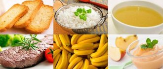
Nutrition correction is a mandatory element of complex therapy. A diet is prescribed for bile in the stomach. Dietary nutrition is aimed at normalizing the motility of the gastroduodenal organs. The main nutritional requirements for the patient are: small frequent meals, adherence to a meal regimen, exclusion from the diet of spicy and salty foods, canned food, marinades, fatty, smoked and fried foods and dishes, wholemeal bread that reduce the rate of gastric emptying. The consumption of various vegetables and legumes rich in fiber (radishes, legumes, turnips, radishes, asparagus) and fruits with rough skin (gooseberries, grapes, dates, currants), as well as coarse cereals (pearl barley, barley, millet, corn) is limited. Limit the consumption of sugar, honey and jam, chocolate. The diet should be based on low-fat boiled, easily digestible foods: light soups, porridges, chicken meat without rough crust, boiled fish and vegetables/vegetable purees, fruit/oatmeal jelly.
Patients with GHD should avoid physical activity and heavy lifting after eating, which increases intra-abdominal pressure. It is equally important to prevent flatulence / constipation . However, since GHD occurs predominantly against the background of other gastrointestinal diseases, a diet for the underlying disease may be prescribed, for example, Diet No. 5 , Diet for gastric ulcer , Diet for gastritis , Diet for gastroduodenitis , etc.
Classification of chronic duodenitis
Classification of chronic duodenitis:
1) chronic duodenitis , mainly bulbitis , of acidopeptic origin;
2) chronic duodenitis, combined with atrophic gastritis or enteritis;
3) chronic duodenitis that developed against the background of duodenostasis;
4) local duodenitis (papillitis, peripapillary diverticulitis).
According to the endoscopic picture there are:
1) superficial chronic duodenitis;
2) atrophic chronic duodenitis;
3) interstitial chronic duodenitis;
4) erosive-ulcerative chronic duodenitis.
Prevention
Prevention of GHD consists of:
- Timely treatment of gastrointestinal diseases (especially gastroduodenal pathology).
- Correction of body weight.
- Maintaining the correct diet to ensure physiologically normal gastrointestinal motility.
- Quitting smoking and alcohol abuse.
- Normalization of physical activity and psycho-emotional sphere. Elimination of factors that provoke reflux (lifting weights, exercises with strong abdominal tension, avoiding being in a horizontal position after eating, working in an inclined position).
- Exclusion/avoidance of long-term use of medications that worsen bile rheology and gallbladder motility (antibiotics, expectorants, sedatives, NSAIDs, estrogens, antispasmodics, β-blockers, hypnotics, calcium channel blockers).
What contributes to the development of duodenitis?
What diseases contribute to duodenitis? Giardiasis , ascariasis, chronic infection in the oral cavity, pharynx, genitals, gall bladder, and renal failure contribute to the development of duodenitis.
The inflammatory process can be sluggish, manifested by disturbances in appetite and general well-being, and mild dyspeptic symptoms. Patients experience lethargy and mental instability. Children grow up thin and develop poorly.
When infected with Giardia, the clinical picture of duodenitis can develop rapidly. Suddenly, usually after an error in diet, an attack of severe abdominal pain occurs, causing patients to squirm. The pain is not relieved by antispasmodics. The patient's face becomes hyperemic and becomes covered with drops of sweat.
List of sources
- Zvyagintseva T.D., Chernobay A.I. Functional disorders of the motor-evacuation function of the digestive tract and their pathogenetic correction // News of medicine and pharmacy. – 2009.– No. 294. pp. 7–11.
- Zvyagintseva T.D., Chernobay A.I. Duodenogastric reflux in the practice of a gastroenterologist: obvious dangers and hidden threats // Medical newspaper “Health of Ukraine”. 2012.—No. 1. P.11.
- Trukhan D.I., Filimonov S.N. Differential diagnosis of the main gastroenterological syndromes and symptoms. M.: Practical Medicine, 2016. 168 p.
- Trukhan D.I. Rational pharmacotherapy in gastroenterology // Directory of a polyclinic doctor. 2012. No. 10. pp. 18–24.
- Bagnenko G.F., Nazarov E.V., Kabanov M.Yu. Methods of pharmacological correction of motor-evacuation disorders of the stomach and duodenum. // Rus. honey. magazine. Diseases of the digestive system. –2004. – T. 1. – P. 19-23.
Operation
Surgical treatment is used when conservative methods of treatment do not provide the desired result or are ineffective due to the characteristics of the disease. So, when the pylorus gapes, plastic surgery is used, the purpose of which is to plastically reduce it.
Using laparoscopic equipment, the anterior part of the pylorus is placed deep into the duodenal bulb, thus forming a functionally active prepyloric pocket. This pocket takes over the contractile and peristaltic functions of the damaged pylorus.
Video about the disease gastroduodenal reflux
This video will be useful for those who periodically experience abdominal pain.
After watching the video, you will learn why seemingly insignificant symptoms are dangerous: heartburn, stomach pain. Experienced gastroenterologists will talk about such a disease as duodenogastric reflux and what it is. Learn about mistakes in treating gastritis. If you are experiencing discomfort in your digestive tract, then let this video be an impetus to visit a doctor. When is gastroduodenitis reflux diagnosed? The gastrointestinal tract consists of separate sections through which food moves. In them it is digested and absorbed, and waste products are then eliminated from the body naturally. When this process is disrupted and food backflows, reflux occurs. If food from the stomach flows back into the esophagus, a diagnosis of reflux gastritis or gastroesophageal reflux disease (GERD) is made; if the contents of the duodenum flow back into the stomach, reflux gastroduodenitis occurs.

